Journal of Gynecological Research and Obstetrics
A very rare mass in the uterus: Malignant lymphoma
Fatma Devran Bıldırcın*
Cite this as
Bıldırcın FD (2019) A very rare mass in the uterus: Malignant lymphoma. J Gynecol Res Obstet 5(2): 040-041. DOI: 10.17352/jgro.000070Introduction
The female genital area is the first site of malignant lymphoma; is a very rare localization. While most genital lymphomas occur in the vagina or cervix, uterine corpus is very rare [1]. Patients usually present with bleeding or pelvic, low back pain, but very rarely tumors are discovered through a routine examination. In the present case; When the radiological examination was performed for avascular femoral necrosis, it was thought that myoma was the incidental mass. However, the patient had no preoperative diagnosis and the pathology was high grade B-cell lymphoma located in the uterus. We were unable to make a preoperative diagnosis and we thought that it could be lyomyosarcoma. Here we report a case of primary lymphoma of the uterus, which is very rare among postmenopausal masses.
Case
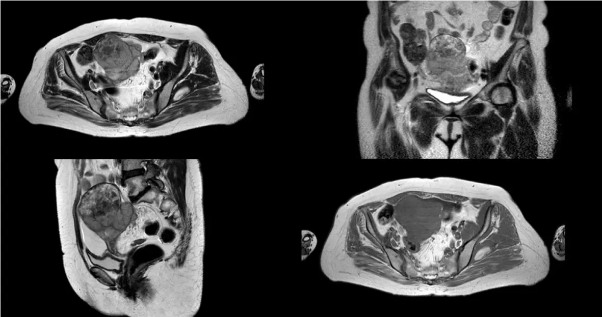
The 60-year-old patient had menopause for 12 years and had two deliveries. When he presented to the orthopedics department for leg pain, radiological examinations were performed for avascular necrosis of the femur. She was admitted to our clinic with a preliminary diagnosis of a mass which was thought to be fibroids. The patient did not complain of any bleeding or pelvic pain. In pelvic examination, the uterus was palpated with immobile and adnexal fixation for 16 weeks. When MRI is performed in our clinic; 87 * 86 * 75 mm hyperintense multilobulated mass was seen in uterus fundus (Figure 1).
Leiomyosarcoma was considered as the preliminary diagnosis. The tumor was operated. It was infiltrated into the cervix and colon wall. The fragile 9 cm mass arising from the uterus was extending into the cervix and adnexa. Hysterectomy, bilateral salpingoopherectomy, intestinal anterior resection, ileostomy, appendectomy and omentectomy were performed due to mass invasion. The pathology examination revealed high grade B cell lymphoma and double expressor immune phenotype (CMYC + BCL-6). The patient was discharged postoperatively and chemotherapy was started by hematology oncology department (Figure 2).
Discussion
Primary malignant lymphomas of the uterus are the rarest of rare non-epithelial tumors. The ratio between primary genital tumors is 0.008% [2]. It is usually well kept in the vagina and cervix. Patients will often consult a doctor with abnormal vaginal bleeding. However, the patient may present with different symptoms such as abdominal pain and urinary obstruction due to compression symptoms.
Endometrial biopsies may be helpful in the diagnosis as patients with primary uterine lymphomas usually present with bleeding complaints [3]. However, our patient had no history of bleeding. Therefore, no biopsy was performed.
A prospective diagnosis of malignant lymphoma of the uterus is almost impossible. In Magnetic Resonance Imagine (MRI), widespread enlargement of the uterus without endometrial or cervical epithelium deterioration is thought to be a specific finding in the diagnosis of malignant lymphoma of the uterus [4]. When MRI images were examined, it was seen that the common features of uterine lymphomas in MRI were large tumors with homogenous signal intensity and multinodular growth [5]. MRI images of our case had similar features.
Conclusion
Uterine lymphomas are very rare and occur with atypical symptoms. Diagnosis is sometimes quite difficult and often mimics the malignancies of the uterus.Accurate histological diagnosis is essential for adequate staging and a correct approach to treatment. As in our case, it would be appropriate not to ignore lymphomas in the differential diagnosis.
- Agrawal A, Ofili G, Allan TL, Mann BS (2000) Malignant lymphoma of uterus: A case report witha review of the literature. ANZJOG 40: 358-360. Link: http://bit.ly/2YYl9RK
- Ekanayake CD, Punchihewa R, Wijesinghe PS (2018) An atypical presentation of an ovarian lymphoma: A case Report. J Med Case Rep12: 338. Link: http://bit.ly/2Z032uH
- Binesh F, Karimi zarchi M, Vahedian H, Rajabzadeh Y (2012) Primary malignant lymphoma of the uterine cervix. BMJ Case Rep 24: 2012. pii: bcr2012006675. Link: http://bit.ly/31E1i8d
- Goto N, Oishi-Tanaka Y, Tsunoda H, Yoshikawa H, Minami M (2007) Magnetic resonance findings of primary uterine malignant lymphoma. Magn Reson Med Sci 6: 7-13 Link: http://bit.ly/2MZaoIu
- Aysen Telce Boza, Evrim Bostanci, Nermin KOC, Semih Tugrul, Selcuk Ayas (2014) The dıagnostıc challange of an uterıne mass: uterıne lymphoma. J Turk Soc Obstet Gynecol 11: 59-63. Link: http://bit.ly/2YHnLUF
Article Alerts
Subscribe to our articles alerts and stay tuned.
 This work is licensed under a Creative Commons Attribution 4.0 International License.


 Save to Mendeley
