Journal of Gynecological Research and Obstetrics
The Importance of Initial Evaluation by Trans Vaginal Sonography in Rudimentary Horn Pregnancy: A Case Report and Literature Review
Asgari Zahra1, Razavi Maryam1, Hoseini Rayhaneh1, Rezaeinejad Mahroo1, Rooholamin Safora1, Javaheri Atieh1 and Ahmadi Farahnazsadat2*
2Department of Gynecologic Obstetric, Yes Hospital, Tehran University of Medical Sciences, Tehran, Iran
Cite this as
Zahra A, Maryam R, Rayhaneh H, Mahroo R, Safora R, et al. (2016) The Importance of Initial Evaluation by Trans Vaginal Sonography in Rudimentary Horn Pregnancy: A Case Report and Literature Review. J Gynecol Res Obstet 2(1): 075-077. DOI: 10.17352/jgro.000025Rudimentary horn pregnancy (RHP) as a rare incidence has been estimated at 1:76,000-1:150,000 pregnancies. It has been also reported that 80-90% of RHP lead to uterine rupture in second trimester. Early diagnosis with the use of 3-dimentional ultrasonography (3-DUS) that is followed by laparoscopic resection of RH and ipsilateral fallopian tube is likely to be considered as the best management strategy that prevents maternal morbidity and mortality. We present a case of 9-week pregnancy in a non-communicating rudimentary horn with positive fetal heart rate (FHR) that was diagnosed by 3-DUS and successfully treated with laparoscopic resection.
Introduction
According to American Society for Reproductive Medicine (ASRM) classification for uterine malformation, unicornuate uterus is described as class II anomaly, which is also divided into 4 following subgroups: (II-a) Unicornuate uterus with communicating rudimentary horn, (II-b) Unicornuate uterus with non-communicating rudimentary horn, (II-c) Unicornuate uterus with noncavitary rudimentary horn, and (II-d) Unicornuate uterus without rudimentary horn. Prevalence rates of Müllerian duct anomalies (MDAs) and unicournuate uterus are estimated as 4.3% and 0.4% of the general population, respectively [1]. Rudimentary horns derived from developmental arrest of one or two Müllerian ducts are accompanied with unicornuate uterus in 74% of cases. Complications of rudimentary horn are reported as follows: (i) hematometra associated with dysmenorrhea and endometriosis, (ii) occurrence of pregnancy and (iii) endometriosis [2]. In 75% of rudimentary horn cases, there is no communication between uterine cavities. Occurrence of rudimentary horn pregnancy (RHP) is likely to be described by peritoneal transmigration of ovum and sperm that has been estimated at 1:76,000-1:150,000 pregnancies [3]. RHP leads to rupture of the pregnant horn in 80-90% of cases in second trimester, indicating that 10% of cases can only reach to term. In a study by Sujata et al., neonatal survival rate was reported 0-13% [4]. In another study by Jayasinghe et al., the sensitivity of ultrasonography for diagnosis of RHP was reported 26%. They also mentioned that only 14% of RHP are diagnosed before occurrence of any related clinical symptoms [5]. The most common differential diagnoses of RHP when using ultrasonography are as follows: tubal pregnancy, cornual pregnancy, abdominal pregnancy and bicornuate uterus pregnancy [6].
Tsafrir et al., have described the following diagnostic criteria for RHP using ultrasonography: pseudo pattern of asymmetrical bicornuate uterus, non-continuity between tissue surrounding the gestational sac and the uterine cervical canal, as well as the presence of myometrial tissue surrounding the gestational sac [7]. Uterine rupture is the most common complication of RHP [5], that leads to life-threatening bleeding, shock, hemoperitoneum, as well as maternal mortality and morbidity. It is noted that early diagnosis before uterine rupture is considered as one of the main steps toward the treatment. Furthermore, laparoscopic management for excision of rudimentary uterine horn and ipsilateral salpingectomy is an easy and feasible approach for treatment of RHP as compared to laparotomy. We used a management strategy including 3-dimentional ultrasonography (3-DUS) and laparoscopic resection of RH and ipsilateral fallopian tube for a case of 9-week pregnancy in a non-communicating rudimentary horn with positive fetal heart rate (FHR).
Case Report
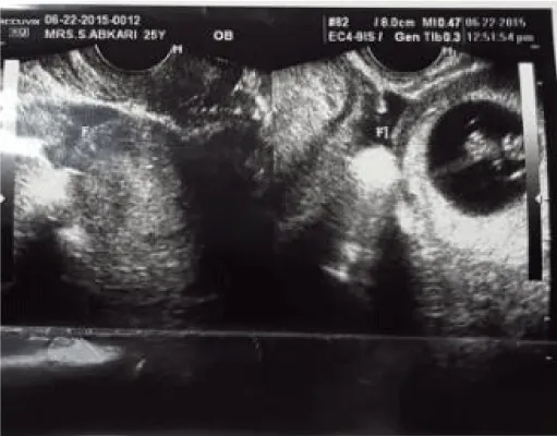
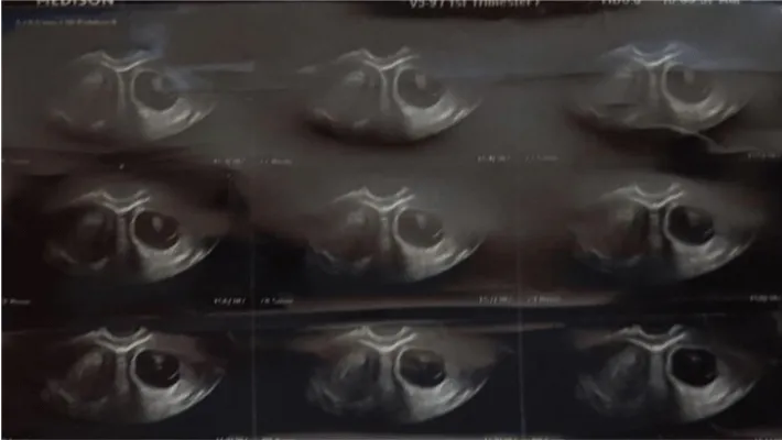
A 25-year-old primigravida woman at 9 weeks of gestation underwent transvaginal ultrasound (TVS) as routine prenatal care at Arash Women’s Hospital affiliated to Tehran University of Medical Sciences, Tehran, Iran, in June 2015. The result of TVS showed a right-sided normal uterine with size of 80 x 47 x 43 cm, normal myometrium, endometrial thickness of 12mm and no gestational sac, while there was a left-sided asymmetrical rudimentary horn containing a gestational sac (GS) that enclosed a 20-mm fetus with crown-rump-length corresponding to 9 weeks of gestational age with normal FHR; however, non-continuity between gestational sac and the uterine cervical canal was observed, indicating that there was possibility of either unicornuate uterus with a rudimentary horn or a bicornuate uterus with an intact pregnancy (Figure 1). Therefore, in order to confirm the primary findings, she underwent 3-dimentional ultrasonography (3-DUS) at the same day. The secondary findings confirmed left RHP with a 9-week live fetus. The differential diagnosis of 3-DUS included a bicornuate uterus with an intact pregnancy (Figure 2).
She had no pain, vaginal bleeding or any other complains. Her abdominal examination was normal. Speculum exam showed one cervix. A bimanual examination revealed a mass on left side of uterus. A blood test showed that her human chorionic gonadotropin (hCG) level was 158 869 IU/L. Subsequently, she underwent elective laparoscopic treatment. The findings revealed that there was a unicornuate uterus with a healthy fallopian tube on right side. On the left side, there was a 6-cm rudimentary horn, connected to the uterus with a fibrotic band, while left fallopian tube attached to the rudimentary horn .Left fallopian tube and ovary were normal. At the same time, we decided to do hysteroscopy. Therefore, the peritoneal cavity gas was emptied and her position was corrected. After performing hysteroscopy, only the right tubal ostium was visible, and RHP was terminated. Then, patient was placed in the Trendelenburg position for performing left non-communicating rudimentary horn resection with left salpingectomy (Figure 3).
Discussion
RHP as a rare condition is accompanied with increased maternal complication and poor obstetrical prognosis. There are increased risks for abortion, malpresentation, intrauterine growth restriction (IUGR), intrauterine fetal death and placenta accreta with such condition [2]. Early diagnosis before uterine rupture can prevent maternal complication. In a systematic review study by Sujata et al., they have suggested ultrasound as the most common technique for the early diagnosis of RHP in order to prevent uterine rupture. They have also indicated that non-continuity of cervical canal with uterine horn containing gestational sac is likely to be detected by ultrasound in these patients; however, vast experience and skills of radiologist is a substantial factor in this regard. In the same study, sensitivity of ultrasound for detection of RHP was reported as 29% to 33% [4]. 3-DUS is also a non-invasive and high accurate procedure that can illustrate uterine anatomy with its details.
In a study by Erika et al., they have reported that transvaginal 3-DUS appears to be extremely accurate for the diagnosis and classification of congenital uterine anomalies as compared with office hysteroscopy and magnetic resonance imaging (MRI) [8]. In a review study regarding the role of 3-DUS in the diagnosis of MDAs by Deutch et al., they have pointed that 3-DUS showed sensitivity of 98%-100% and specificity of 100% in the diagnosis of MDAs [9]. In our case, TVS indicated non-continuity of cervical canal with GS that led to the pseudo pattern of a bicornuate uterus, as mentioned by Tsafrir et al., whereas RHP was confirmed by 3-DUS that was followed by laparoscopic resection of RH and ipsilateral fallopian tube. The first report of laparoscopic resection of RHP was published by Yahata et al. (1998), while it has increased since then [10]. It is noteworthy to mention that laparoscopy results in shorter hospital stay, lower risk of infection, less bleeding, less adhesion formation and less peritoneal damage as compared to laparotomy; therefore, laparoscopic removal of horn and ipsilateral salpingectomy is considered as the best management treatment of RHP. In our case, 3-DUS was carried out after application of TVS in a patient with suspected RHP in order to confirm diagnosis. In conclusion, our findings showed that TVS accompanied with 3DUS is considered as the best combined diagnosis technique for the patients with suspected MDAs, especially RHP, during the first trimester. Furthermore, the best treatment management is termination of pregnancy via laparoscopic resection of RHP. However, further study is required to determine effective criteria of TVS for normal GC.
- Buntugu K, Ntumy M, Ameh E, Obed S (2008) Rudimentary horn pregnancy: Pre-rupture diagnosis and management. Ghana Med J 42: 92-94 .
- Kadan Y, Romano S (2008) Rudimentary horn pregnancy diagnosed by ultrasound and treated by laoaroscopy. A case report and review of the literature. J of Minim Invas Gynecol 15: 527-530 .
- Nahum GG, Stanislaw H, McMahon C (2004) Preventing ectopic pregnancies: how often does transperitoneal transmigration of sperm occur in effecting human pregnancy? BJOG 111: 706-714.
- Sujata S, Reeti M, Dilpreet KP, Anju H (2013) Rudimentary horn pregnancy: a 10-year experience and review of literature. Arch Gynecol Obestet 287: 687-695 .
- Jayasinghe Y, Rane A, Stalewski H, Grover S (2005) The presentation and early diagnosis of the rudimentary uterinhorn. Obstet Gynecol 105: 1456-1467 .
- Tonguc A, Ergun B, M Baki S, Nese Y (2009) Rudimentary uterin horn pregnancy: a mastery diagnosis. FertilSteril 92: 2037. e1-3 .
- Tsafrir A, Rojansky N, Sela HY, Gomori JM, Nadjari M (2005) Rudimentary horn pregnancy: first trimester prerupture sonographic diagnosis and confirmation by magnetic resonance imaging. J Ultrasound Med 24: 219-223 .
- Faivre E, Fernandez H, Deffieux X, Gervaise A, Frydman R, et al. (2012) Accuracy of three-dimensional ultrasonography in differential diagnosis of septate and bicornuate uterus compared with office hysteroscopy and pelvic magnetic resonance imaging. J Minim Invasive Gynecol 19: 101-106 .
- Deutch TD, Abuhamad AZ (2008) The role of 3-dimensional ultrasonography and magnetic resonance imaging in the diagnosis of mullerian duct anomalies: a review of literature. J Ultrasound Med 27: 413-423 .
- Yahata T, Kurabayashi T, Ueda H, Kodama S, Chihara T, et al. (1998) Laparoscopic management of rudimentary horn pregnancy. A case report. J Reprod Med 43: 223-336 .
Article Alerts
Subscribe to our articles alerts and stay tuned.
 This work is licensed under a Creative Commons Attribution 4.0 International License.


 Save to Mendeley
