Journal of Gynecological Research and Obstetrics
The role of the three-dimensional ultrasound in uterine evaluation in patients with reproductive failure: A comparative study
Abdulrahman Yehia Elkashef*, Hossam El-Din Shawki, Ahmed Sameer Sanad and Ahmad Ezz El-Din Mahran
Cite this as
Elkashef AY, El-Din Shawki H, Sanad AS, El-Din Mahran AE (2020) The role of the three-dimensional ultrasound in uterine evaluation in patients with reproductive failure: A comparative study. J Gynecol Res Obstet 6(2): 054-059. DOI: 10.17352/jgro.000087Background: Congenital Mullerian duct anomaly is a common cause of infertility, preterm labor, first trimester abortion. Its estimated percentage varies between 0.1% - 3% in the general population and between 3% to 38% in patients with recurrent spontaneous pregnancy loss or with infertility. The use of 3-D ultrasound with image reconstruction in the diagnosis of congenital uterine anomalies has already been described [1].
Objective: To test the validity of 3D ultrasound in the diagnosis of different anatomical and structural lesions of the uterus in patients presented with reproduction failure, taking the combined diagnostic laparoscopy & diagnostic hysteroscopy as a gold standard method for diagnosis.
Material and methods: Patients who came to our hospital with uterine structural anomalies, suspected by 2D ultrasound or Hysterosalpingogram (HSG), were subjected to 3D ultrasound before undergoing combined laparoscopy and hysteroscopy.
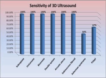
Results: This study included 42 patients selected from 463 cases that presented to Minia University Hospital, Egypt. Sensitivity of 3D Ultrasound was 100% for Double system, submucous fibroid, Subseptae uterus, Unicornuate uterus, Bicornuate uterus, Arcuate uterus. As regards Asherman’s syndrome it was 50% sensitivity , As regards polypi it was 67%
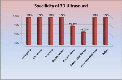
While the specificity of 3D Ultrasound was: 100% in Double system, Subseptae uterus, Unicornuate uterus, Bicornuate uterus, Asherman’s syndrome, polypi . As regards Arcuate uterus it was 95.2%, As regards Submucous fibroid it was 92%. The age of patients ranged from 21-35 years (mean age 28.4±4.3), 52.4% of the patients had primary infertility while 47.6% had secondary infertility.
Conclusion: Three Dimensional Ultrasound is a reliable diagnostic tool in cases of reproductive failure; it is superior to HSG in the diagnosis of all intracavitary lesions or anomalies, but not yet superior to laparoscopy & hysteroscopy.
Introduction
Approximately more than 85% of healthy young couples conceive within one year. Hence, infertility affects approximately 10-15% of couples and it is a crucial part of clinical practice for many clinicians. The female factors contribute most (i.e. 40-55%) in the etiologies of infertility followed by male factors (30-40%), both partners (10%) and unexplained (10%) [2].
Primary reproductive failure: is defined by the World Health Organization (WHO) as “a disease of the reproductive system defined by the failure to achieve a clinical pregnancy after 12 months or more of regular unprotected sexual intercourse”[3].
Congenital Mullerian duct anomaly is a common cause of infertility, first trimester abortion, and fetal malpresentations [2]. Its estimated prevalence varies between 0.1% - 3% [4], in the general population and between 3% to 38% in patients with repeated spontaneous miscarriages or with infertility [5]. This discrepancy results from the heterogeneous population samples, the clinical diversity of Mullerian anomalies and the different diagnostic techniques used [5].
Several techniques are available for the evaluation of Mullerian duct anomaly, among which the traditional hysterosalpingography is the most world-widely used, with its known restrictions resulting from its inability to detect the external surface of the uterus. Thus, an invasive method which combines hysteroscopy and laparoscopy has been suggested as the gold standard for establishing a final diagnosis [4].
Recently, noninvasive tools such as three-dimensional (3-D) ultrasound and Magnetic Resonance Imaging (MRI) have been added to our diagnostic tools, with their ability to demonstrate both the uterine cavity and external contours, and consequently to improve the detection and differentiation Mullerian anomalies such as septate and bicornuate uteri.
The present study aims at testing the validity of 3D ultrasound in the diagnosis of different anatomical and structural lesions of the uterus in patients presented with reproduction failure through comparing the results of the three dimensional ultrasound with combined diagnostic laparoscopy and hysteroscopy.
Patient and methods
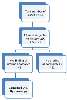
This prospective study was done in the Department of Obstetrics and Gynecology at Minia Maternity University Hospital, Egypt. From Nov.2013 to March 2015. A total number of 463 patients presented with reproductive failure (primary or secondary) were subjected to thorough history taking (name, age, detailed menstrual history, regularity of marital life, obstetric history of previous preterm labour or recurrent miscarriages), clinical examination, HSG, 2D ultrasound, 3D ultrasound scanning of the uterus, combined diagnostic hysteroscopy and diagnostic laparoscopy (STORZ 1.2 06-2015). Trans-vaginal ultrasonography was done using Voluson S8 (RIC5-9 RS 5 to 10 MHZ Trans-vaginal probe) All scans were done at post menstrual period. Regarding the hysteroscopy, All patients underwent an office hysteroscopy (Verscope of Johnson and Johnson) angle of vision 0 and diameter 2.7, without dilatation of the cervix while they were under general anaesthesia 42 patients from this group were suspected to have uterine structural anomalies as evident by different imaging techniques. Thus, this group of 42 patients had performed combined diagnostic laparoscopy& hysteroscopy to confirm the diagnosis. Prior to the procedures all benefits, risks and possible complications of the procedure were clearly explained to the patients, and informed consents were taken from all patients. This study was approved by the local ethical committee of research department of obstetrics & gynecology, Minia University. Patients with reproductive failure primary infertility, secondary infertility, recurrent miscarriage & recurrent pregnancy loss (PTL) were recruited regarding the following criteria: Age 18-35y, Normal semen analysis, Normal ovulatory function, Suspected abnormality by 2 dimensional ultrasound (suspected subeptate uterus, bicornuate uterus, submucous fibroid, endometrial polyp), HSG: showing either anatomical or structural abnormality. Whereas, patients with age <18 or > 35y., abnormal semen analysis, abnormal ovulatory function, were excluded. The classification used for describing the anomalies was the American Fertility Society classification.
The data were coded, entered and processed on an IBM-PC compatible computer using software called Statistical Package for Social Science (SPSS) for windows version 13. Quantitative data were expressed as range and mean± SD; qualitative data were expressed as number and percent. The level P <0.05 was considered the cut-off value for significance.
Results
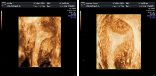
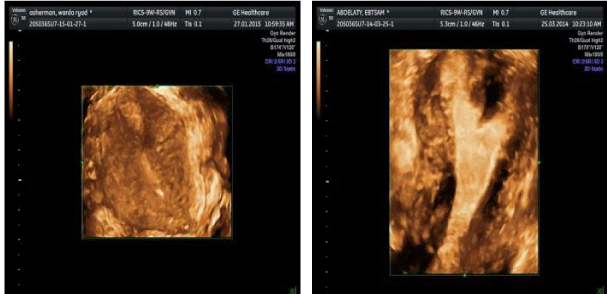
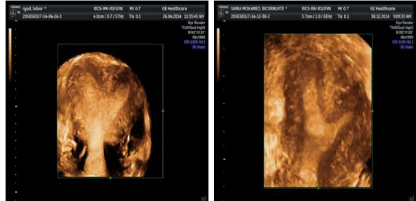
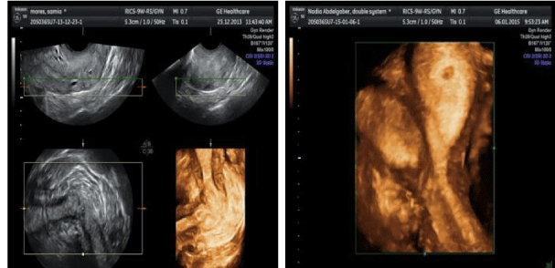
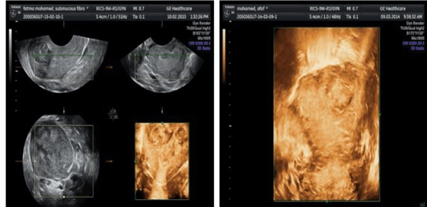
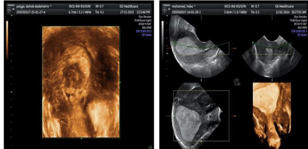
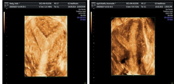
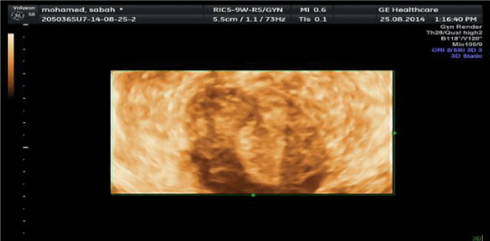
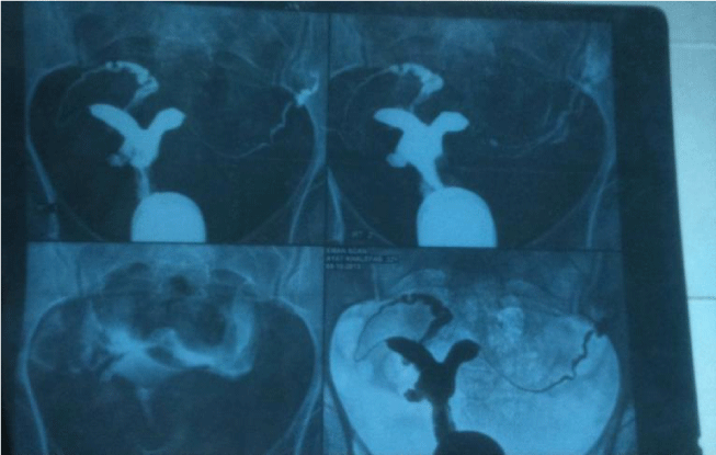
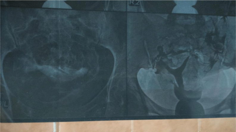
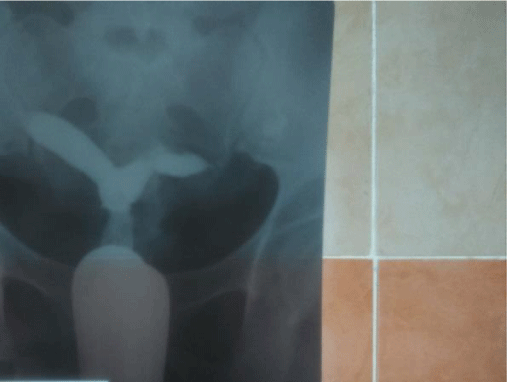
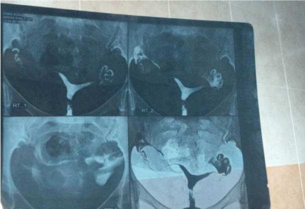
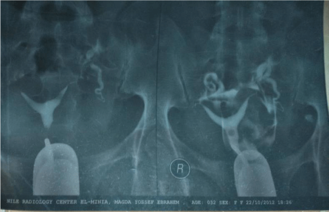
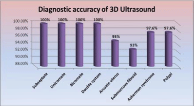
This study included 42 patients selected from 463 case presented to our outpatient clinic or admitted to our hospital for an infertility workup. The age of patients ranged between 21-35 years (mean age 28.4±4.3), 52.4% of the patients had primary infertility while 47.6% had secondary infertility. 3D ultrasound showed high sensitivity and specificity regarding the diagnosis of anatomical uterine abnormalities in comparison to combined laparoscope and hysteroscope Tables 1,2 Figures 1-13.
Discussion
Uterine anomalies are associated with both normal and adverse reproductive outcomes; they occur in approximately 3–4% of fertile and infertile women, 5–10% of women with recurrent early pregnancy loss, and up to 25% of women with late first or second-trimester pregnancy loss or preterm delivery [6], Overall, uterine anomalies are associated with difficulty maintaining a pregnancy, and not an impaired ability to conceive [7], The proper management of infertile women with uterine anomalies is controversial.
Primary infertility, secondary infertility and recurrent pregnancy loss due to uterine structural abnormalities are prevalent in Egypt. We aimed at testing the ability of the 3D ultrasound in comparison to combined diagnostic laparoscopy and hysteroscopy, in assessment of cases with reproductive failure secondary to uterine abnormities. This study was conducted on cases presented to Minia University Hospital during the period of study. In this study 42 infertile cases were recruited, 52.4% of them had primary infertility while 47.6% had secondary infertility. They were subjected to HSG, 2D ultrasound, 3D ultrasound, diagnostic laparoscopy& hysteroscopy.
In current study Sensitivity, Specificity and diagnostic accuracy of 3D ultrasound was as follow
Another Egyptian study performed at Obstetrics and Gynaecology Department, Tanta University, Egypt 2014 on 36 patients, named (Septate or bicornuate uterus: Accuracy of three-dimensional trans-vaginal ultrasonography and pelvic magnetic resonance imaging) stated that; HSG showed sensitivity of 77.4%, specificity of 60% and overall accuracy of 75% in the differentiation between the septate and bicornuate uterus. MRI showed sensitivity of 93.5%, specificity of 80%, PPV of 96.6% and negative predicative value of 66.6%, with overall accuracy of 91.6%. The 3D ultrasound showed the highest diagnostic parameters, with sensitivity of 96.7%, specificity of 100%, PPV of 100% and negative predicative value of 83.3%, with overall accuracy of 97.2% [8] Figures 13-16.
In Khaled, et al. study sensitivity & specificity of HSG were lower in comparison to the current study, MRI was used as a gold standard of diagnosis, but in our study combined diagnostic laparoscopy & diagnostic hysteroscopy was used.
Another study by Ashraf Moini, et al. on 2012 was conducted on 214 women with fertility problems who were suspected to have Müllerian anomalies at Arash Hospital from April 2010 to April 2011. All patients underwent 3D sonography to assess the uterine abnormalities during the luteal phase of the spontaneous cycle. Sonography was performed by one radiologist, after which one surgeon performed a hysteroscopy and a laparoscopy in each of the patients. The 3D sonographic results were concealed from the surgeon. Finally, the results were compared to determine the sensitivity, specificity, and accuracy of 3D sonography.
They concluded that final evaluations of 204 patients showed uterine anomalies in 84.3% of the patients. For diagnosis of uterine anomalies, the sensitivity of 3D sonography was 86.6%, and specificity was 96.9%. The positive predictive value was 99.3%, and the negative predictive value was 54.4%, with an 88.2% accuracy rate. For classification, the positive predictive value of 3D sonography was 82.3%, and accuracy was 76%; without short septa and arcuate uteri, accuracy was 95% [9].
Current study and Ashraf’s study almost agreed despite of larger number of patients recruited by Ashraf, et al.
According to Momtaz and Ebrashy 2007 Department of Obstetrics and Gynecology, Kasr El Aini Medical School, Cairo University, they examined 123 cases by transvaginal 2D,HSG, 3D TVS and hysteroscopy and laparoscopy. Twenty-one cases had normal uterine cavity and no uterine pathology where 102 patients had a uterine cavity abnormality or pathology on the final diagnosis. Their results were: congenital anomalies (N = 38), 2D TVS (21/38), HSG(36/38), 2D+HSG (36/38), 3D TVS (37/38). Sensitivity was 0.55, 0.95, 0.95, 0.97 respectively. Specificity was 0.95, 0.78, 0.94, 0.96 respectively. Positive predictive value was 0.84, 0.65, 0.90, 0.92 respectively. Negative predictive value 0.83, 0.97, 0.95, 0.99 respectively. Positive Likelihood Ratio 11.7, 4.24, 14.7, 27.6 respectively. Negative Likelihood Ratio 0.47, 0.07, 0.08, 0.03 respectively [10].
The Sensitivity of 3D ultrasound in current study (100%) was so close to Momtaz & Ebrashy (97%). Sensitivity of HSG in Momtaz & Ebrashy was 94.7%, in current study sensitivity of HSG was variable (from zero to 100%) according to type of anomaly (Momtaz didn’t categorize types of anomalies).Specificity of 3D ultrasound was 96% in Momtaz, but ranged from 92 to 100% in our study according to type of Mullerian duct anomaly.
Specificity of HSG was 78% in Momtaz study, and ranged from 86.8 to 100% in our study according to type of anomaly. 2D TVS. HSG and 3D TVS in cases of Group II (Myomas and Polyps). Myomas and Polyps (N = 52), 2D TVS (41/52), HSG (34/52), 2D+HSG (44/52), 3D TVS (50/52). Sensitivity 0.79, 0.65, 0.85, 0.96 respectively. Specificity 0.96, 0.91, 0.96, 0.97 respectively. Positive predictive value 0.95, 0.85, 0.94, 0.96 respectively. Negative predictive value 0.81, 0.78, 0.89, 0.97 respectively. Positive Likelihood Ratio 19.7, 7.7, 20.0, 34.1 respectively. Negative Likelihood Ratio 0.22, 0.38, 0.16, 0.04 respectively [10].
In current study sensitivity of HSG was 100% to myomas & 0% to polyps. While 3D ultrasound sensitivity was 100% to myomas & 66.7% to polyps. Regarding specificity, in Momtaz specificity of HSG was 91% & specificity of 3D ultrasound was 97%, while in current study specificity of HSG was 86.8% to myomas & 100% to polyps [10].
In Momtaz study he included 12 cases of intrauterine adhesions, Sensitivity of HSG was 58%, sensitivity of 3D ultrasound was 67%, Specificity of HSG was 95%, and specificity of 3D ultrasound was 94%. In current study we had 2 cases with Asherman’s syndrome or intrauterine adhesions; Sensitivity of HSG was 50%, for 3D ultrasound was 50%. Specificity of HSG was 100%, for 3D ultrasound was 100%.
The result of present study was hand in hand with results of similar studies that ensure the validity of 3D ultrasound examination as a simple convenient non-invasive outpatient procedure for examination of uterine anomalies in patients with reproductive failure.
Strengths and limitations
Uterine structural anomalies are a quite common etiology for uterine factor of infertility, and usually found during invasive procedures as laparoscopy and hysteroscopy. Thus, providing a non-invasive technique which has a comparable sensitivity, specificity and accuracy to invasive maneuvers would certainly reduce the complications resulting from the anesthesia and surgery. Moreover, 3D doesn’t indicate hospital admission and it is widely accepted as a diagnostic technique. Limitations could be that 3D ultrasound is only diagnostic method, whereas laparoscopy and hysteroscopy are both diagnostic and therapeutic. In addition, three-dimensional ultrasound is not available in all hospitals and it may be done with a relatively high cost.
Conclusion
Three Dimensional Ultrasound is a reliable diagnostic tool in cases of reproductive failure; it is superior to Hysterosalpingogram (HSG) in diagnosis of all intracavitary lesions or anomalies, but not yet superior to laparoscopy & hysteroscopy. Three Dimensional Ultrasound is superior to Hysterosalpingogram (HSG) in diagnosis of all intracavitary lesions or anomalies because of being Non-invasive, high sensitivity & specificity in comparison with HSG, Three Dimensional Ultrasound delivers a better image of the lesion with its outlines making the diagnosis of submucous fibroid, endometrial polypi, or intrauterine adhesions more accurate, also it can easily differentiate between bicornuate uterus & subseptate uterus. Three Dimensional Ultrasound is highly accurate when compared with combined diagnostic laparoscopy & hysteroscopy, but low rate of fallacies are still present.
Recommendations
Any case of primary or secondary infertility is recommended to do Three Dimensional Ultrasound, especially when Mullerian duct anomaly or intrauterine lesion (fibroid, polyp or adhesions) are suspected by HSG or 2D ultrasound.
- Graupera B, Pascual MA, Hereter L, Browne JL, Úbeda B, et al. (2015) Accuracy of three‐dimensional ultrasound compared with magnetic resonance imaging in diagnosis of Müllerian duct anomalies using ESHRE–ESGE consensus on the classification of congenital anomalies of the female genital tract. Ultrasound Obstetrics & Gynecol 46: 616-622. Link: https://bit.ly/3bd5FNC
- Kantharia N (2018) Evaluation of Infertile Couple Having Normal Male Factor (Doctoral dissertation, Sumandeep Vidyapeeth). Link: https://bit.ly/3b8pyVW
- Zegers-Hochschild F, Adamson GD, de Mouzon J, Ishihara O, Mansour R, et al. (2009) International Committee for Monitoring Assisted Reproductive Technology (ICMART) and the World Health Organization (WHO) revised glossary of ART terminology. Fertil Steril 92:1520–1524. Link: https://bit.ly/32Gzl1G
- Bhagavath B, Ellie G, Griffiths KM, Winter T, Alur-Gupta S, et al. (2017) Uterine malformations: an update of diagnosis, management, and outcomes. Obstet Gynecol Surv 72: 377-392. Link: https://bit.ly/2G7I70R
- van Dijk MM, Kolte AM, Limpens J, Kirk E, Quenby S, et al. (2020) Recurrent pregnancy loss: diagnostic workup after two or three pregnancy losses? A systematic review of the literature and meta-analysis. Hum Reprod Update 26: 356-367. Link: https://bit.ly/2YOS3TL
- Bala B, Ellie G, Kara MG, Tom W, Snigdha AG, et al. (2017) Uterine malformations: an update of diagnosis, management, and outcomes. Obstetrical & Gynecological Survey 72: 377-392. Link: https://bit.ly/2GgsswB
- Agenor A, Bhattacharya S (2015) Infertility and miscarriage: common pathways in manifestation and management. Women's Health 11: 527-541. Link: https://bit.ly/2YOvL4d
- Abo Dewan KAA, Hefeda MM, ElKholy DGE (2014) Septate or bicornuate uterus: accuracy of three-dimensional trans-vaginal ultrasonography and pelvic magnetic resonance imaging. Egyptian Journal of Radiology and Nuclear Medicine 45: 987-995. Link: https://bit.ly/2DhACUe
- Moini A, Sedigheh M, Reihaneh H, Bita E, Ahmadi F (2013) Accuracy of 3‐Dimensional Sonography for Diagnosis and Classification of Congenital Uterine Anomalies. Journal of Ultrasound in Medicine 32: 923-927. Link: https://bit.ly/2EEt1zy
- Momtaz MM, Marzouk AA, Ebrashy AM (2007) Three-dimensional ultrasonography in the evaluation of the uterine cavity. Middle East Fertil Soc J 12: 41-46. Link: https://bit.ly/3hK0ya2
Article Alerts
Subscribe to our articles alerts and stay tuned.
 This work is licensed under a Creative Commons Attribution 4.0 International License.

 Save to Mendeley
