Journal of Gynecological Research and Obstetrics
Evaluation of the histopathology results of patients operated due to Ovarian Mass
Pervin Karlı1* and Asuman Kilitci2
2Ahi Evran University Faculty of Medicine, Department of Pathology, 40 100, Kırşehir, Turkey
Cite this as
Karlı P, Kilitci A (2019) Evaluation of the histopathology results of patients operated due to Ovarian Mass. J Gynecol Res Obstet 5(1): 001-004. DOI: 10.17352/jgro.000060Introduction: Ovarian masses quite frequently appear before us in gynecological practice. In the present study, our purpose was to determine the types and age range of masses detected in ovarian surgeries in and around the city of Kırsehir.
Material and Methods: The study was conducted in the retrospective fashion on the pathology reports of 93 patients, who had histopathological examination in the pathology clinic due to ovarian mass between January 2015 and July 2018. The diagnoses of the patients were divided in pathological terms into 5 groups as the epithelial tumors, sex-cord stromal tumors, germ-cell tumors, other primary tumor groups, and ovarian pathologies that were not neoplastic.
Results: Epithelial tumors were determined to be 38.7%, sex-cord stromal tumors were 8.6%, germ-cell tumors were 4.3%, other primary tumor group was 1.1%, and ovarian pathologies that were not neoplastic were 47.3%. When the age groups were evaluated, it was determined that the rates were 9.6% in the 10-20 age range; 12.9% in the 21-30 age range; 35.4% in the 31-40 age range; 30.1% in the 41-50 age range; and 11.8% in the 51+ age group.
Conclusion: In surgeries carried out due to reasons that are not malign in the ovary, approximately half of these surgeries are carried out because of the temporary ovarian pathologies.
Introduction
Ovarian masses occur depending on functional, congenital, inflammatory and neoplastic processes and are common in women of all age groups [1]. Nearly 5-10% women receive surgery for adnexal masses in the United States. The rate of adnexal masses in general population vary between 0.17% and 5.9% in asymptomatic women; and between 7.1% and 12% in symptomatic women [2,3].
Materials and Methods
The present study was conducted between January 2015-July 2018 in Ahi Evran University, Education and Research Hospital for the purpose of determining the types of the masses of 93 patients who were examined and operated in the pathology unit due to ovarian mass according to their age groups. It was also aimed to determine the age group in which these masses were detected, to determine the most frequent ovarian pathological lesion, the types that led us to operate the patients, and to define the most frequent mass types that might cause regional differences. The patients were divided into 5 main groups. Those who had preoperative diagnosis of malignancy were not included in the study. The epithelial tumors, sex-cord stromal tumors, germ-cell tumors, other primary ovarian tumors and non-neoplastic ovarian pathologies constituted the study groups. Ovarian cysts (follicle cyst, corpus luteum cysts, hemorrhagic cysts), endometriotic cyst, ovarian abscess, paraovarian cyst, oophoritis, adnexal torsion, simple serous cyst and pregnancy luteoma were included in the scope of the non-neoplastic ovarian cysts. Ovarian leiomyoma was included in the “other” primary tumor group.
According to preoperative tumor characteristics by ultrasound or magnetic resonans imaging patients with echogenic areas of more than 6 cm, patients with ascites in peritoneal cavitiy, the cyst wall having more than 3 mm cysts, suspicion of malignancy in the doppler measurements and exophytic protrusions in the cyst were not operated in our clinic. Surgical procedures were planned for patients with severe symptomatic conditions or patients who need emergency surgery in our clinic. The age groups consisted of 10-20, 21-30, 31-40, 41-50 and 51 and over. Statistical analyses were carried out by using the SPSS15 for Windows Program. The definitive statistics were given as median, minimum, maximum and percentage. This study was conducted upon the approval of Local Ethics Committee of Ahi Evran University, Faculty of Medicine (with the decision number 2018-19/166).
Results
There were a total of 93 patients with no malign suspicions in the study. The epithelial tumors were 38.7%; sex-cord stromal tumors were 8.6%; germ-cell tumors were 4.3%; other primary tumor group (leiomyoma) was 1.1% and the ovarian pathologies that were not neoplastic were 47.3% (Table 1). When the age groups were evaluated, it was determined that the rates were 9.6% in the 10-20 age range; 12.9% in the 21-30 age range; 35.4% in the 31-40 age range; 30.1% in the 41-50 age range; and 11.8% in the 51+ age group (Table 2).
Discussion
Ovarian Masses (OMs) are the ones observed by clinicians in a frequent manner in both reproductive and postmenopausal and premenopausal periods [1]. It is associated with the size and age as well as symptomatic presentation origins. One of the most precious methods is ultrasonography for diagnosis. Computed Tomography (CT) and Magnetic Resonance Imaging (MRI) must also be employed in differential diagnosis.
Ovarian cancers constitute the second most common gynecological cancer type that has the highest mortality rates [4,5]. The CA-125 level may be elevated in half of the patients, particularly if the mass is malignant [6]. The predictive value of the CA-125 levels is associated with ultrasound imaging, the age of the patient, and menopausal status in the diagnosis of ovarian masses. Ultrasonography is a sensitive method; however, it is also nonspecific. The CT and MRI may help the diagnosis [7-9]. Using the Doppler Method does not give any clue about the diagnosis [10.11].
It is already known that ovarian masses exhibit various clinical and pathological characteristics. The possible causes of the pelvic masses that are detected in physical and radiological examinations vary in prepubertal, adolescence and postmenopausal periods, are mostly ovarian-originated, and are benign. Pelvic mass may originate from gynecological area or from urinary or gastrointestinal system [12,13]. Adnexal masses might have benign or malignant characteristics. Although physiological follicle cysts and corpus luteum cysts are the adnexal masses that are most commonly detected in premenopausal period, ectopic pregnancy or adnexal torsion must be absolutely considered in the differential diagnosis since they require urgent intervention [14,15]. In addition, endometrioma, polycystic ovaries, tuboovarian abscesses and benign neoplasia must be taken into consideration in reproductive age group. Malignant neoplasia increase with furthering age. Ovarian neoplasia is classified under 4 main groups as epithelial, sex cord-stromal, germ-cell tumors and other primary tumors [16]. Among the benign neoplastic group patients who were included in the present study, epithelial tumors were determined at a rate of 38.7% in 36 patients; sex cord-stromal tumors were determined at a rate of 8.6% in 8 patients; germ-cell tumors were determined at a rate of 4.3% in 4 patients; the other group, which also included those with ovarian leiomyoma, were determined at a rate of 1.1% in 1 patient, respectively.
The frequency of the subtypes of the 36 patients, who had epithelial tumors, were as follows; 22 of them were determined to be serous cystadenoma, 7 were determined to be mucinous cystadenoma, 6 were determined to be serous cystadenofibroma and 1 was determined to be borderline serous neoplasia, respectively. The diagnosis of the tumors in the sex cord-stromal group was fibroma in 3 cases, granulosa cell tumor in 2 cases, thecoma in 1 case, fibrothecoma in 1 case, and sclerosing stromal tumor in 1 case. The diagnosis of the 4 patients, who had germ-cell tumors, was mature cystic teratoma. Our case series did not have any malignant diseases as the patients, who had clinically malignant masses, were referred to a central oncology hospital. Surprisingly, no histopathologically malignant patients were detected.
It is believed that the most prevalent type is the epithelial one when the situation is considered in a classification; however, there are several studies claiming that mature cystic teratoma alone and serous cystadenoma are more frequent. A total of 25% of all ovarian tumors consist of serous cysts. A total of 70% of these are benign; 5-10% are borderline; and 20-25% are malignant.
Myoma and diverticulitis are among the conditions that must be distinguished from adnexal masses particularly in the postmenopausal period [17]. Although the definitive diagnosis is possible in a histopathological manner after surgery, the diagnosis of most of ovarian malignancies might be made preoperatively with pelvic examination, ultrasonography and tumor markers [18].
Ovarian mucinous cystadenomas, on the other hand, are the ones that is mostly detected in middle-aged women, and constitute 15% of all ovarian tumors, which tend to extend very large sizes because of the surface epithelium of the ovary. A total of 80% of mucinous tumors are benign, 10% are borderline, and 10% are malignant [19-21]. In the present study of ours, the most common ovarian masses that were not malignant constituted 47.3% of the patients included in the study with functional ovarian cysts with ovarian pathologies that were not neoplastic [23], endometriotic cyst [11], ovarian abscess [3], paraovarian cyst [1], oophoritis [1], adnexal torsion [1], simple serous cyst [1], pregnancy luteoma [1]’.
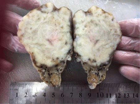
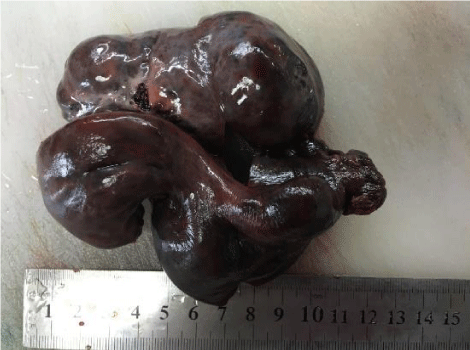
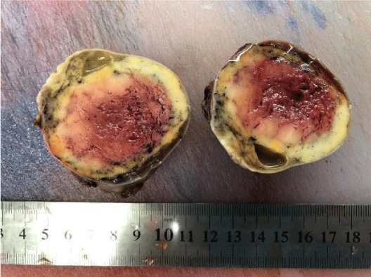
The most interesting ovarian mass was determined as a 7 cm-diameter Ovarian Leiomyoma (OL), which was detected accidentally during cesarean in the 37th week of the gestation in a 41 year old asymptomatic pregnant woman. (Figure 1). We considered it within the other primary tumor group (1.1%). OL may be considered among the rarest ovarian solid masses. Among all ovarian masses, it is detected as 0.5-1%. Approximately 70 cases were reported in the literature. A small number of cases were reported in pregnant women [22,23]. Our patient was 41 years old. The age range that was reported in the literature was the age range of 20-65 [22]. It is mostly detected in premenopausal women. It is believed that OLs mostly stem from the smooth muscles of the hilar vessels, from smooth muscle metaplasia of the stromal cells, or from hormonal stimulations [23,24]. It was determined that IVF method was applied twice to our patient; and it was predicted that the tumor developed and/or grew because of this. Our second interesting case was a 12-year-old girl with adnexal torsion. She was apply with severe abdominal pain. Unilateral adnexal site were seen completely necrotic after pathological examination. It was completely necrosis (Figure 2). The third interesting case was 56-years-old woman with granulosa cell tumor (Figure 3). She was apply for abdominal pain. In the present study of ours, the most common ovarian masses that were not malignant constituted 47.3% of the patients included in the study with functional ovarian cysts with ovarian pathologies that were not neoplastic [23], endometriotic cyst [11], ovarian abscess [3], paraovarian cyst [1], oophoritis [1], adnexal torsion [1], simple serous cyst [1], pregnancy luteoma [1].
Conclusion
In the present study, it was determined that in nearly half of the ovarian surgeries carried out for reasons that are not malignant, these surgeries were carried out because of functional and temporary ovarian lesions, followed by epithelial ovarian neoplasms, which are from neoplastic ovarian pathologies. In this study, it was concluded that in approximately half of the ovarian surgeries that were carried out because of pathology that was not malignant, there was in fact ovarian pathologies that were benign and that were not neoplastic. Based on this outcome, we believe that unless an additional indication like urgent intervention (i.e. adnexal torsion, etc.) or abscess drainage is involved, it may be more accurate to avoid surgery in such pathologies.
- Ayhan A, Durukan T, Günalp S, Gürgan T, Önderoğlu LS, et al, (2008) Temel Kadın Hastalıkları ve Doğum Bilgisi. Güneş Tıp Kitabevi. İstanbul. 987. Link: https://goo.gl/zgcNPi
- Hilger WS, Magrina JF, Magtibay PM (2006) Laparoscopic Management of Adnexal Masses. Clin Obstet Gynecol 49: 535-548. Link: https://goo.gl/LV8TUf
- Padilla LA, Radosevich DM, Milad MP (2000) Accuracy of thepelvic examination in detecting adnexal masses. Obstet Gynecol 96: 593-598. Link: https://goo.gl/8RwLJ9
- Sassone AM, Timor-Tritsch IE, Artner A, Westhoff C, Warren WB (1991) Transvaginal sonographic characterization of ovarian disease: evaluation of a new scoring system to predict ovarian malignancy. Obstetrics and gynecology 78: 70-76. Link: https://goo.gl/QmZj68
- Andolf E, Joergensen C (1988) A prospective comparison of clinical ultrasound and operative examination of the female pelvis. Journal of ultrasound in medicine 7: 617-620. Link: https://goo.gl/hZaT9u
- Gorp TV, Veldman J, Calster BV (2012) Subjective assesment by ultrasound is superior to the risk of malignancy index (RMI) or the risk of ovarian malignancy algorithm (ROMA) in discriminating benign from malignant adnexal masses. European Journal of Cancer 48: 1649-1656. Link: https://goo.gl/YpBvtN
- Rieber A, Nüssle K, Stöhr I, Grab D, Fenchel S, et al. (2001) Preoperative diagnosis of ovarian tumors with MR imaging: comparison with transvaginal sonography, positron emission tomography, and histologic findings. American Journal of Roentgenology 77: 123-129. Link: https://goo.gl/izeh5Z
- Taner CE, Başoğul Ö, Semih MU, Mizra T, Deri G (2005) Overin Müsinöz Kistadenokarsinomları ve Sassone Skorlama Sistemi. Kocatepe Tıp Dergisi 6. Link: https://goo.gl/94JqfB
- Zarcone R, Bellini P, Carfora E, Monarca M, Longo M, et al. (1997) Role of ultrasonography in the early diagnosis of ovarian cancer. European journal of gynaecological oncology 18: 418-419. Link: https://goo.gl/QmYTqv
- Teneriello MG, Park RC (1995) Early detection of ovarian cancer. CA: a cancer journal for clinicians 45: 71-87. Link: https://goo.gl/ipZ5k3
- Goff BA, Mandel L, Muntz HG, Melancon CH (2000) Ovarian carcinoma diagnosis: Cancer 89: 2068-2075. Link: https://goo.gl/mD7cqd
- Disaia, PJ, Creasman WT (2012) Clinical Gynecologic Oncology, Elsevier Saunders published, Eighth Edition. 259.
- Atasü T, Şahmay S (1996) Adneksiyal kitlelerin değerlendirilmesi. Jinekoloji, Üniversal Dil Hizmetleri ve Yayıncılık İstanbul. 37.
- Drake J (1998) Diagnosis and management of the adnexial mass. Am Fam Physician 57: 2471-2476: 2479-2480. Link: https://goo.gl/6tHQEP
- Bayer AI, Wiskind AK (1994) Adnexial torsion: can the adnexa be saved? Am J Obstet Gynecol 171: 1506-1510. Link: https://goo.gl/71Y1r9
- Mülayim B, Gürakan H, Dagli V, Mülayim S, Aydin O, et al. (2006) Akkaya H. Unaware of a giant serous cyst adenoma: a case report. Arch Gynaecol Obstet 273: 381–383. Link: https://goo.gl/dnnGJM
- Akercan F, Çırpan T, Yıldız PS, Özşener S, Karadadaş N, et al. (2005) Benign adneksiyal kitlelerde tanı ve tedavi yaklaşımları. Ege Tıp Dergisi 44: 151-154. Link: https://goo.gl/nPzbzU
- Chechia A, Kaubaa A, Bahri N, Terras K, Makhlouf T (1999) Retrospective study of 167 ovarian tumors, Tunis Med 77: 551-557. Link: https://goo.gl/RWFJAs
- Tomas D, Lenicek T, Tuckar N, Puljiz Z, Ledinsky M, et al. (2009) Kruslin B. Primary ovarian leiomyoma associated with endometriotic cyst presenting with symptoms of acute appendicitis: a case report. Diagn Pathol 4: 25. Link: https://goo.gl/edXmKy
- Vizza E, Galati GM, Corrado G, Atlante M, Infante C, et al. (2005) Voluminous mucinous cystadenoma of the ovary in a 13-year-old girl. J Ped Adoles Gynecol 18: 419–422. Link: https://goo.gl/8tuhif
- Mittal S, Gupta N, Sharma A, Dadhwal V (2008) Laparoscopic management of a large recurrent benign mucinous cystadenoma of the ovary. Arch Gynecol Obstet 277: 379–380. Link: https://goo.gl/pD1Hnn
- Taskin MI, Ozturk E, Yildirim F, Ozdemir N, Inceboz U (2014) Primary ovarian leiomyoma: A case report. International journal of surgery case reports 5: 665-668. Link: https://goo.gl/gfR2nf
- Agrawal R, Kumar M, Agrawal L, Agrawal KK (2013) A huge primary ovarian leiomyoma with degenerative changes: an unusual. J Clin Diagn Res 7: 1152–1154. Link: https://goo.gl/T6bkmQ
- Blue NR, Felix JC, Jaque J (2013) Primary ovarian leiomyoma in a premenarchal adolescent: first reported case. J Pediatr Adolesc Gynecol 27: e87-88.
Article Alerts
Subscribe to our articles alerts and stay tuned.
 This work is licensed under a Creative Commons Attribution 4.0 International License.


 Save to Mendeley
