Journal of Gynecological Research and Obstetrics
The role of Umbilical Cord thickness, Interventricular Septum thickness and HbA1c levels in the prediction of Fetal Macrosomia in patients with Gestational Diabetes Mellitus
Rehab Mohamed Abdelrahman* and Mostafa Mohamed Salama
Cite this as
Abdelrahman RM, Salama MM (2018) The role of Umbilical Cord thickness, Interventricular Septum thickness and HbA1c levels in the prediction of Fetal Macrosomia in patients with Gestational Diabetes Mellitus. J Gynecol Res Obstet 4(3): 039-043. DOI: 10.17352/jgro.000057Objective: To assess and evaluate the role of measuring umbilical cord thickness, interventricular septum thickness and HbA1c level in predictability of fetal macrosomia development in cases having gestational DM
Methods: A prospective case-control research study that recruited 80 pregnant study subjects. They were categorized into two research groups: 40 pregnant women as cases research group having gestational DM and 40 non-diabetic pregnant Study subjects as control research group. Sonographic examination was conducted in which the sonographic cross sectional area of umbilical cord. Umbilical arteries and the umbilical vein measured from a free loop of the umbilical cord, implementing the software of the sonographic machine. The cross-sectional area of Wharton’s jelly have been obtained by subtracting the cross sectional area of the vessels from that of the umbilical cord and the interventricular septum thickness was measured. HbA1c level have been assayed for diabetic cases recruited.
Results: Umbilical cord diameter was raised in study subjects with gestational diabetes more than the control group (3.03±1.26) cm. Increase in interventricular septal thickness (0.85±51) cm have been correlated and linked with fetal macrosomia in diabetic cases. HbA1c levels in patients with GDM (7.0±1.2) % showed more observed fetal macrosomia cases.
Conclusion: The research study revealed and displayed the sonographic value of measuring umbilical cord thickness, interventricular septum thickness and HbA1c as a prediction tool for fetal macrosomia in cases with gestational DM.
Introduction
Glucose intolerance newly diagnosed during pregnanacy despite any degree of severity is clinically defined as gestational DM. Maternal hyperglycemia Cause subsequently fetal hyperglycemia, that triggers pancreatic islet cells in an abnormal manner causing pathological development of fetal hyperinsulinemia . That pathophysiological triggering factor causes abnormally excessive fat tissue deposition and increases total fetal size that is proven by various researchers to be the cornerstone cause of unfavourable perinatal clinical outcomes due to fetal growth acceleration and macrosomia [1-3].
Antenatal detection and predictability of fetal macrosomia is considered one of the major challenging clinical scienarios in every day obstetric practice .Sonographic measurements such as AC,EFW are extensively researched and are popularly used among obstetricians and fetal medicine specialists .However more sensitive and clinically presenting parameters should be implemented to aid in early detection ,prediction and diagnosis of those case scienarios to hinder or prevent the full developmental pathway of fetal macrososmia with its subsequent maternal and fetal clinical issues such as post-partum hemorrhage and shoulder dystocia .HbA1c a commonly used marker to determine and reflect the glucose levels over a period of 3 months and is used as tool to reveal the risk of anomaly development in diabetic cases within the first trimester .However research efforts should in addition involve other sonographic markers requiring the expertise of fetal echocardiographic specialists such as interventricular septal thickness that is used to determine the functional and developmental prognosis of the fetal cardiac muscle among other cardiac indices and parameters [4-6].
Sonograpohic e evaluation and assement of umbilical cord is frequently restricted, To Doppler blood flow studies and counting the number of vessels. However its crucial role in fetal development and physiology denotes that its thickness and componenets should be thourly investigated as a possible sonographic tool reflecting the fetal growth development in integration to biochemical markers such as HbA1C particularly in diabetic gestations that are at high risk of developing fetuses with macrosomic issues [7-9].
Methodology
A prospective case-control research study was carried out at Ain shams University maternity hospital between April 2015 and October 2015 on 80 pregnant study subjects. Categorized into two research groups: 40 pregnant women as case research group with gestational DM and 40 non-diabetic pregnant women as Research control group after being approved by the local hospital ethical and research committee. A verbal consent have been obtained from each study subject. Inclusive research criteria involved singleton gestation, gestational age above 27 gestational weeks, intact membranes, normal umbilical morphology (two arteries and one vein) and diagnosis of gestational DM Exclusive research criteria was the presence of congenital anomalies, Multifetal gestation , maternal chronic medical diseases (e.g hypertension, renal disease, cardiac and pulmonary disease, etc.) and cases with diagnosis of oligohydramnios, preeclampsia and intrauterine growth retardation.
Sonographic examination was conducted in all study subjects at 36-37 gestational weeks with Voluson E6 equipped with a 3.5 Hz transabdominal probe. Involving the following parameters and indices fetal biometry (biparietal diameter, abdominal circumference, femur length) and estimated fetal weight calculated according to hadlock’s formula by the sonographic machine software .The sonographic cross sectional area of umbilical cord, the umbilical arteries and the umbilical vein were measured in a free loop of the umbilical cord using the software of the ultrasound device.The cross-sectional area of Wharton’s jelly was computed by subtracting the cross-sectional area of the vessels from that of the umbilical cord. The interventricular septum thickness was measured. HbA1c levels was measured for diabetic cases. Cases have been followed up till time of delivery. The neonates were weighed and fetal macrsomia was diagnosed if fetal weight is 4 kg or more.
Results
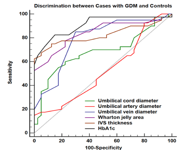
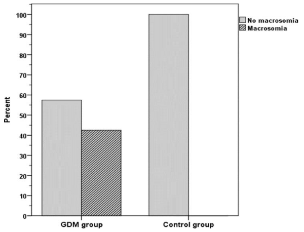
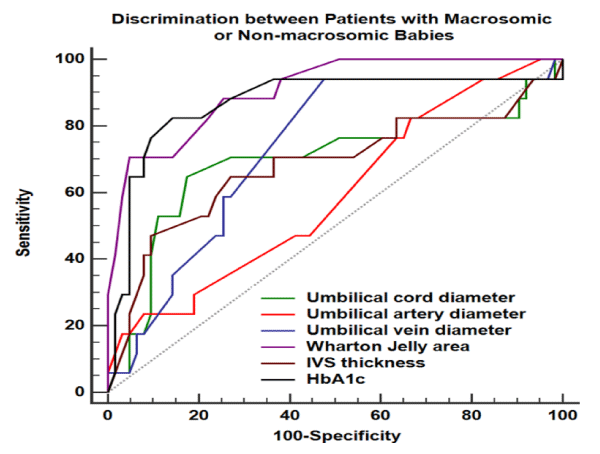
This study was conducted on 80 pregnant women with their mean age was (31±8) years, mean parity (1-3), mean previous abortions from (0-2) gestational age by LMP ranges from (27 to 39 wks). 17 among the 40 diabetic patients had macrosomic neonates (higher or equal to 4 kg) (42.5%) while there was no macrosomic neonates for the non-diabetic pregnant women (0%) (Figure 2). Large umbilical cord diameter was measured, among the diabetic group with mean (2.77±1.19) cm, among the control group (2.06±0.77) cm with specificity, positive and negative predictive values of sonographic large umbilical cord in the prediction of birth weight > 4000 g were 82.5%, 50%, 89.7% respectively. Umbilical artery measures were (0.67±0.47) cm among diabetic group and (0.51 ± 0.16) among non-diabetic group, with specificity, positive and negative predictive values of sonographic. Large umbilical artery diameter in prediction of fetal macrosomia were 33.3%, 25%, 87.5% respectively. Umbilical vein diameter varied from (0.83 ± 0.41) cm among diabetic women and (0.53 ± 0.2) cm among non-diabetic women, with specificity, positive and negative predictive values in the prediction of fetal macrosomia (52.4, 34.8%, 97.1%) respectively. Large Wharton’s Jelly area was measured in diabetic group (65.03±17.56) mm2 and (44.6±8.99) mm2 in non-diabetic group with specificity, positive and negative predictive values of fetal macrosomia (95.2%, 80%, 92.3% respectively). Mean IVS (0.86±0.33) among macrosomic fetuses and (0.65±0.24) among non macrosomic fetuses. IVS has sensitivity, specificity, positive predictive value and negative predictive value 64.7%, 73%, 39.3%, 88.5% respectively. HbA1c is more sensitive (82.4%) than umbilical cord diameter (64.7%) (Tables 1,2 and Figures 1-3).
Discussion
Fetal macrosomia exists in around 20-30% of gestational DM pregnancies. Delivery of a macrosomic fetus is correlated various unfavouable clinical issues as regrds fetal clinical wellbeing e,g shoulder dystocia causing brachial plexus injuries with raised rates of morbidity and mortality of macrosmic fetuses in comparison to normal weight fetuses. Operative vaginal delivery interventions required in some case scenarios could result in post-partum haemorrage, third and fourth degree vaginal lacerations [10-12].
In the current research study conducted, the recruited 80 gestations have been assessed and evaluated that there was statistically significant difference between both research groups(normal cases versus Gestational DM) as regards umblical cord, atery,vein,wharton jelly area ,IVS diameter ,HbA1C,birth weight,macrosomic babies (p values = 0.002,0.050,<0.001, <0.001, <0.001, <0.001, <0.001, <0.001,consecutively).Furthermore the current research study revealed that the outcomes in cases with and without macrosomia had statistical significant difference as regards Wharton jelly area ,HbA1c birth weight (p values<0.001).
A prior research study performed prospectively investigated the correlation and linkage between umbilical cord thickness (diameter or area) and HbA1c in the predictive value for development of macrosomic fetus, 103 gestations in the study group and 57 gestations in the control group. Percentage of fetal macrosmia observed was 14% in the study research group and 10% in the control research group. The relative risk statistical estimation of fetal macrosomia in the study research group was 1.5 times more than in the control reserch group. The preliminary assessment and examination at 27–28 gestational weeks, the mean surface area of the umbilical cord was 214 mm2 for the study research group and 210 mm2 for the control research group. Whilst comparison between macrosomic fetuses with normal weight fetuses in both research groups, have displayed that the umbilical cord and Wharton’s jelly surface area indices have been statistically significantly variable by comparing between study group and control group [13-15].
Another issue revealed by investigators in a prior similar research study to the current research is that by comparing between macrosomic fetuses with non macrosomic fetuses have shown that umbilical artery area, vein and diameter indices are not different statistically those findings show great contradiction to the current research study findings [1,4,7].
Another research group of investigators have revealed and displayed that HbA1c serum levels were statistically significantly greater in macrsomic fetuses in comparison to non macrosomic fetuses furthermore the investigators have shown that HbA1c indices are more statistically sensitive (82.4%) than umbilical cord diameter indices (64.7%). There was strong correlation between HbA1c serum levels and development of fetal macrosomia [3,8,9].
Interestingly a prior research study performed observing fetal myocardial muscle changes have revealed and displayed that fetuses of diabetic gestations are at risk of developing hypertrophic myocardial changes due to fetal hyperglycemia and hyper insulinism regardless of glycemic control. These apparently temporary changes chiefly impact the fetal cardiac interventricular septum furthermore another research group revealed and displayed that IVS thickness is the most crucial sonographic measurement mirroring tightness of glycemic control and its influence on the fetal cardiac development [10,11].
Conclusions and recommendations
The current research denotes that there is great value to integrate the IVS thickness, glycosylated HbA1cand umblical cord thickness indices to predict possible pathological macrosomia development in gestational DM. However future research studies are recommended to be conducted in multicentric fashion with larger sample sizes to determine the normal and abnormal ranges of values according to racial and ethnic differences that could affect those parameters. Future guidelines could be innovated by extensive research to determine the possible effective algorithm to detect those cases before full development and to enhance the level of care offered to cases with gestational DM.
- Gracia-Flores J, Jan˜ez M, Gonzalez MC, Martinez N, Espada M, et al. (2011) Fetal myocardial morphologica l and functional changes associated with well-controlled gestational diabetes. European Journal of Obstetric, Gynecology and ReproductiveBiology 154: 24-26. Link: http://tinyurl.com/y5xxc8sc
- AhmedB, AbushamaM, KhraishehM, Dudenhausen J (2015) Role ofultrasoundin the managementof diabetesin pregnancy.JMaternFetal NeonatalMed 28: 1856-1863. Link: http://tinyurl.com/y5okvxa7
- Binbir B, Yeniel AO, Ergenoglu AM, Kazandi M, Akercan F, et al. (2012) The role of umbilical cord thickness and HbA1c levels for the prediction of fetal macrosomia in patients with gestational diabetes mellitus Arch Gynecol Obstet 285: 635–639. Link: http://tinyurl.com/y23nagoj
- Roman AS, Rebarber A, Fox NS, Klauser CK (2011) The effect ofmaternal obesity on pregnancy outcomes in women with gestation-al diabetes. J Matern Fetal Neonatal Med 24: 723-727. Link: http://tinyurl.com/yywq7ga5
- Nohr EA, Villamor E, Vaeth M (2012) Mortality in infants of obesemothers: is risk modified by mode of delivery? Acta ObstetGynecol Scand 91: 363-371. Link: http://tinyurl.com/y2kfryez
- Colomiere M, Permezel M, Lappas M (2010) Diabetes and obesity during pregnancy alter insulin signalling and glucose transporter expression in maternal skeletal muscle and subcutaneous adipose tissue. J Mol Endocrinol 44: 213-223. Link: http://tinyurl.com/yxpdhmso
- Wojcik M, Chmielewska-Kassassir M, Grzywnowicz K, Wozniak L, Cypryk K (2014) The relationship between adipose tissue-derived hormones and gestational diabetes mellitus (GDM). Endokrynol Pol 65: 134-142. Link: http://tinyurl.com/y4x6ttch
- Gabbay-Benziv R, Reece EA, Wang F, Yang P (2015) Birth defects in pregestational diabetes: Defect range, glycemic threshold and pathogenesis. World J Diabetes 6: 481-488. Link: http://tinyurl.com/y5qyj572
- López-Tinoco C, Roca M, García-Valero A (2013) Oxidative stress and antioxidant status in patients with late-onset gestational diabetes mellitus. Acta Diabetol 50: 201-208. Link: http://tinyurl.com/yxpedsop
- Vural M, Camuzcuoglu H, Toy H (2012) Evaluation of the future atherosclerotic heart disease with oxidative stress and carotid artery intima media thickness in gestational diabetes mellitus. Endocr Res 37: 145-153. Link: http://tinyurl.com/y5s3lsvd
- El-Ganzoury MM, El-Masry SA, El-Farrash RA, AnwarM, Abd Ellatife RZ (2012) Infants of diabetic mothers: echocardiographic measurements and cord blood IGF-I and IGFBP-1. Pediatric Diabetes 13: 189-196. Link: http://tinyurl.com/y55gdgfr
- Corrigan N, Brazil DP, McAuliffe F (2009) Fetal cardiac effects of maternal hyperglycemia during pregnancy. Birth Defects Res A 85: 523-530. Link: http://tinyurl.com/yxvey6cy
- Ghezzi F, Raio L, Cromi A, Dinaro E, Siesto G, et al. (2007) Large cross-sectional area of the umbilical cord as a predictor of fetal macrosomia. Ultrasound Obstet Gynecol 30: 861-866. Link: http://tinyurl.com/y58scbt4
- Bethune M, Bell R (2003) Evaluation of the measurement of the fetal fat layer, interventricular septum and abdominal circumference percentile in the prediction of macrosomia in pregnanciesaffected by gestational diabetes. Ultrasound Obstet Gynecol 22: 586-590. Link: http://tinyurl.com/yykpeqya
- Wang HS, Hung SC, Peng ST, Huang CC, Wei HM, et al. (2004) Mesenchymal stem cells in the Wharton's jelly of the human umbilical cord. Stem cells 22: 1330-1337. Link: http://tinyurl.com/y6feah6t
Article Alerts
Subscribe to our articles alerts and stay tuned.
 This work is licensed under a Creative Commons Attribution 4.0 International License.


 Save to Mendeley
