Journal of Gynecological Research and Obstetrics
Prune Belly Syndrome: A Special Case
G Bassir, I Maali*, A Gotni, A Assal, M Bensouda, M Jalal, A Lamrissi and N Samouh
Abderrahim Harouchi Mother and Child Hospital, Casablanca, Morocco
Cite this as
Bassir G, Maali I, Gotni A, Assal A, Bensouda M, Jalal M, et al. Prune Belly Syndrome: A Special Case. J Gynecol Res Obstet. 2024;10(2):044-046. Available from: 10.17352/jgro.000130Copyright License
© 2024 Bassir G, et al. This is an open-access article distributed under the terms of the Creative Commons Attribution License, which permits unrestricted use, distribution, and reproduction in any medium, provided the original author and source are credited.Prune Belly Syndrome (PBS) is an extremely rare anatomo-radiological syndrome that combines aplasia of the muscles of the anterior wall of the abdomen, dilatations of the urinary tract, and testicular malformations, forming the classic triad of the syndrome. However, up to 75% of patients with PBS present with pulmonary, skeletal, cardiac, and gastrointestinal malformations. We report the case of a male neonate born at term to a mother with no particular pathological history, in whom the clinical examination at birth revealed hypoplasia of the muscles of the anterior abdominal wall and congenital dislocation of the hip. Hypoplasia of the abdominal wall muscles was confirmed by abdominal ultrasound and abdominal CT scan, and cardiac malformations were also found on transthoracic ultrasound. The outcome can vary widely, from stillbirth due to major renal and respiratory dysplasia to a virtually normal child. All this explains the great diversity of opinions on the attitude to adopt when faced with this syndrome.
Introduction
Prune Belly Syndrome (PBS) or Eagle-Barrett syndrome is a congenital disorder that typically combines aplasia or large hypoplasia of the muscles of the anterior wall of the abdomen, urinary malformations, and bilateral cryptorchidism [1,2]. However, other malformations may be associated, such as pulmonary, skeletal, cardiac, and gastrointestinal malformations. There are also so-called incomplete or partial forms, more common in females, where hypoplasia of the abdominal wall is partial or unilateral, with or without associated renal, testicular, and osteoarticular malformations, which are generally on the same side. These partial forms are known as Pseudo-Prune Belly Syndrome (PPBS) [3,4]. The clinical forms can be highly variable, ranging from stillbirths with major renal and respiratory dysplasia to practically normal children. 95% of carriers of this syndrome are male, although it is generally more severe in girls due to a higher incidence of urethral atresia. This explains the diversity of opinion on how to deal with this syndrome. We report a rare case of the incomplete form of Prune Belly syndrome discovered in a male newborn at birth.
Case report
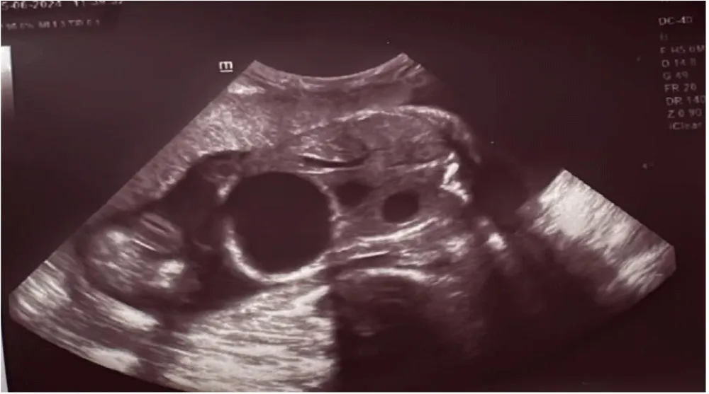
This was a male newborn from an unattended pregnancy carried to term, vaginal delivery, cephalic presentation, Apgar 8/10/10. Mother aged 34, G5P5, 4 healthy children, no particular pathological history, negative infectious anamnesis, with notion of consanguinity with morphological echography: GMFE AC present, cephalic presentation, male sex, fundic placenta, LA of normal quantity: GC at 4 cm, Mega bladder, Bilateral major UHN, Cryptorchidism, Hypoplasia of the anterior abdominal wall, short long bones, doubt about the boot hand, the rest of the morphological examination is without anomaly with an EPF: 1775 ± 259 g (Figure 1).
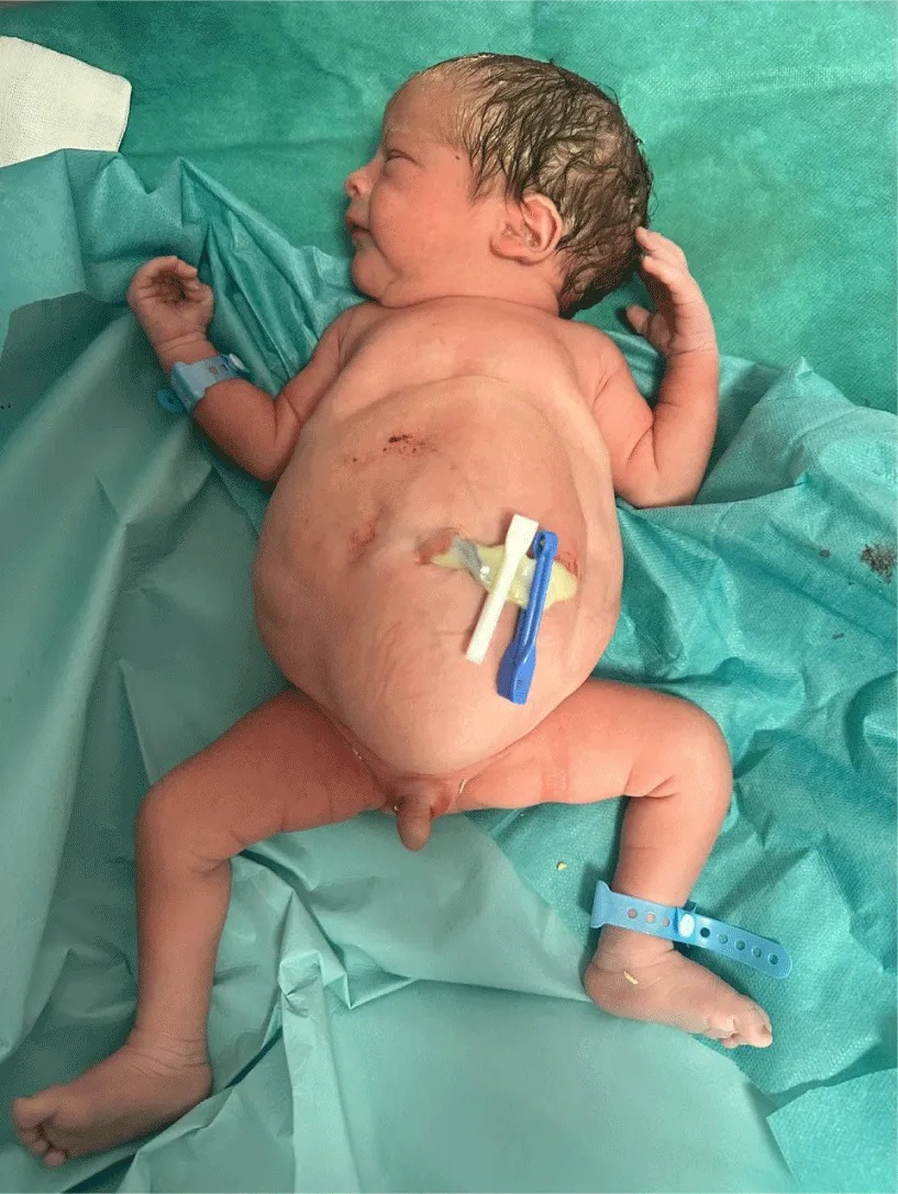
Clinical examination revealed a pink, toned, responsive newborn with a sucking reflex, a weight of 2090 g, a height of 49 cm, and a head circumference of 34 cm. The Dubowitz was estimated at 38 weeks’ gestation. Abdominal examination revealed a thinned and wrinkled abdominal wall, with subcutaneous palpation of the intestinal ansae (Figure 2). Osteoarticular examination revealed a dislocated hip. The cardiovascular, pleuropulmonary, and abdominal examinations were unremarkable. Paraclinical examinations included: a thoraco-abdominal X-ray, which showed thoracic and spinal deformity, with an appearance of delayed resorption, and intestinal ansae deviated to the left. Abdominal ultrasound showed aplasia of the deep abdominal wall muscles, with bilateral major UHN. Trans-thoracic ultrasound revealed a patent foramen ovale with a patent ductus arteriosus and a tiny IVC, and sub-systemic PAH of respiratory origin. Transfontanellar ultrasound was unremarkable. An ultrasound scan of the hips showed a congenital dislocation of the left hip with an anomaly of the long bones 5os courts° (Figure 2). An abdominopelvic CT scan showed agenesis of the abdominal musculature and costovertebral anomalies consistent with a constitutional poly-malformation syndrome. Biological tests were unremarkable, particularly renal function, which was normal (Table 1).
Discussion
The term Prune Belly syndrome was first described in 1839 by Fröhlich. Later in 1895, Parker made the first description associating abnormalities of the urinary tract. It was not until 1950 that Osler named it ‘Prune Belly’ because of the appearance of the abdominal wall [1,2]. According to various studies, the incidence of Prune Belly syndrome is estimated at one in 40,000 births [2]. This condition is marked by a clear male predominance, with more than 95% of patients being male, and by the rarity of complete forms in female children, who generally do not present urinary malformations [1,3-5].
The exact etiology of PBS is unknown. There are 3 main theories: firstly, prenatal obstruction of urine; secondly, embryologically based failure of primary mesodermal differentiation between the 6th and 10th week of gestational age, leading to defective muscular development of the abdominal wall and urinary tract; and thirdly, bladder bag dysgenesis and allantois [2,6]. Clinically, the main components of this syndrome are Urinary malformations, namely megavessia, dilated urethras, and ureters, which may also be stenotic or atretic, with a higher incidence in girls, polycystic kidney disease, hydronephrosis, and sometimes a diverticulum near the vesicoureteral and urethral junction [1,2,4,6].
Renal function is an important prognostic factor [1,2]. Our case, with an incomplete form of the syndrome, had no vesico-renal malformations and normal renal function. However, up to 75% of patients with PBS have other malformations, including pulmonary, cardiac, skeletal, gastrointestinal, and genital malformations. These malformations were reported by Routh, et al. with an incidence of 25% for cardiovascular, 24% for gastrointestinal, 23% for musculoskeletal, 58% for respiratory, and 15% for genital [2,6]. Respiratory malformations include pulmonary hypoplasia and adenomatoid cystic malformation, which can lead to varying degrees of respiratory failure, the main cause of neonatal mortality.
Gastrointestinal malformations such as mesenteric malrotation, atresia, stenosis, volvulus, anal imperforation, splenic torsion, Hirschsprung’s disease, and gastroschisis. Osteoarticular malformations such as clubfoot, hip dysplasia, vertebral malformations, and scoliosis. The osteoarticular malformations found in our patient were clubfoot with homolateral hip dislocation and costovertebral malformations. Cardiovascular malformations such as patent ductus arteriosus and tetralogy of Fallot [1]. Our patient has a patent ductus arteriosus. Genital malformations such as cryptorchidism are present in almost all male patients, although anomalies of the corpus cavernosum and prostatic hypoplasia have also been reported. In females, genital malformations include vaginal atresia, bicornuate uterus and urogenital sinus. Infertility has never been reported in either women or men [7,8]. In rare cases, abdominal wall muscle hypoplasia is unilateral, and other associated malformations, such as kidney, testis, and bone, are usually found on the same side as the abdominal muscle hypoplasia, thus describing the incomplete form generally more common in females, also known as Pseudo Prune Belly Syndrome (PPBS) [9-11].
Our patient presents with an incomplete form of the syndrome with unilateral involvement of the abdominal wall associated with bone malformations on the same side. Prenatal diagnosis is based on obstetric ultrasound, which can detect abnormalities of the urinary tract associated with the typical appearance of the abdominal wall. Hosbino reported a case of PBS diagnosed at 12 weeks gestation [1,2,6]. Postnatal diagnosis is based on abdominopelvic ultrasound with abdominopelvic CT scan, trans-thoracic ultrasound to look for cardiac malformations, a renal workup to assess renal function, ultrasound of the hips with a skeletal X-ray to look for skeletal malformations and a karyotype to look for deletion of nuclear factor 1-beta (HNF1beta) [1,12,13]. Treatment is mainly surgical: abdominoplasty, orchidopexy, and urinary tract reconstruction [1-4,6,14,15]. For patients with mild abdominal wall dysplasia, postures are acceptable and do not require abdominoplasty.
However, in severe cases, surgical treatment is discussed on a case-by-case basis. Pyelostomies, ureterostomies, and cystostomies are also undertaken to shunt urine temporarily in certain unstable infants who cannot tolerate the surgical procedure. Sometimes renal transplantation is unavoidable for patients with renal failure. In any event, whether or not surgery is undertaken, patients with PBS require ongoing multidisciplinary medical care and close monitoring. In our patient, postures were proposed given the mild dysplasia he presented, with multidisciplinary follow-up. The prognosis for patients with PBS varies according to the severity of pulmonary hypoplasia and urinary tract anomalies. Pulmonary hypoplasia is the main cause of mortality in the neonatal period. The severity of urinary tract anomalies and renal function determine not only mortality but also long-term prognosis [1-4].
Conclusion
Prune Belly syndrome is rare and mainly affects males. Renal failure and pulmonary hypoplasia are the main causes of mortality. In the absence of the classic triad, it is imperative to look for other malformations, given the existence of atypical forms, to undertake appropriate and immediate management. The clinical forms can be highly variable, ranging from stillbirths with major renal and respiratory dysplasia to practically normal children. 95% of carriers of this syndrome are male. This explains the diversity of opinion on how to deal with this syndrome. We report a rare case of the incomplete form of Prune Belly syndrome discovered in a male newborn at birth.
Ethical declaration
We had the patient’s consent for this study.
- Fette A. Associated rare anomalies in prune belly syndrome: a case report. J Pediatr Surg Case Rep. 2015;3(2):65-71. Available from: https://doi.org/10.1016/j.epsc.2014.12.007
- Xu W, Wu H, Wang DX, Mu ZH. A case of prune belly syndrome. Pediatr Neonatol. 2015;56(3):193-6. Available from: https://doi.org/10.1016/j.pedneo.2013.03.014
- Seidel NE, Arlen AM, Smith EA, Kirsch AJ. Clinical manifestations and management of prune belly syndrome in a large contemporary pediatric population. Urology. 2015;85(1):211-5. Available from: https://doi.org/10.1016/j.urology.2014.09.029
- Diao B, Diallo Y, Fall PA, Ngom G, Fall B, Ndoye AK, et al. Syndrome de prune belly: aspects épidémiologiques, cliniques et thérapeutiques. Prog En Urol. 2008;18:470-4. Available from: https://doi.org/10.1016/j.purol.2008.04.003
- Lopes RI, Baker LA, Dénes FT. Modern management of and update on prune belly syndrome. J Pediatr Urol. 2021;17(4):548-554. Available from: https://doi.org/10.1016/j.jpurol.2021.04.010
- Fernández-Bautista B, Angulo JM, Burgos L, Ortiz R, Parente A. Surgical approach to prune-belly syndrome: A review of our series and novel surgical technique. J Pediatr Urol. 2021;17(5):704.e1-704.e6.
- Tibor-Denes F, Tavares A, Machado M, Giron A, Srougi M. Gonadal function and reproductive system anatomy in postpuberal prune belly syndrome patients. J Urol. 2017;197(4S).
- Gallo C, Costa W, Favorito L, Sampaio F. Prune belly syndrome: is penile structure similar to normal fetuses? In: State University of Rio de Janeiro, Urogenital Research Unit, Rio de Janeiro, Brazil. London, United Kingdom. 2017.
- Zugor V, Schott GE, Labanaris AP. The prune belly syndrome: urological aspects and long-term outcomes of a rare disease. Pediatr Rep. 2012;4(2):e20. Available from: https://doi.org/10.4081/pr.2012.e20
- Grover H, Sethi S, Garg J, Ahluwalia AP. Pseudo prune belly syndrome: diagnosis revealed by imaging? A case report and brief review. Pol J Radiol. 2017;82:252-7. Available from: https://doi.org/10.12659/pjr.899743
- Amado NG, Nosyreva ED, Thompson D, Egeland TJ, Ogujiofor OW, Yang M, et al. PIEZO1 loss-of-function compound heterozygous mutations in the rare congenital human disorder prune belly syndrome. Nat Commun. 2024;15:339. Available from: https://doi.org/10.1038/s41467-023-42634-9
- Dénes FT, Park R, Lopes RI, Moscardi PRM, Srougi M. Abdominoplasty in prune belly syndrome. J Pediatr Urol. 2015;11(5):291-2. Available from: https://doi.org/10.1016/j.jpurol.2015.06.006
- Kidney function and transplants in prune belly syndrome: a scoping review. Systematic Review/Meta-analysis. November 2023.
- Granberg CF, Harrison SM, Dajusta D, Zhang S, Hajarnis S, Igarashi P, et al. Genetic basis of prune belly syndrome: screening for HNF1? Gene. J Urol. 2012;187(1):272-8. Available from: https://doi.org/10.1016/j.juro.2011.09.036
- Ngwanou DH, Ngantchet E, Moyo GPK. Prune-belly syndrome, a rare case presentation in neonatology: about one case in Yaoundé, Cameroon. Pan Afr Med J. 2020;36:102. Available from: https://doi.org/10.11604/pamj.2020.36.102.24062
Article Alerts
Subscribe to our articles alerts and stay tuned.
 This work is licensed under a Creative Commons Attribution 4.0 International License.


 Save to Mendeley
