Journal of Gynecological Research and Obstetrics
Hamman Syndrome during Labor. Report of a Case
Diana Mayela Esquivel Muñoz1, Diego Fernando López Salinas1, Alexander Porter Magaña1 and Victor Manuel Vargas-Hernández2,3*
1Department of Gynecology and Obstetrics, Regional General Hospital No.1 of the IMSS, Mérida Yucatán, Mexico
2Mexican Academy of Surgery, Mexico
3National Academy of Medicine of Mexico, Mexico
Cite this as
Esquivel Muñoz DM, López Salinas DF, Magaña AP, Vargas-Hernández VM. Hamman Syndrome during Labor. Report of a Case. J Gynecol Res Obstet. 2024;10(2):040-043. Available from: 10.17352/jgro.000129Copyright License
© 2024 Esquivel Muñoz DM, et al. This is an open-access article distributed under the terms of the Creative Commons Attribution License, which permits unrestricted use, distribution, and reproduction in any medium, provided the original author and source are credited.Hamman Syndrome is defined as a self-limiting syndrome, characterized by the coexistence of free air in the mediastinum without an identifiable cause or secondary to trauma, intrathoracic infections, or medical procedures. Clinical manifestations commonly present abruptly and consist mainly of substernal chest pain of a pleuritic nature (forced inspiration), dyspnea, facial pain, odynophagia, cough, dysphagia, and others. A case of Hamman syndrome during labor in a 21-year-old woman is presented.
Background
The first documented cases occurred in 1618 and 1883; the first of these being commented on by the midwife of the queen of France; However, it was in 1939 when Louis Hamman recognized it as a syndrome, commenting at this time on the report with seven cases; referring to spontaneous pneumomediastinum associated with subcutaneous emphysema produced in the postpartum period [1]. Unfortunately, it is a disease diagnosed in an unusual way, approximately in 1/7,000 to 1/44,000; Cases of young patients being the most frequent, with no comorbidity mentioned; except asthma [2].
Pathophysiology
Overall, it consists of the rupture of marginal alveoli secondary to the increase in intraalveolar pressure with sustained Valsalva maneuvers (forced expiration against the glottis that at that moment remains closed) associated with cough, vomiting, straining, and speaking (second period) (during labor); sometimes increasing intrathoracic pressure up to 50 cm of water [3]. After alveolar rupture, air passes from the interstitium (pulmonary capillary hypotension and alveolar wall defects), towards the hilum and then to the mediastinum (due to the pressure difference between it and the pulmonary periphery: Macklin effect); and this to subcutaneous planes; It can extend to the cervical subcutaneous tissue, pleura, pericardium, peritoneal cavity and the epidural space [1,3]. Some cases have also been associated with recreational drug use (cocaine or marijuana), lung function tests, and asthma.
Diagnosis
Physical examination is usually up to 30% normal [4]; however; The clinical manifestations commonly present abruptly and consist mainly of substernal chest pain of a pleuritic nature (forced inspiration), dyspnea, facial pain, odynophagia, cough, dysphagia, and others such as pain or inflammation of the neck, torticollis, dysphonia or abdominal pain [4]. Other signs found are subfebrile temperature, or hemodynamic changes (30 to 50% increase in cardiac output and blood volume) [3]. Hamman described the “mediastinal crack or snap” sign, a sound obtained over the left hemithorax in the anterior part and present in approximately 50% of patients [4].
In case of strong suspicion, the initial (less invasive) technique is a chest x-ray; considering gold standard to computed tomography (detection of up to 30% cases not detected by radiography; Macklin effect (thoracic CT) linear collections of air adjacent to the bronchovascular sheaths [1]; however, lateral radiographs can be added, achieving an accuracy of almost 100% [4]. Among the radiological signs found may be subcutaneous emphysema, thymic veil sign, pneumopericardium sign of the ring around the artery, double bronchial wall sign, continuous diaphragm sign, air in the pulmonary ligament and presence of extrapleural air.
Differential diagnosis
Differential diagnoses include Boerhaave syndrome (esophageal rupture), Mallory-Weiss syndrome (laceration of the gastrointestinal tract mucosa secondary to hyperemesis), acute myocardial infarction, tracheal rupture, pericarditis, aortic dissection [3].
Treatment
For such a pathology, the initial management is conservative; since the majority of patients respond favorably; which consists of analgesia on demand, oxygen therapy, and rest [1]. When it is due to an event secondary to pathology, therapy should be included with a special focus on underlying causes such as bronchospasm, infection, detection, and removal of aspirated foreign bodies if any. Supplemental oxygen therapy may help with the earlier absorption of air from the mediastinum and subcutaneous tissues [4]. Observation depends on the clinical evolution and can last from hours to days (not beyond the fourth day); the criteria for hospital discharge are the disappearance of symptoms and pneumomediastinum with serial control x-rays [3]. The prognosis is good since recurrence is rare and surgical intervention (mediastinotomy) is rarely required [2,3].
Clinical case
A 21-year-old woman, without a significant family history, blood group A Rh+, originally from Escárcega Campeche, resident of Mérida Yucatán for 7 years, completed high school education, Catholic Religion, denies history of smoking, alcoholism, or use of illicit drugs. She denies chronic non-communicable diseases, surgeries, trauma, or transfusions. Gynecological and obstetric history, menarche at 12 years of age, regular cycles, begins sexual relations at 18 years of age, 3 sexual partners, uses condom, has a pregnancy of 38.5 weeks of gestation with good prenatal control, prenatal laboratory studies and ultrasound normal Negative rapid COVID-19 test upon admission; her current condition: she enters the hospital, presenting with moderately intense cramping pain, with cervical modifications compatible with labor in the active phase, so it was decided to admit her to labor for surveillance and delivery. During her stay in labor with a duration of 8 hours until the expulsion period, she gave birth to a male newborn weighing 2970 grams, Capurro 40 weeks of gestation, APGAR 8/9, clear amniotic fluid, simple circular from cord to neck, placenta and complete chorioamniotic membranes, no postpartum hemorrhage.
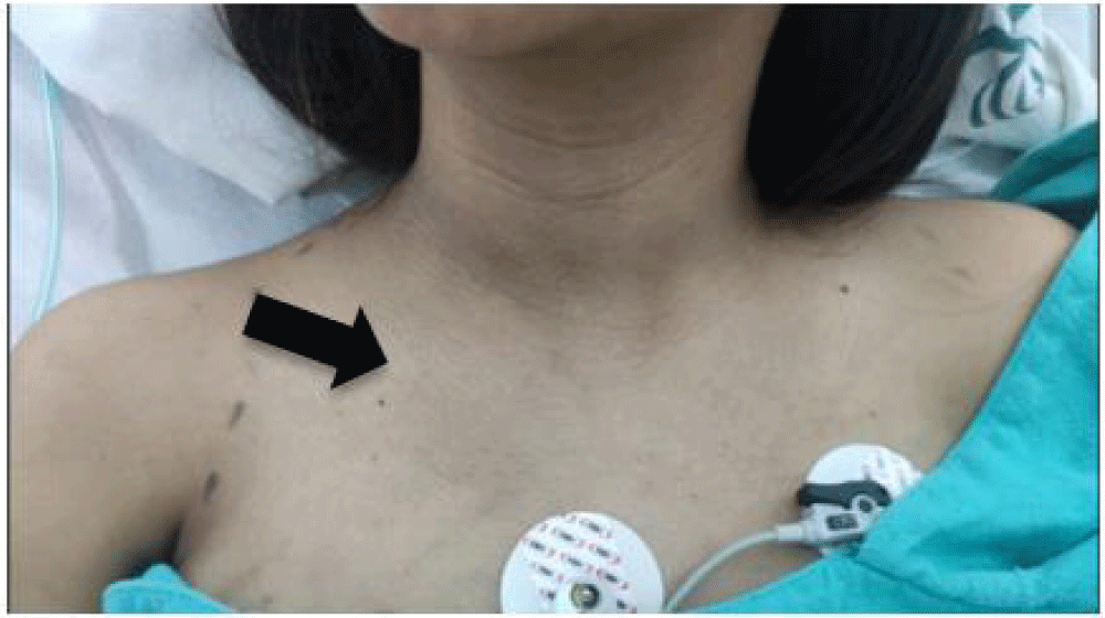
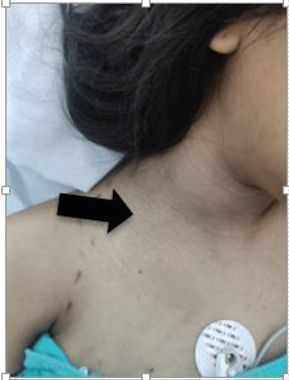
During labor care, nasal tips at 3L/min, without improvement in symptoms, after that, she reported a sensation of crepitation in the mandibular region. In addition, the physical examination identified the presence of crepitation on deep palpation in the region of upper hemithorax with distribution towards the right cervical region as shown in Images 1 and 2.
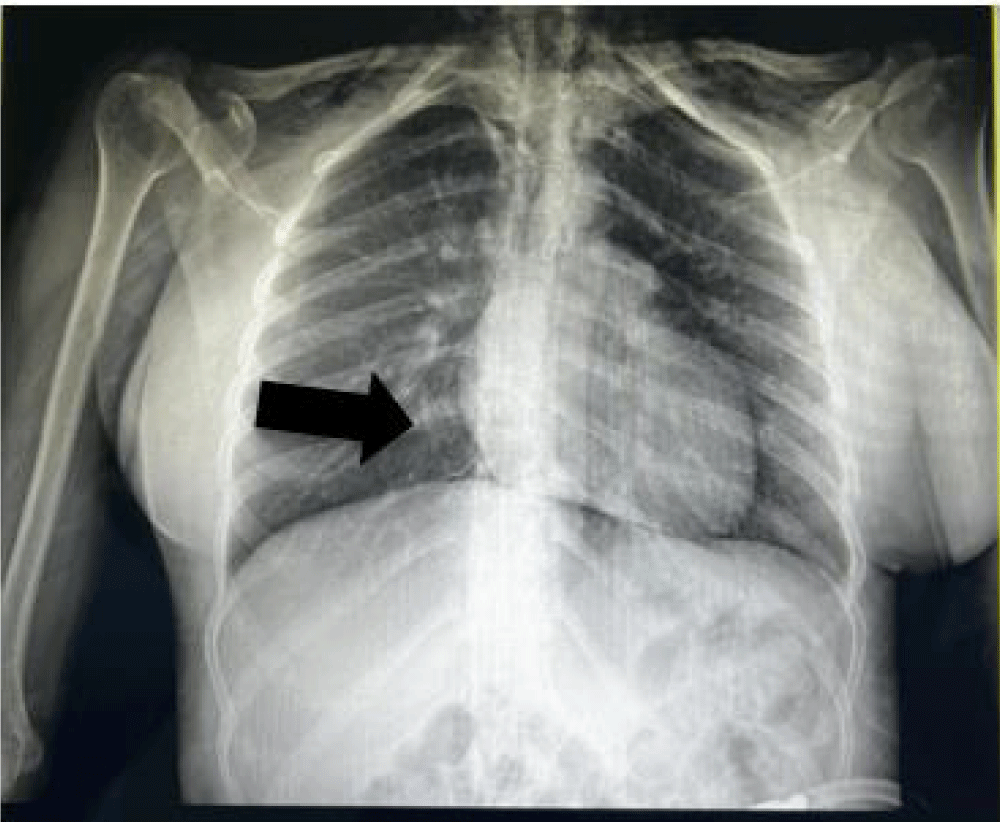
An intensive care evaluation was requested, which indicated admission to the intensive care unit for monitoring and management. A chest x-ray was performed (Image 3) in which thepresence of subcutaneous emphysema in the upper hemithorax was reported, requiring computed tomography without contrast.
As part of the complementary studies, a simple chest CT was performed which reported: soft tissues with the presence of subcutaneous emphysema that dissects the soft tissues of the neck, the vascular structures, and the subcutaneous cellular tissue in the supraclavicular region, coming from the mediastinum, with a separation of up to 14 mm from the anterior chest wall to the heart, no signs of pneumothorax are identified, no pleural dissection. Bone structures without alterations, central trachea without displacements or lesions, lung parenchyma with adequate aeration, without evidence of nodular lesions or areas of consolidation, pleura preserved without thickening. Both hemidiaphragms without evidence of alterations (Image 4).
The laboratories upon admission reported within normal parameters; as shown by days of hospital stay in comparative Table 1. The blood gasometry result is presented in Table 2.
Case analysis and discussion
Hamman Syndrome is defined as a self-limiting syndrome, characterized by the coexistence of free air in the mediastinum without an identifiable cause or secondary to trauma, intrathoracic infections, or medical procedures; according to a study realized by Peña et al its incidence is estimated at 1 case per 2000 or 100,000 inhabitants [3]; However, in another study realized by Campbell concludes that, truly spontaneous pneumomediastinum should not have any agent that causes it; defined as the presence of air in the mediastinum in healthy subjects without an obvious causal factor. With both definitions, the classification of it into two categories or groups is discussed: spontaneous (when there is no triggering cause that causes it) and secondary (when a causal or predisposing factor is identified). The secondary category could also be divided into iatrogenic and non-iatrogenic trauma; or congenital, hereditary, or genetic alterations in lung anatomy such as bronchiectasis, cystic fibrosis, surfactant disorders, etc.); or acquired, mainly asthma, COPD, interstitial lung disease, SARS-CoV-2 pneumonia [5].
In our patient, we don’t have any obvious causal factor to classify as secondary, thus, if we have in mind this syndrome as a differential diagnosis, we can give a prompt diagnosis and treatment, excluding other severe pathologies.
Conclusion
As obstetricians, it is important to know about the existence of this syndrome, although there is not so much information available, or it can easily be confused with other pathologies, as we are the companions and monitors of hundreds of young patients in labor.
We have to teach and give a correct follow-up about how they are behaving and breathing during labor and how we can help them and use this information in our favor to prevent these kinds of complications.
- García-García A, Parra-Virtoa A, Galeano-Vallea F, Demelo-Rodríguez P. Spontaneous pneumomediastinum and subcutaneous emphysema: Hamman's syndrome. Elsevier Spain. 2019 SEPAR. S.L.U. Available from: https://dx.doi.org/10.1016/j.arbres.2019.05.011
- Álvarez Z C, Jadue Ta A, Rojas Ra F, Cerda Cb C, Ramírez Vc M, Cornejo S C. Spontaneous pneumomediastinum (Hamman syndrome): A benign disease that is misdiagnosed. Rev Med Chile. 2009;137:1045-1050. Available from: http://dx.doi.org/10.4067/S0034-98872009000800007
- Peña Vega CJ, Buitrón-García R, ZavalaBarrios B, Aguirre-García R. Postpartum Hamman's syndrome (pneumomediastinum): Literature summary and case report. Gynecol Obstet Mex. 2023;91(3):197-209. Available from: https://doi.org/10.24245/gom.v91i3.3711
- Sánchez DC, Aguilar Aguilar J, Afanador A, Carralero I, Andrea L. Hamman's syndrome, case report. Dr. Federico Abete Trauma and Emergency Hospital, Buenos Aires, Argentina. 20th September 2020. Available from: http://revista.sacd.org.ar/sindrome-de-hamman-reporte-de-un-caso/
- Campbell-Silva S. Spontaneous pneumomediastinum: Order from chaos. Rev Esp Pathol Thorac. 2023;35(2):167-168. Available from: https://www.rev-esp-patol-torac.com/files/publicaciones/Revistas/2023/35.2/Carta%20al%20director.pdf
Article Alerts
Subscribe to our articles alerts and stay tuned.
 This work is licensed under a Creative Commons Attribution 4.0 International License.


 Save to Mendeley
