Journal of Clinical Research and Ophthalmology
Paradoxical worsening of presumed tubercular serpiginous like choroiditis on anti-tubercular therapy
Priyanka1*, Amber Kumar2, Mahesh Maheshwari3 and Jyotirmay Biswas4
2Assistant Professor, Department of Pediatrics, AIIMS, Bhopal, India
3Associate Professor, Department of Pediatrics, AIIMS, Bhopal, India
4Director of Uveitis and Ocular Pathology department, Sankara Nethralaya, Chennai, India
Cite this as
Priyanka, Kumar A, Maheshwari M, Biswas J (2019) Paradoxical worsening of presumed tubercular serpiginous like choroiditis on anti-tubercular therapy. J Clin Res Ophthalmol 6(2): 028-030. DOI: 10.17352/2455-1414.000060Choroidal tuberculosis may present as multifocal progressive or serpiginous like choroiditis. It is important to recognize these presentations as these eyes show good response to systemic antitubercular therapy. Paradoxical reaction in tuberculosis can occur in patients receiving anti-tuberculous therapy (ATT). It consists of clinical or radiological worsening of pre-existing tuberculous lesions or the development of new lesions in patients who initially improved on treatment. Intraocular tuberculosis presents most commonly as posterior uveitis, either as choroidal tubercles, choroidal tuberculoma, subretinal abscess or serpiginous-like choroiditis; however, it can also present, in order of decreasing prevalence, as granulomatous anterior uveitis, panuveitis and intermediate uveitis. We report, a case of paradoxical worsening of presumed tubercular serpiginous like choroiditis on antitubercular therapy. Through this interesting case we try to highlight the fact that continued progression of lesions may be noted inspite of ATT and systemic steroids but it is important to be aware of paradoxical worsening, so that one doesn’t interrupt ATT.
Introduction
Serpiginous like choroiditis (SLC) and serpiginous choroiditis (SC) are uveitic entities on the same spectrum but with different clinical morphologic features. Serpiginous choroiditis is believed to be an autoimmune disease and responds well to systemic steroids; in contrast, treating serpiginous-like choroiditis with steroids can lead to serious local and systemic complications if not accompanied by anti-tubercular therapy (ATT).Paradoxical reaction to anti‑tubercular therapy has been observed in various forms of ocular tuberculosis including serpiginous‑like choroiditis, granulomatous anterior uveitis, intermediate uveitis, panuveitis, retinal vasculitis and has been documented frequently in extrapulmonary tuberculosis (TB) [1‑3]. It is believed to be mediated by the host’s immune system due to an enhanced delayed hypersensitivity of the host, decreased suppressor mechanisms, and as a response to mycobacterial antigens.
Case Report
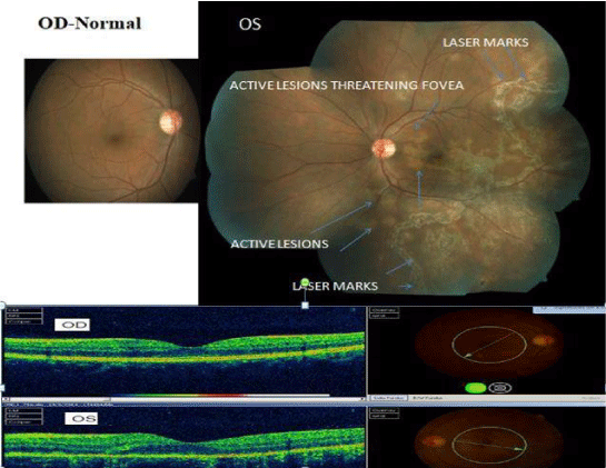
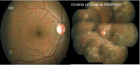
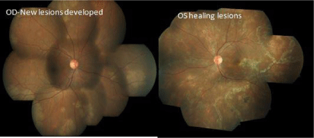
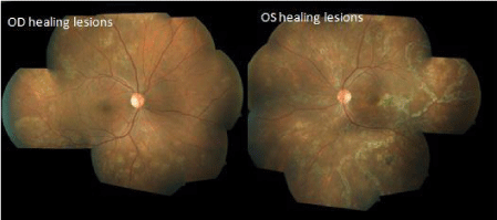
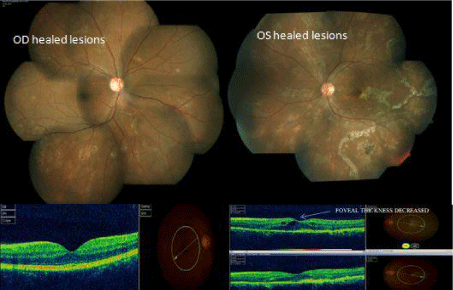
A man of age 37 year presented with complaints of diminution of vision in left eye since 15 days. He was previously diagnosed and treated by his local ophthalmologist, as acute retinal necrosis, had received 600 mg Acyclovir intravenous thrice daily for 1 week followed by oral Valacyclovir 1 gm thrice daily and oral steroid (60 mg for first week followed by weekly tapering) along with laser barrage. On examination he had best corrected visual acuity of 6/6 in right eye and 6/9 in his left eye. Slit lamp examination revealed quiet anterior chamber in right eye and AC cells 1+ along with vitreous cells 1+ in left eye. Intraocular pressure measured was normal in both eyes. Fundus examination showed active choroidal lesion threatening fovea along with laser mark in left eye. Right eye was within normal limit (Figure1). His investigation revealed strongly positive tuberculin skin test with ulceration (24 mm induration); positive Quantiferon TB-Gold Test and contrast enhanced computed tomography chest revealed mediastinal lymphadenopathy (necrotic lymph nodes without calcification suggestive of tubercular etiology). Biopsy of lymphnode for confirmation was not attempted in this case in view of positive tuberculin test and Quantiferon TB-Gold test. However, other investigations for syphilis, toxoplasmosis, HIV was negative. Left eye anterior chamber tap was done for PCR of CMV/HSV/VZV/MTB but it was negative. He was started with intravenous methyl prednisolone (1 gm) for 3 days followed by systemic steroid (60 mg prednisolone with weekly tapering). The patient was referred to a chest physician, who initiated 4 drugs ATT (Isoniazide, rifampicin, pyrazinamide, ethambutol). After 1 week left eye fundus examination showed healing lesion. At 1 month of follow up, slit lamp examination revealed quiet anterior chamber with no vitreous cell in both eye. Fundus examination showed healing lesions at centre with peripheral active lesion. Right eye was within normal limit (Figure 2). Dose of systemic steroid was increased to 60 mg from 30 mg and advised to continue ATT. Steroid was tapered again after 1 week. At 2 month of follow up, slit lamp examination revealed quiet anterior chamber with no vitreous cell in both eye. Fundus examination showed healing choroidal lesion in left eyebut active new lesion in right eye (Figure 3). He was advised to continue ATT along with tapering dose of oral steroid. Azathioprine 50 mg thrice daily was added. At 3 month of follow up, fundus examination revealed healing lesion of choroiditis in both eyes (Figure 4). He was advised to continue same treatment. At 5 month of follow up, he had best corrected visual acuity 6/6 in both eyes. Slit examination revealed quiet anterior chamber with fundus showing healed choroidal lesion in both eyes (Figure 5). ATT completed at 6 months. Steroid and immunosuppressive were also stopped at 6 month. At one year follow-up, there was no recurrence.
Discussion
Choroidal tuberculosis may present as multifocal progressive or serpiginous like choroiditis. Tubercular serpiginous-like choroiditis is considered as an immune-mediated hypersensitivity reaction to the tubercular bacilli sequestrated in the retinal pigment epithelium layer. These eyes show good response to ATT [4]. Resolution of lesions in patients with serpiginous choroiditis can occur with combination therapy of systemic steroids and immunosuppressive agents [5]. In patients with tuberculous choroiditis, ATT accelerates theprocess of healing and reduces recurrence risk by decreasing the number of bacilli. In some patients, the rapid destruction of bacilli after initiation of ATT may cause existing lesions to worsen and new lesions to form. This paradoxical phenomenon occurs more often during the treatment of systemic TB infections, but has also been found in ocular TB. Paradoxical worsening may even occur in patients initiated on simultaneous ATT and systemic steroid. One reason for this may be very severe inflammation and inadequate suppression. Another reason may be that rifampicin increases steroid metabolism [6]. In that case, increasing the steroid dose or adding immunosuppressive therapy may resolve the issue. The factor that could have contributed to the paradoxical reaction in our case was that our patient was also on rifampicin, which is reported to reduce the bioavailability of corticosteroids. Hawkey et al. found that a higher bacillary load or a persistent antigenic stimulus that is poorly cleared from the diseased site may be responsible for the development of paradoxical worsening [7]. The RPE shares many features with the macrophages, including phagocytosis of bacteria and expression of toll-like and complement receptors and perturbed RPE produces a variety of inflammatory substances, including interleukin-1 and granulocyte-macrophage colony-stimulating factor [8]. Therefore, the RPE cells that have engulfed Mycobacteria may release cytokines triggered by mycobacterial LAM (lipoarabinomannan) thus causing continued progression.
Conclusion
The possibility of the paradoxical phenomenon upon ATT initiation should be kept in mind, so that one does not interrupt anti-tubercular treatment and think of drug-resistant tubercular serpiginous-like choroiditis or investigate for other possible non tubercular aetiologies. To monitor the disease progression, recurrences, and involvement of the other eye, serial examination at regular intervals is recommended. Systemic steroid and/or immunosuppressive therapy is useful to suppress inflammation and control progression.
- Gupta V, Gupta A, Arora S, Bambery P, Dogra MR, et al. (2003) Presumed tubercular serpiginous like choroiditis: Clinical presentations and management. Ophthalmology 110: 1744‑1749. Link: http://bit.ly/2G86wke
- Basu S, Nayak S, Padhi TR, Das T (2013) Progressive ocular inflammationfollowing anti‑tubercular therapy for presumed ocular tuberculosisin a high‑endemic setting. Eye (Lond) 27: 657‑662. Link: http://bit.ly/2JGmMJZ
- Gupta M, Bajaj BK, Khwaja G (2003) Paradoxical response in patientswith CNS tuberculosis. J Assoc Physicians India 51: 257‑260. Link: http://bit.ly/2XzQCd0
- Rao NA, Saraswathy S, Smith RE (2006) Tuberculous uveitis: distribution of Mycobacterium tuberculosis in the retinal pigment epithelium. Arch Ophthalmol 124: 1777–1779. Link: http://bit.ly/2XJQurK
- Abrez H, Biswas J, Sudharshan S (2007) Clinical profile, treatment, and visual outcome of serpiginous choroiditis, Ocular Immunology and Inflammation 15: 325-335. Link: http://bit.ly/2G8wns9
- McAllister WA, Thompson PJ, Al-Habet SM, Rogers HJ (1983) Rifampicin reduces effectiveness and bioavailability of prednisolone. Br Med J (Clin Res Ed) 286: 923-925. Link: http://bit.ly/2XJEEsG
- Hawkey CR, Yap T, Pereira J, Moore DA, Davidson RN, et al. (2005)Characterization and management of paradoxical upgrading reactions in HIV‑uninfected patients with lymph node tuberculosis. Clin Infect Dis 40: 1368‑1371. Link: http://bit.ly/2G8yRa3
- Planck SR, Huang XN, Robertson JE, Rosenbaum JT (1993) Retinal pigment epithelial cells produce interleukin-1and granulocyte-macrophage colony-stimulating factor in response to interleukin-1. Curr Eye Res 12: 205-212. Link: http://bit.ly/2YNb1YT

Article Alerts
Subscribe to our articles alerts and stay tuned.
 This work is licensed under a Creative Commons Attribution 4.0 International License.
 Save to Mendeley
