Imaging Journal of Clinical and Medical Sciences
The Fleming Method for Tissue and Vascular Differentiation and Metabolism-Finding breast cancer in women with breast implants
Richard M Fleming1*, Matthew R Fleming1, Tapan K Chaudhuri2 and William C Dooley3
2MD, Eastern Virginia Medical School, Norfolk, Virginia, USA
3MD, University of Oklahoma Health Sciences Center, Oklahoma, USA
Cite this as
Fleming RM, Fleming MR, Chaudhuri TK, Dooley WC (2019) The Fleming Method for Tissue and Vascular Differentiation and Metabolism-Finding breast cancer in women with breast implants. Imaging J Clin Medical Sci 6(1): 097-098. DOI: 10.17352/2455-8702.000129Women with both dense breasts and breast implants present a special challenge for consideration due to the increased incidence of false-positives and false-negatives with mammography [1]. Molecular Breast Imaging [MBI] has provided limited proven benefit until recently, when improvements in camera calibration, enhancement and quantification [2,3], resulted in the ability to differentiate tissue based upon measurable changes in regional blood flow and metabolism [4].
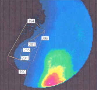
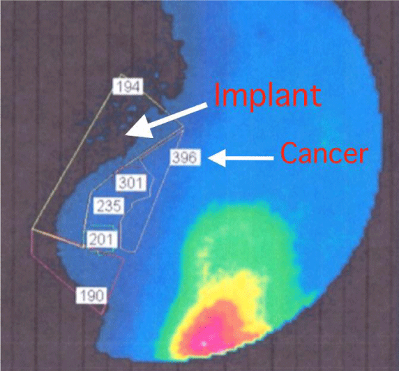
In a 54-year old Caucasian woman with sub-mammary breast implants and a family history of breast cancer, molecular breast imaging using *FMTVDM was performed and is shown in Figure 1, including FMTVDM obtained measurements. The majority of the breast tissue reveals either normal range FMTVDM values or inflammatory changes. However, cancer is also present with measured values of 301 and 396. These measured FMTVDM values are measurements of the tissue regional blood flow and metabolism. The breast implant is neither metabolically active nor has blood flow. Consequently it qualitatively appears as a blackened area on the imaging reflecting no isotope presence. Figure 2 includes arrows establishing the breast implant and cancer regions.
Teaching point: Non-invasive assessment and measurement of breast tissue looking for evidence of inflammation or cancer can be obtained in women with dense breasts and/or breast implants using FMTVDM. Elevated measurements reveal both inflammation and cancer in this patient.
*FMTVDM: The Fleming Method for Tissue and Vascular Differentiation and Metabolism
*FMTVDM: The Fleming Method for Tissue and Vascular Differentiation and Metabolism issued to first author. The patient signed informed consent at the Nevada Imaging location for the procedure and release of results for publication.
- Miller AB, Wall C, Baines CJ, Sun P, To T, et al. (2014) Twenty-five-years follow-up for breast cancer incidence and mortality of the Canadian National Breast Screening Study: randomized screening trial. BMJ 348: g366. Link: http://bit.ly/2Pxw1QX
- The Fleming Method for Tissue and Vascular Differentiation and Metabolism (FMTVDM) using same state single or sequential quantification comparisons. Patent Number 9566037.
- Fleming RM, Dooley WC (2002) Breast enhanced scintigraphy testing distinguishes between normal, inflammatory breast changes, and breast cancer: a prospective analysis and comparison with mammography. Integr Cancer Ther 1: 238-245. Link: http://bit.ly/2RZAOMO
- Fleming RM, Fleming MR (2019) The Importance of Thinking about and Quantifying Disease like Cancer and Heart Disease on a “Health-Spectrum” Continuum. J Compr Cancer Rep 3: 1-3. Link: http://bit.ly/36MaYQr
Article Alerts
Subscribe to our articles alerts and stay tuned.
 This work is licensed under a Creative Commons Attribution 4.0 International License.


 Save to Mendeley
