Imaging Journal of Clinical and Medical Sciences
Acute Myocardial Infarction on Contrast-Enhanced Computed Tomography
Department of Radiology, Tokyo Dental College Ichikawa General Hospital, Chiba, Japan
Author and article information
Cite this as
Baba A, Okuyama Y, Yamazoe S, Mogami T (2016) Acute Myocardial Infarction on Contrast-Enhanced Computed Tomography. Imaging J Clin Medical Sci. 2015; 3(1): 4-5. Available from: 10.17352/2455-8702.000023
Copyright License
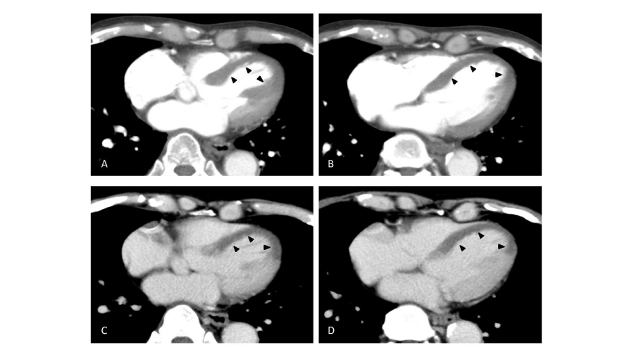
© 2015 Baba A, et al. This is an open-access article distributed under the terms of the Creative Commons Attribution License, which permits unrestricted use, distribution, and reproduction in any medium, provided the original author and source are credited.A 79-year-old man presented to our emergency department with sudden back pain. Laboratory results showed CK 2836 U/L (normal range 30-160), CK-MB 393 U/L (<25), D-dimer 1.4 μg/ mL (<1), BNP 37.9 pg/mL (<19.5), cTnl 0.133ng/mL (<0.028) and electrocardiogram showed ST elevation in V1~6. Acute myocardial infarction (AMI) was suspected. Because of the back pain, acute aortic dissection (AAD) and pulmonary thromboembolism (PE) needed to be ruled out and contrast-enhanced computed tomography (CECT) was performed.
Acute myocardial infarction; Computed tomography; Myocardial perfusion
Text
A 79-year-old man presented to our emergency department with sudden back pain. Laboratory results showed CK 2836 U/L (normal range 30-160), CK-MB 393 U/L (<25), D-dimer 1.4 µg/mL (<1), BNP 37.9 pg/mL (<19.5), cTnl 0.133ng/mL (<0.028) and electrocardiogram showed ST elevation in V1~6. Acute myocardial infarction (AMI) was suspected. Because of the back pain, acute aortic dissection (AAD) and pulmonary thromboembolism (PE) needed to be ruled out and contrast-enhanced computed tomography (CECT) was performed. CECT images showed no findings of AAD or PE. Instead, the images revealed the decreased enhancement in the interventricular septum and apex of the left ventricle (Figure A,B, arterial phase, Figure C,D, delayed phase). With the diagnosis of ST elevated MI (STEMI), emergent invasive coronary angiography was carried out to reveal total occlusion of LAD, corresponding to the CT findings. Percutaneous cardiac intervention was performed for LAD #6 first with plain old balloon angioplasty (POBA) and then thrombus aspiration, completed with drug-eluted stent (DES). The final coronary blood flow was TIMI III. The patient developed mild sub-acute pulmonary edema, which soon subsided with intravenous diuretics. The patient was released without complications.
When a patient presents with acute back pain, CECT is often ordered to evaluate aorta and pulmonary arteries. There have been cases reported where CECT revealed decreased perfusion in myocardium in patients with AMI, without increased cardiac biomarkers [1]. Assessing myocardial enhancement with CECT can be useful in detecting MI [2].
- Mano Y, Anzai T, Yoshizawa A, Ohki T (2015) Role of non-electrocardiogram-gated contrast-enhanced computed tomography in the diagnosis of acute coronary syndrome. Heart Vessels 30: 1-8 .
- Arnett JH, Mohajer K, Okon SA (2007) Evidence of acute myocardial infarction on CT. Br J Radiol 80: e219-221 .


 Save to Mendeley
Article Alerts
Subscribe to our articles alerts and stay tuned.
 This work is licensed under a Creative Commons Attribution 4.0 International License.

