International Journal of Clinical Endocrinology and Metabolism
Childhood Gynecomastia: A Mini Review
Professor and Senior Consultant Pediatric Endocrinologist, Endocrine Division, Department of Pediatrics, College of Medicine and King Khalid University Hospital, King Saud University, Riyadh, Saudi Arabia
Author and article information
Cite this as
Al Jurayyan NA (2016) Childhood Gynecomastia: A Mini Review. Int J Clin Endocrinol Metab. 2016; 2(1): 12-15. Available from: 10.17352/ijcem.000015
Copyright License
© 2016 Al Jurayyan NA. This is an open-access article distributed under the terms of the Creative Commons Attribution License, which permits unrestricted use, distribution, and reproduction in any medium, provided the original author and source are credited.Gynecomastia, referred to enlargement of the male’s breast tissue is a common finding in boys during childhood. Although most cases are benign and self-limited, it may be a sign of an underlying systemic disease or even drug induced. Rarely, it may represent male breast cancer. Understanding its pathogenesis is crucial to distinguish a normal developmental variant from pathological cases.This review will highlight the pathophysiology, etiology, diagnosis and various medical and surgical therapies.
Gynecomastia, referred to enlargement of the male breast tissue (Figure 1), it is a common finding in childhood reported to be between 30 and 60%. Although, most cases are benign and self-limited, it may be a sign of an underlying systemic disease or drug-induced. It is usually bilateral but sometimes unilateral, and results from proliferation of the glandular component of the breast. It is defined clinically by the presence of a rubbery or firm mass extending con-centrically from the nipple. Gynecomastia should be differentiated from peusogynecomastia (lipomastia), which is characterized by desposition of fat without, glandular proliferation [1-6].
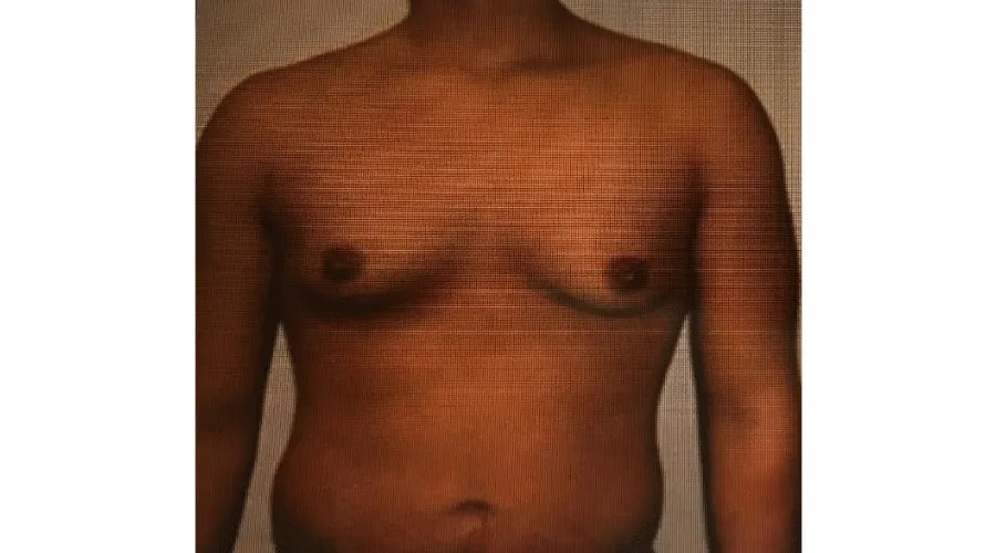
In this brief review we highlight the pathophysiology, etiology, diagnosis and discuss the various modalities of therapy (medical and surgical).
Pathophysiology
The mechanism and pathophysiology of gynecomastia are not really clear (Figure 2). However, the major cause in believed to be an altered imbalance between estrogen and androgen effects, absolute increase in estrogen effects either an increase in estrogen production, relative decrease in androgen production, or a combination of both. Estrogen acts as a growth hormone of the breast and, therefore, excess of estradiol in men leads to breast enlargement by inducing ductal epithelial hyperplasia, dual elongation and branching, and the proliferation of periductal fibroblasts and vascularity. Local tissue factors in the breast can also be important, for example, increased aromatic activity that can cause excessive local production of estrogen, decreased estrogen degradation and changes in the levels or activity of estrogen, and androgen receptors.
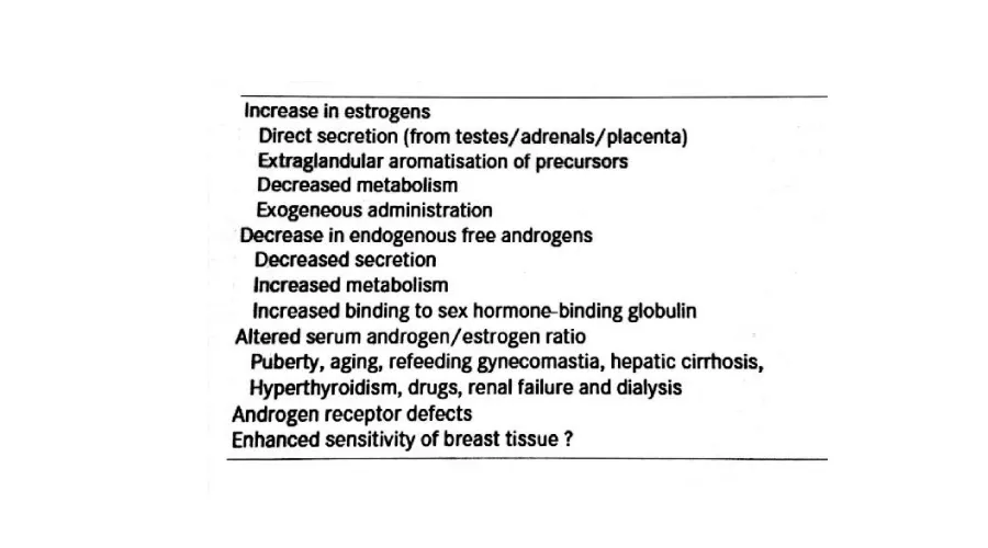
Although prolactin (PRL) receptors are present in male breast tissue, hyperprolacticemia may lead to gynecomastia through effects on the hypothalamia causing central hypogonadism. Activation of PRL, also, leads to decreased androgens and increased estrogen and progesterone receptors. The role of PRL, progesterone and other growth factors such as insulin like growth factors (IGF-1) and epidermal growth factor (EGF), in the development of gynecomastia need to be clarified [5-10].
Classification
The spectrum of gynecomastia severity has been categorized into a grading system [11]:
Grade I Minor enlargement, no skin excess
Grade II Moderate enlargement, no skin excess
Grade III Moderate enlargement, skin excess
Grade IV Marked enlargement, skin excess
Etiology of gynemastia
Generally can be subdivided, according to the cause, into; (Table 1) [12-24].
PPhysiological: Estrogen levels rise during neonatal and pubertal period, which leads to an elevated estrogen/testosterone ration and, hence, gynecomastia the condition usually regress within two years of onset.Pathological: Gynecomastia can occur at any age, as a result of a number of medical conditions such as liver cirhossis primary hypogonadism and trauma.
Medicational: Medication induced gynecomastia is the most common cause. Agents associated with gynecomastia are listed in Table 2.
Diagnosis of Gynecomastia
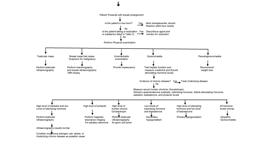
The history and physical examination should direct the laboratory and radiological imaging studies (Figure 3).
Clinical Evaluation: (History and physical examinations)
All boys with gynecomastia should be evaluated thoroughly by an experienced clinician. A detailed history should include the onset and duration of the breast enlargement, pain or tenderness, weight loss or gain, nipple discharge, virilization, medication history, and family history of gynecomastia which may suggest androgen insensitivity syndrome, familial aromatase excess or sestoli cell tumours. Physical examination should differentiate between true gynecomastia and pseudogynecomastia and should include signs of tomours, liver and kidney diseases, or hyperthyroidism and should also include genital examination [5,6,25-31].
Diagnostic testing
In cases without a clear cause, laboratory investigations should be pursued and must include liver, kidney and thyroid function tests as well as hormonal tests, estrogen, and free testosterone, luteinizing hormone (LH) , follicle-stimulating hormone (FSH), prolactin, human chorlonic gonadtrophin (HCG), dehydro epiandrosterone sulphate (DHEA-SO4) and α Feto protein (α FP). If testes are small the patients Karyotype should be obtained to roll out Klinefelter’s Syndrome. Mammography (MMG) is the primary imaging when there is any suspicion of malignancy. Breast ultrasonography (USG), scrotal USG and abdominal computerized tomography (CT) of the abdomen can also be used as well as magnetic resonance imaging of the pituitary. A percutaneous biopsy should be taken, at times, when it is difficult to differentiate gynecomastia and breast cancer [5,6,32-36].
Treatment of gynecomastia
Most cases of gynecomastia regress overtime without treatment. However, if gynecomastia is caused by an underlying conditions such hypogonadism, malnutrition or Cirrhosis, that condition may need treatment. If the patient taking medications that can cause gynecomastia, the clinician may recommend stopping or substituting them with other medication. In adolescents with no apparent cause, gynecomastia often goes away without treatment in less than 2 years. Reassurance and frequent follow up 3-6 months that’s what needed. However, treatment may be necessary if gynecomastia does not improve on its own or if it causes significant pain tenderness or embarrassment [1,6,37].
Medical treatment
Although no medical treatment cause complete regression of gynecomastia, they may provide partial regression, or symptomatic relief. Several agents regulate the hormonal imbalance. The major medical intervention options are androgens, anti-estrogens and aromatase inhibitors. Androgen testosterone replacement can be used to improve gynecomastia secondary to hypogonadism. Topical preparations are preferable as they lead to more steady state levels of testosterone in the body as compared with the injectable forms, which can worsen breast enlargement by aromatizing to estardiol. In recent years, anti-estrogens such as tamoxifen and anastrozole has been shown to be effective. More studies are needed to assess the effectiveness of aromatose inhibitors such as anastrozle and testalactone, which are powerful agents that block estrogen [32,38-43].
Surgical treatment
Surgical treatment should be individualized to each patient. Numerous techniques have been described for the correction of gynecomastia and the surgery is forced with a wide range of excisional and liposuction procedures. The most frequently encountered complication was a residual subareolar lump. Although skin excess remains a challenge, it can be satisfactorily managed without excessive scarring. A practical approach to the surgical management of gynecomastia, should take into account breast size, consistency, skin excess and skin quality. However generally it is not recommended until the testis has reached adult size, because if surgery is performed before achieving puberty, breast tissue may regrow [44-47].
If pseudogynecomastia is suspected no work up is needed and the patient can be reassured that weight loss will lead to resolution of pseudogynecomastia. If necessary liposuction procedures can reduce breast enlargement secondary to subareolar fat accumulation.
Acknowledgement
The author would like to thank Ms. Cecile Sael for typing the manuscript, and extend his thanks and appreciation to Miss Hadeel N. Al Jurayyan for her help in preparing this manuscript.
- Glenn D, Braunstein MD (2007) Gynecomastia. N Engl J Med 357:1229-1237 .
- Lazala C, Saenger P (2002) Pubertal Gynecomastia. J Pediatr Endocrinol Metab 15: 553-560 .
- De Sanctis V, Bernasconi S, Bona G, Bozzola M, Buzi F, et al. (2002) Pubertal gynecomastia. Minerva Pediatr 54: 357-361 .
- Nordt CA, DiVasta AD (2008) Gynecomastia in adolescents. Curr opin Pediatr 20: 375-382 .
- Cuhaci N, Polat SB, Evranos B, Ersoy R, Cakir B (2014) Gynecomastia: Clinical evaluation and management. Indian J Endocrinol Metab 18: 150-158 .
- Dickson G (2012) Gynecomastia. Am Fam Physician 85: 716-722 .
- Barros AC, Sampaio MD (2012) Gynecomastia: pathophysiology, evaluation and treatment. So Paulo Med J 130: 187-197 .
- Narula HS, Carlson HE (2014) Gynecomastia-pathophysiology, diagnosis and treatment. Nat Rev Endocrinol 10: 684-698 .
- Farthing MJ, Green JR, Edwards CR, Dawson AM (1982) Progesterone, prolactin and gynecomastia in men with liver disease. Gut 23: 276-279 .
- Ladizinski B, Lee KC, Nutan FN, Higgins HW, Federman DG (2014) Gynecomastia; Etiologies, Clinical Presentation, Diagnosis and Management. South Med J 107: 44-49 .
- Wollina U, Goldman A (2011) Minimally invasive esthetic procedures of the male breast. J Cosmetic dermatol 10: 150-155 .
- Bhasin S (2008) Testicular disorders. in; Kroneberg HM, Palonsky KS, Larson PR editors. William textbook of Endocrinology. 11th ed, Philadelpia Saunders Elsevier .
- Nuttall FQ (2010) Gynecomastia. Mayo Clin Proc 85: 961-962 .
- Lanfranco F, Kamischke A, Zitzmann M, Nieschlag E (2004) Klinefelter's Syndrome. Lancet 364: 273-283 .
- Handelsman DJ, Dong Q (1993) Hypothalamo-pituitary gonadal axis in chronic renal failure. Endocrinol Metab Clin North Am 22: 145-161 .
- Karagiannis A, Harsoulis F (2005) Gonadal dysfunction in systemic diseases. Eur J Endocrinol 152: 501-513 .
- Layman LC (2007) Hypogonadotropic hypogonadism. Endocrinol Metab Clin North Am 36: 283-296 .
- Fentiman IS, Fourquet A, Hortobagyi GN (2006) Male breast cancer. Lancet 367: 595-604 .
- Eckman A, Dobs A (2008) Drug-induced gynecomastia. Expert Opin Drug Saf 7: 691-702 .
- Thompson DF, Carter JR (1993) Drug -induced gynecomastia. Pharmacotherapy 13: 37-45 .
- Goldman RD (2010) Drug-induced gynecomastia in children and adolescents. Canadian Family physician 56: 344-345 .
- Deepinder F, Braunstein GD (2012) Drug-Induced Gynecomastia: an evidence based review. Expert Opin Drug Saf 11: 779-795 .
- Basaria S (2010) Androgen abuse in athletes: detection and consequences. J Clin Endocrinol Metab 95: 1533-1543 .
- Goh SY, Loh KC (2001) Gynecomastia and the herbal tonic "Dong Quai" Singapore Med J 42:115-116 .
- Bembo SA, Carlson HE (2004) Gynecomastia: it's features, and when and how to treat it. Cleve Clin J Med 71: 511-517 .
- Gikas P, Mokbel K (2007) Management of gynecomastia: an update. Int J Clin Pract 61: 1209-1215 .
- Carlson HE (2011) Approach to the patients with gynecomastia. J Clin Endocrinol Metab 96: 15-21 .
- Dobs A, Darkes MJ (2005) Incidence and management of gynecomastia in men treated for prostate cancer. J Urol 174: 1737-1742 .
- Derkacz M, Chmiel-Perzynskal, Nowakoski A (2011) Gynecomasti- a difficult diagnostic problem. Endokrynol Pol 62: 190-202 .
- Biro FM, Lucky AW, Huster GA, Morrison JA (1990) Hormonal studies and physical examination in adolescents gynecomastia. J Pediatr 116: 450-455 .
- Hassan HC, Cullen IM, Casey RG, Rogers E (2008) Gynaecomastia: an endocrine manifestation of testicular cancer. Andrologia 40: 152-157 .
- Plourde PV, Kulin HE, Santner SJ (1983) Clomiphene in the treatment of adolescent gynecomastia, Clinical and endocrine studies. AM J Dis Child 137: 1080-1082 .
- Sansone A, Romanelli F, Sansone M, Lenzi A, Di Luigi L (2016) Gynecomastia and hormone. Endocrine PMID 27145756 .
- La Franchi SH, Parlow AF, Lippe BM, Coyotupa J, Kaplan SA (1975) Pubertal gynecomastia and transient elevation of serum estradiol level. Am J Dis Child 129: 927-931 .
- Bridenstein M, Bell BK, Rothman MS (2003) Gynecomastia in McDermott MT,ed Endocrine secrets Am ed Philadelphia, PA elserver sanders Chig 40.
- Munoz CR, Alvarez BM, Munoz GE, Raya PJL, Martinez PM (2010) Mammography and ultrasound in evaluation of male breast disease. Eur Radiol 20: 2797-2805 .
- Ruth EJ, Cindy AK, M Hassan M (2011) Gynecomastia evaluation and current treatment options. Ther Clin Risk Manag 7: 145-148 .
- Lapid O, van Wingerden JJ, Perlemuter L (2013) Tamoxifen therapy for the management of pubertal gynecomastia: a systematic review. J Pediatr Endocrinol Metab 26: 803-807 .
- Khan HN, Rampaul R, Blamey RW (2004) Management of physiological gynaecomastia with tamoxifen. Breast 13: 61-65 .
- Lawrence SE, Faught KA, Vethamuthu J, Lawson ML (2004) Beneficial effects of raloxifene and tamoxifen in the treatment of pubertal gynecomastia. J Pediatr 145: 71-76 .
- Mauras N, Bishop K, Merinbaum P, Emeribe U, Agbo F, et al. (2009) Pharmacokinetics and Pharmacodynamics of anastrozole in prepubertal boys with recent-onset gynecomastia. J Clin Endocrinol Metab 94: 2975-2978 .
- Wit JM, Hero M, Nunez SB (2011) Aromatase inhibitors in Pediatrics . Nat Rev Endocrinol 8: 135-147 .
- Braustein GD (1999) Aromatase and gynecomastia. Endocr Relat Cancer 6: 315-324 .
- Fruhstorfer BH, Malata CM (2003) A systematic approach to the surgical treatment of gynecomastia. Br J Plast Surg 56: 237-246 .
- Brown RH, Chang DK, Siy R, Friedman J (2015) Trends in surgical correction of gynecomastia. Semin Plast Surg 29: 122-130 .
- Handschin AE, Biertry D, Husler R, Banic A, Constaninescu M (2008) Surgical Management of gynecomastia: a 10 years analysis. World J Surg 32: 38-44.
- Kasielska A, Antoszewski B (2013) Surgical management of gynecomastia: an outcomeanalysis. Ann Plast Surg 71: 471-475 .



 Save to Mendeley
Article Alerts
Subscribe to our articles alerts and stay tuned.
 This work is licensed under a Creative Commons Attribution 4.0 International License.