International Journal of Clinical Endocrinology and Metabolism
Killian-Jamieson Diverticulum Mimicking a Thyroid Tumor
1Department of Endocrinology and Metabolism, Hirosaki University Graduate School of Medicine, 5 Zaifu-cho, Hirosaki, Aomori 036-8562, Japan
2Department of Endocrinology and Metabolism, Odate Municipal General Hospital, 3-1 Yutaka-cho, Odate 017-8550, Japan
3Endo Clinic, 15-3 Kaerimichi, Takanosu, Kitaakita 018-3331, Japan
Author and article information
Cite this as
Kageyama K, Watanuki Y, Endo K (2015) Killian-Jamieson Diverticulum Mimicking a Thyroid Tumor. Int J Clin Endocrinol Metab. 2015; 1(1): 7-8. Available from: 10.17352/ijcem.000002
Copyright License
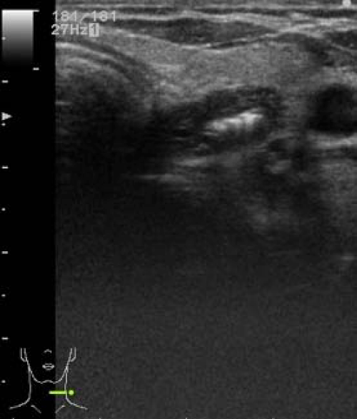
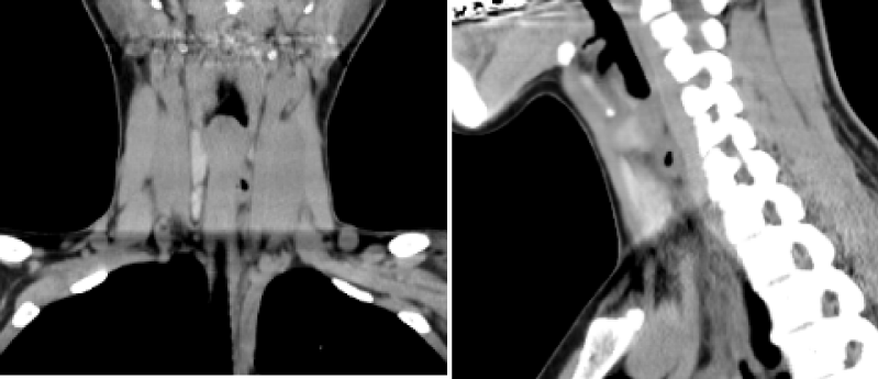
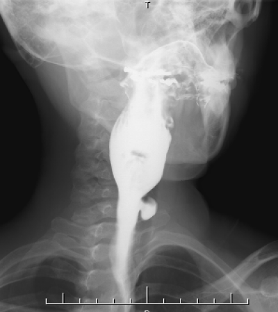
© 2015 Kageyama K, et al. This is an open-access article distributed under the terms of the Creative Commons Attribution License, which permits unrestricted use, distribution, and reproduction in any medium, provided the original author and source are credited.A 42-year-old woman was referred to us for evaluation of a suspicious mass in her left thyroid gland. She had experienced left anterior neck pain and odynophagia for a few weeks. Ultrasonography (US) demonstrated a heterogenous and hypoechoic mass with bright internal hyperechoic foci and a partial surrounding halo involving the posterior aspect of the left thyroid lobe (Figure 1). Computed tomography (CT) of the neck with contrast enhancement demonstrated a soft tissue mass with internal air between the left back side of the thyroid and esophagus (Figure 2). Barium swallow pharyngoesophagography showed a barium-filled sac protruding from the left anterolateral wall of the cervical esophagus (Figure 3).
A 42-year-old woman was referred to us for evaluation of a suspicious mass in her left thyroid gland. She had experienced left anterior neck pain and odynophagia for a few weeks. Ultrasonography (US) demonstrated a heterogenous and hypoechoic mass with bright internal hyperechoic foci and a partial surrounding halo involving the posterior aspect of the left thyroid lobe (Figure 1). Computed tomography (CT) of the neck with contrast enhancement demonstrated a soft tissue mass with internal air between the left back side of the thyroid and esophagus (Figure 2). Barium swallow pharyngoesophagography showed a barium-filled sac protruding from the left anterolateral wall of the cervical esophagus (Figure 3).
We herein report a case of Killian-Jamieson diverticulum mimicking a thyroid tumor. Killian-Jamieson diverticulum is a rare form of hypopharyngeal pulsion diverticulum resulting from herniation of mucosa and submucosa through an area of weakened musculture [1]. The diverticulum is caudal to that of the more common Zenker’s diverticulum [2]. These hypopharyngeal diverticula that cause dysphagia sometimes mimic a thyroid tumor incidentally detected on neck US [3,4], because it looks like a hypoechoic mass with calcifications in the thyroid. For a differential diagnosis, it may be important to show mobility of the mass by swallowing and moving the head. Kim et al. proposed to show changes in the shape of a mass after drinking soda [5].
In the present case, diagnosis by pharyngoesophagography and CT images, together with US, prevented an unnecessary fine needle-aspiration biopsy. Such a fine needle-aspiration biopsy could have been potentially harmful in the context of Killian-Jamieson diverticulum. Clinicians should pay attention to the presence of the diverticulum.
Disclosure
None of the authors have anything to disclose.
- Shanker BA, Davidov T, Young J, Chang EI, Trooskin SZ (2010) Zenker's diverticulum presenting as a thyroid nodule. Thyroid 20: 439-440 .
- Rodgers PJ, Armstrong WB, Dana E (2000) Killian-Jamieson diverticulum: a case report and a review of the literature. Ann Otol Rhinol Laryngol 109: 1087-1091 .
- Lee F, Leung CH, Huang WC, Cheng SP (2012) Killian-Jamieson diverticulum masquerading as a thyroid mass. Intern Med 51: 1141-1142.
- Pang JC, Chong S, Na HI, Kim YS, Park SJ, et al. (2009) Killian-Jamieson diverticulum mimicking a suspicious thyroid nodule: sonographic diagnosis. J Clin Ultrasound 37: 528-530 .
- Kim TH, Kim S, Chang KS (2015) Simple method of using soda for distinguishing Killian-Jamieson diverticulum from a thyroid nodule. Endocrine 48: 351-352 .


 Save to Mendeley
Article Alerts
Subscribe to our articles alerts and stay tuned.
 This work is licensed under a Creative Commons Attribution 4.0 International License.