Journal of Clinical Research and Ophthalmology
Bilateral iris, lens and Chorioretinal Coloboma: A Case Report
Mohamed Abdallahi Ould Hamed*, Abdoulsalam Youssoufou Soulay, Karim Reda and Abdelbarre Oubaaz
Cite this as
Ould Hamed MA, Soulay AY, Reda K, Oubaaz A (2018) Bilateral iris, lens and Chorioretinal Coloboma: A Case Report. J Clin Res Ophthalmol 5(1): 012-012. DOI: 10.17352/2455-1414.000047Colobomas are genetic malformations due to lack of closure of the embryonic fissure. These are rare malformations that can sit at any level of the eye. Colobomas can be uni or bilateral, sporadic or hereditary. It may be associated with other ocular manifestations and extra-ocular malformations involving a general, clinical and radiological examination.
We report the case of a 28 year old young man with no significant pathological history whose ophthalmological examination revealed a coloboma affecting the iris, lens and choroid.
Case Report
Colobomas are genetic malformations due to lack of closure of the embryonic fissure [1]. These are rare malformations that can sit at any level of the eye. Colobomas can be uni or bilateral, sporadic or hereditary [2]. It may be associated with other ocular manifestations and extra-ocular malformations involving a general, clinical and radiological examination.
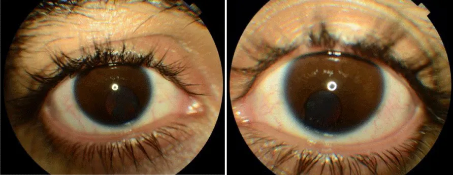
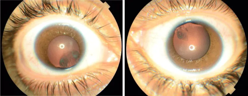
We report the case of a 28 year old young man, with no notable pathological antecedents showing a progressive decline in visual acuity. On examination, his visual acuity is 7/10 not improvable with the optical correction. Bio microscopic examination revealed bilaterally iris coloboma (Figure 1A,B) and lens cataract and coloboma (Figure 2A,B).
Intraocular pressure was normal in the right eye and the left eye.
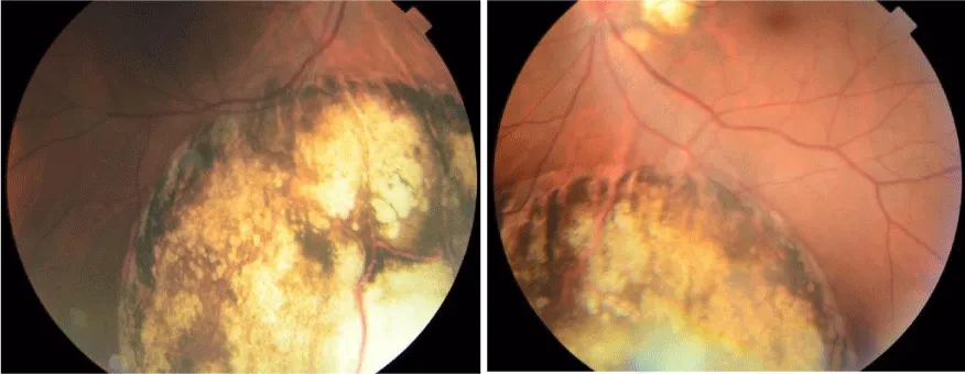
At the eye’s fundus there is the presence of a bilateral choroidal coloboma (Figure 3A,B). The neurological examination and brain MRI have been performed are without abnormalities.
The patient is monitored regularly for possible complications including retinal detachment or choroidal neovascularization [3].
- Duvall J, Miller SL, Cheatle E, Tso MO (1987) Histopathologic study of ocular changes in a syndrome of multiple congenital anomalies. Am J Ophthalmol 103: 701-705.Link: https://goo.gl/H3DoKG
- Pavan-Langston D (1996) Manual of Ocular Diagnosis and Therapy. Boston: Little Brown and Co 350.Link: https://goo.gl/BS7dAZ
- Spitzer M, Grisanti S, Bartz-Schmidt KU, Gelisken F (2006) Choroidal neovascularization in retinochoroidal coloboma: thermal laser treatment achieves long-term stabilization of visual acuity. Eye (London) 20: 972-1069.Link: https://goo.gl/sdyRBd

Article Alerts
Subscribe to our articles alerts and stay tuned.
 This work is licensed under a Creative Commons Attribution 4.0 International License.


 Save to Mendeley
