Open Journal of Trauma
Ankle Fractures in Children
Samena Chaudhry1* and K Dehne2
2Senior Clinical Fellow, Royal Manchester Childrens Hospital, UK
Cite this as
Chaudhry S, Dehne K (2019) Ankle Fractures in Children. Open J Trauma 3(1): 018-021. DOI: 10.17352/ojt.000022Ankle Fractures in the paediatric population are among the most common physeal injuries. Trauma around the ankle often results in distal tibial metaphyseal fractures in the very young child, medial and lateral malleolus fractures in middle childhood (McFarland fractures) and transitional fractures in adolescence (Tillaux and Triplane fractures).
Factors affecting treatment decisions include the Salter Harris classification, the age of the child and intra-articular involvement.
Factors affecting management include the mechanism of injury, the position of the foot after injury and the information obtained from a CT scan. The ability to achieve satisfactory reduction determines need for open reductions and internal fixation. Growth plate injuries can result in complications requiring further surgery.
Introduction
A child with an ankle fracture should be assessed and managed according to ATLS principles. Any life threatening injuries should be identified and treated and the limb should be carefully examined to rule out open injuries, the presence of compartment syndrome and the neurovascular status. A full assessment of sensation and motor function of the foot should be undertaken. The entire limb should be examined and ipsilateral proximal injuries should be recorded. The skin over the ankle is examined for bruises, ecchymosis and tenting which will require a reduction manoeuvre urgently. Most fractures are temporarily treated with a splint. Many ankle fractures in children will require a CT-scan for further assessment and planning for treatment [1].
What classification system is helpful in treating ankle fractures in children?
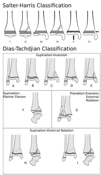
The Salter- Harris (SH) classification is helpful in guiding treatment but its prognostic value is debated with reports of higher complication rates in type II fractures [2]. The Tillaux fracture is a Salter Harris type III fracture of the distal tibial lateral physis and a Triplane fracture is best described as a complex type lV fracture often a two three or four part fracture with components in all planes. McFarland fractures are usually seen in mid- childhood (age 8-12 years) and they can be a Salter Harris Type III or IV injuries of the medial Malleolus.
The Classification system of Diaz and Tachdjian is based on the adult Lauge Hansen classification [3]. Fractures are classified according to the foot position and the direction of force applied to the foot. Fractures are classified into supination-inversion, consisting of a physeal plate fibula injury combined with a Salter Harris Type I to IV injury of the tibial physeal plate. Pronation/eversion–external rotation results in High Fibula fracture. Supination–plantar flexion injuries mainly cause Salter Harris type II fractures of the distal Tibia. Supination–external rotation is usually associated with a low fibula fracture.Unfortunately, this system is rather complex and not particularly helpful in guiding the choice of treatment. It can however, assist in choosing the reduction manoeuvre required.
From a prognostic point of view, these fractures have been classified into low risk injuries which include avulsion fractures and non-displaced Salter Harris Type I fractured and II injuries, and high risk ones which include Salter Harris type III and IV and displaced Salter Harris Type I and II injuries, and transitional fractures [4] (Figure 1).
It is better to classify these fractures based on the chronological age of the child and the grade of the physeal injury and the categorise them as low risk and high risk, as treatment and prognosis is related to these factors (Figure 2).
Radiological investigations
In recent years, significant efforts have been undertaken to reduce the number of unnecessary investigations in accident and emergency. The Ottawa Ankle Rules in children have been validated in children, and their use can reduce the number of Xrays obtained [5].
An AP, Lateral and Mortise view is required to assess the fracture. The mortise view is particularly important when there is no obvious deformity as it can delineate the mortise and show small avulsion fractures of the lateral side of the tibial plafond or an occult Mcfarland fracture [6]. If a reduction manoeuvre was carried out, a post reduction X-Ray is required.
Surgeons vary widely in their use of other imaging modalities; The role of CT-Scanning is well established for intra-articular fractures, complex transitional fractures and in planning for surgery [7]. One should always offset the benefit of the extra information gained by ct scan against the radiation dose required. Once a decision to scan the child is made, limiting the region of Scanning, and adjusting individual settings based on the body area scanned, indication, and size of the child should help in order to limit the exposure to radiation.
MRI scans have been used to diagnose occult fractures and in cases of incomplete ossification of malleoli to establish displacement [8]. In a recent study of 18 Children with a suspected Salter Harris Type I injury, an MRI scan demonstrated ligamentous injuries in most of these patients [9].
Relevant anatomy
The ossification centre of the epiphysis of the distal tibia appears between 6 and 24 months of age. In 80% of cases it extends into medial malleolus at 7 to 8 years old. In 20% of cases a separate ossification centre exists and should not be confused with a fracture. The distal tibial physis accounts for 45% of overall tibial growth, and closes over 18 months period at 14-15 years in girls and 16-17 years in boys. The central part of the physis closes first, followed by the medial side and lastly the lateral side.
The ankle joint is a hinged synovial joint stabilised with ligaments. The combined ankle and subtalar joints can be considered as universal joint providing extra levels of freedom of movement. The syndesmotic ligaments include the anterior inferior tibiofibular ligament, the posterior inferior tibiofibular ligament, and the interosseous ligament.
The lateral ligamentous complex includes the anterior and posterior talofibular ligaments and the calcaneofibular ligament. The medial ligamentous structures include the superficial deltoid ligament, between the medial malleolus and the medial calcaneus and medial aspect of the navicular, and the deep deltoid ligament, which connects the medial malleolus to the talus.
The physis
The physis is a cartilaginous structure that is divided into four zones: germinal, proliferative, hypertrophic and provisional calcification. The germinal and proliferative zones provide cellular proliferation while the latter two zones are where matrix production, cellular hypertrophy, apoptosis and matrix calcification occur.
This unique structure is often weaker than bone and therefore makes it more vulnerable to injury.
There are two important areas in the physeal region: the zone of Ranvier and the ring of LaCroix. The zone of Ranvier is responsible for the peripheral growth of the physis and the ring of LaCroix is an overlying fibrous structure that stabilises the epiphysis on the metaphysis.
What is the treatment of choice for non-displaced ankle fractures in children?
A walking below knee cast is prescribed [10]. The length of treatment depends on the fracture type and age of the child. While toddler fractures are commonly immobilised for three weeks, complex adolescent fractures will require six weeks in plaster. It is wise to use a long leg above knee cast where compliance is an issue. This cast can be reduced to below the knee at three weeks.
When should displaced ankle fractures treated?
Petratos et al. [11], reviewed 20 McFarland fractures, They found that delays in operative treatment is associated with increased incidence of premature physeal plate closure. Their findings are of significant interest, as paediatric orthopaedic services are increasingly centralised to secondary and tertiary referral centres and treatment delays should be minimised when the care of the child is passed from a district general hospital to a central tertiary hospital. The longer the fracture is left untreated, the more difficult it is to reduce it (increasing the potential for growth plate damage) and increasing the need for operative intervention.
What is the treatment for displaced paediatric ankle fractures?
Following pre-operative planning and appropriate investigations as detailed above, an attempt of closed reduction under general anaesthesia and adequate relaxation is advocated. Open reduction and internal fixation is reserved for fractures that cannot be reduced closed or when the fracture is intra-articular as in Salter Harris Type III and IV injuries.
Salter Harris (S-H) Type I injuries are managed by closed reduction and Plaster immobilization. The potential for growth arrest is 3%. In contrast, growth arrest has been reported in up to 60% of S-H II injuries. In these particular injuries, growth arrest is related to interposition of periosteum [12].
The presence of incarcerated soft tissue is usually periosteum, although different tissues including tendons and the anterior tibial artery have been reported [13].
The presence of a fracture gap larger than 2-3 mm following a manipulation will require open reduction and removal of the interposed periosteum or soft tissue [14]. The periosteum is delivered from the separation site and not excised as it is in continuity with the perichondrial ring of Lacroix and excising it would risk injury to it.
How are McFarland fractures treated?
Mcfarland Fracture is a Salter Harris Type III OR IV injury of the medial Malleolus, it is usually caused by a Supination-inversion injury. These fractures are particularly tricky because they have the highest reported potential for growth arrest [11] and can lead to significant angular deformity. The choice of open reduction and internal fixation is supported in the literature, with many authors confirming superiority over closed methods [15]. A trans physeal fixation is ideal but not always possible and restoring joint congruity is paramount even if growth to be sacrificed [16]. Caterini and Colleagues followed 68 patients with a distal physeal injury to the tibia and/or the fibula with an average follow-up of 27 years; outcome was related to the type of Salter-Harris lesion, the amount of the initial displacement and the quality of reduction.
Tillaux fractures
Sir Astley Cooper was the first to describe fracture of the lateral portion of the distal tibial epiphysis, commonly reffered to as Tillaux fracture. This injury is usually the result of avulsion injuries of the lateral epiphyseal plate caused by external rotation of the ankle. The lateral part of physeal plate is usually the last to close [17].
Should all tillaux fractures have open reduction and internal fixation?
It is a common misconception that these fractures should be treated surgically to prevent growth arrest [18]. Many authors have demonstrated low complication rates where the articular congruity is maintained. If this is achievable by closed methods, the choice is between above knee cast, and percutaneous fixation. Although percutaneous screw fixation is common practice; there is no evidence to support it.
Triplane fractures
The term Triplane Fracture was first coined by Lynn in [19]. Triplane fractures of the distal tibia are relatively uncommon [20]. Children are typically slightly younger than those with Tillaux fracture. Plain X-rays will show a Salter Harris Type III on the AP and a Salter Harris type II fracture on the lateral View. These fractures are usually evaluated by a Ct-scan and the availability of 3D reconstruction will improve the understanding of the fracture pattern and assist in planning reduction manoeuvre [21].
What is the evidence for restoring articular congruity in a triplane fracture?
Numerous studies have demonstrated the importance of articular congruity in triplane fractures. Ertl et al., followed twenty-three patients with a triplane fracture, and found that a favourable outcome was related to articular congruity of the weight bearing part of the distal tibia [22]. Rapariz found the outcome was generally good and that the development of Osteoarthritic changes after 5 years. This was related to the presence of articular incongruity [23].
Follow-up
Prolonged follow-up is recommended in most paediatric ankle fractures. Initial discussion with the parents should include an explanation regarding the potential for the fracture to cause complications related to growth arrest. High risk fractures and those treated by surgery are followed for at least one year.
Park-Harris lines caused by temporary growth arrest of the physis should be examined closely for symmetry and tethering or tilting, as this could be a sign that growth arrest has occurred.
Growth arrests
Complete or partial growth arrests are potential complications and can lead to limb length discrepancies or angular deformities. These complications can result from complete or partial loss of the physis or bony formation across the physis resulting in a tether known as a physeal bar. Surgical correction of these defects is often necessary. Resection of physeal bars can allow for normal growth to resume [10].
In some cases osteotomies must be performed to correct for length discrepancies. These are relatively uncommon complications but it is necessary to monitor these patients for early signs of growth arrest so early intervention can be carried out. The options include bar excision in the younger children and physeal ablation in older children.
- Kay RM, Matthys GA (2001) Pediatric Ankle Fractures: Evaluation and Treatment. J Am Acad Orthop Surg 9:268-278. Link: https://tinyurl.com/y4e76luj
- Leary JT, Handling M, Talerico M, Yong L, Bowe JA (2009) Physeal fractures of the distal tibia: Predictive factors of premature physeal closure and growth arrest. J Pediatr Orthop 29: 356-361. Link: https://tinyurl.com/yykto8tm
- Dias LS, Tachdjian MO (1978) Physeal injuries of the ankle in children: classification. Clin Orthop Relat Res 136: 230-233. Link: https://tinyurl.com/y4fadjsc
- Spiegel PG, Cooperman DR, Laros GS (1978) Epiphyseal fractures of the distal ends of the tibia and fibula. A retrospective study of two hundred and thirty-seven cases in children. J Bone Joint Surg Am 60: 1046-1050. Link: https://tinyurl.com/y3m3fgrf
- Plint AC, Bulloch B, Osmond MH, Stiell I, Dunlap H, et al. (1999) Validation of the Ottawa Ankle Rules in children with ankle injuries. Acad Emerg Med 6: 1005-1009. Link: https://tinyurl.com/y34v253l
- Symeonidis PD, Konstantinidis GA, Dionellis PS, Ousantzopoulos J, Givissis PK (2014) Late diagnosis of a McFarland fracture: imaging and treatment. Skeletal Radiol 43: 65-69. Link: https://tinyurl.com/y42syu4x
- Jones S, Phillips N, Ali F, Fernandes JA, Flowers MJ, et al. (2003) Triplane fractures of the distal tibia requiring open reduction and internal fixation. Pre-operative planning using computed tomography. Injury 34: 293-298. Link: https://tinyurl.com/y56hpy87
- Lohman M, Kivisaari A, Kallio P, Puntila J, Vehmas T, et al. (2001) Acute paediatric ankle trauma: MRI versus plain radiography. Skeletal Radiol 30: 504-511. Link: https://tinyurl.com/yyjy5od6
- Boutis K, Narayanan UG, Dong FF, Mackenzie H, Yan H, et al. (2010) Magnetic resonance imaging of clinically suspected Salter-Harris I fracture of the distal fibula. Injury 41: 852–856. Link: https://tinyurl.com/y4px8qjj
- Blackburn EW, Aronsson DD, Rubright JH, Lisle JW (2012) Ankle fractures in children. J Bone Joint Surg Am 94: 1234–1244. Link: https://tinyurl.com/y2y4kfqy
- Petratos DV (2013) Prognostic factors for premature growth plate arrest as a complication of the surgical treatment of fractures of the medial malleolus in children. Bone Joint J 95: 419-423. Link: https://tinyurl.com/y3wtg9cx
- Phieffer LS, Meyer RA, Gruber HE, Easley M, Wattenbarger JM (2000) Effect of interposed periosteum in an animal physeal fracture model. Clin Orthop Relat Res 376: 15-25. Link: https://tinyurl.com/y49hj4b5
- Grace D (1983) Irreducible fracture-separations of the distal tibial epiphysis. J Bone Joint Surg Br 65: 160–162. Link: https://tinyurl.com/y3ye45gq
- Rohmiller MT, Gaynor TP, Pawelek J, Mubarak SJ (2015) Salter-Harris I and II fractures of the distal tibia: does mechanism of injury relate to premature physeal closure? J Pediatr Orthop 26: 322-328. Link: https://tinyurl.com/yyygmubk
- Kling TF, Bright RW, Hensinger RN (1984) Distal tibial physeal fractures in children that may require open reduction. J Bone Joint Surg Am 66: 647-657. Link: https://tinyurl.com/yxznqart
- Caterini R, Farsetti P, Ippolito E (1991) Long-term followup of physeal injury to the ankle. Foot Ankle 11: 372-383. Link: https://tinyurl.com/yyg2ahnx
- Kleiger B, Mankin HJ (1964) Fracture of the Lateral Portion of the Distal Tibial Epiphysis. J Bone Joint Surg Am 46: 25-32. Link: https://tinyurl.com/y35gvrlu
- Barmada A, Gaynor T, Mubarak S (2003) Premature Physeal Closure Following Distal Tibia Physeal Fractures. J Pediatr Orthop 23: 733–739. Link: https://tinyurl.com/y3hy8l8b
- Lynn MD (2015) The triplane distal tibial epiphyseal fracture. Clin Orthop Relat Res 86: 187-190. Link: https://tinyurl.com/y2rfzpsz
- Dias LS, Giegerich CR (1983) Fractures of the distal tibial epiphysis in adolescence. J Bone Joint Surg Am 65: 438-444. Link: https://tinyurl.com/y4ssqown
- Brown SD, Kasser JR, Zurakowski D, Jaramillo D (2004) Analysis of 51 tibial triplane fractures using CT with multiplanar reconstruction. AJR. Am J Roentgenol 183: 1489-1495. Link: https://tinyurl.com/y3alfcnh
- Ertl JP, Barrack RL, Alexander AH, VanBuecken K (1988) Triplane fracture of the distal tibial epiphysis. Long-term follow-up. J Bone Joint Surg Am 70: 967-976. Link: https://tinyurl.com/yxldkc3w
- Rapariz JM, Ocete G, González-Herranz P, López-Mondejar JA, Domenech J, et al. (2015) Distal tibial triplane fractures: long-term follow-up. J Pediatr Orthop 16: 113-118. Link: https://tinyurl.com/y6aqauro
Article Alerts
Subscribe to our articles alerts and stay tuned.
 This work is licensed under a Creative Commons Attribution 4.0 International License.


 Save to Mendeley
