Open Journal of Pain Medicine
Navigating the landscape of periodontal and peri-implant microsurgery: A contemporary review
Devadharshini Chandrasekar1, Burnice Nalina Kumari2*, Jaideep Mahendra3, Nikita Ravi4 and Vijayalakshmi Rajaram5
2Associate Professor, Department of Periodontology, Meenakshi Ammal Dental College and Hospital, Maduravoyal, Chennai, Tamil Nadu, India
3Professor and Head, Department of Periodontology, Meenakshi Ammal Dental College and Hospital, Maduravoyal, Chennai, Tamil Nadu, India
4Assistant Professor, Department of Periodontology, Meenakshi Ammal Dental College and Hospital, Maduravoyal, Chennai, Tamil Nadu, India
5Professor, Department of Periodontology, Meenakshi Ammal Dental College and Hospital, Maduravoyal, Chennai, Tamil Nadu, India
Cite this as
Chandrasekar D, Kumari BN, Mahendra J, Ravi N, Rajaram V (2024) Navigating the landscape of periodontal and peri-implant microsurgery: A contemporary review. Open J Pain Med 8(1): 001-005. DOI: 10.17352/ojpm.000037Copyright License
© 2024 Chandrasekar D, et al. This is an open-access article distributed under the terms of the Creative Commons Attribution License, which permits unrestricted use, distribution, and reproduction in any medium, provided the original author and source are credited.Periodontal and per-implant microsurgery have revolutionized the field of dental surgery, enabling precise and minimally invasive procedures with improved outcomes. The advent of microsurgical instruments, enhanced magnification systems, and specialized suturing techniques has allowed clinicians to achieve superior aesthetic and functional results while minimizing patient discomfort and morbidity. Furthermore, emerging technologies such as digital planning and guided surgery have complemented microsurgical techniques, enabling more predictable outcomes and patient-centered treatment approaches. The review aims to discuss the principles of microsurgery, including flap design, tissue handling, and suture placement, and explores their applications in the management of mucogingival defects and periimplantitis. Continued research and clinical advancements in this field are essential to further enhance treatment outcomes and refine surgical protocols for the benefit of patients and practitioners.
Introduction
Periodontal and peri-implant microsurgery has emerged as a transformative approach in the field of Periodontology and Implantology, offering precise and minimally invasive solutions for the management of soft tissue defects around natural teeth and dental implants. Over the years, significant strides have been made in understanding the principles and applications of microsurgical techniques, leading to improved clinical outcomes and patient satisfaction.
The American Academy of Periodontology (AAP) defines periodontal plastic surgery as encompassing procedures aimed at preventing or correcting defects of the gingiva, alveolar mucosa, and alveolar bone [1]. This definition underscores the broad scope of interventions involved in addressing both soft and hard tissue deficiencies in the oral cavity.
Peri-implant surgical procedures, an integral component of contemporary implant dentistry, encompass non-augmentative and augmentative therapy approaches. Non-augmentative techniques such as Open Flap Debridement (OFD) and resective treatment are indicated in cases of horizontal bone loss, particularly in aesthetically non-demanding areas. Augmentative strategies, on the other hand, aim to enhance the peri-implant soft and hard tissue architecture to optimize esthetic and functional outcomes [2].
The advent of microsurgical techniques has revolutionized various aspects of periodontal and peri-implant surgery, enabling clinicians to achieve optimal outcomes with minimal trauma and improved patient comfort. The concept of periodontal microsurgery, pioneered by Shanelec and Tibbetts, represents a paradigm shift in conventional surgical approaches by integrating magnification and specialized microinstruments [3]. Microsurgery, as elucidated by Serafin, leverages magnification to refine existing surgical methods, thereby facilitating superior visualization, precise tissue manipulation, and accelerated wound healing [4] (Table 1).
Moreover, the evolution of microsurgical approaches has facilitated the development of innovative strategies for addressing peri-implant esthetic complications, including the absence of keratinized tissue, alterations in papillary architecture, soft tissue thickness deficiencies, and exposure of prosthetic components. The integration of microsurgical principles in implant dentistry holds promise for minimizing crestal bone loss, reducing soft tissue inflammation, and optimizing long-term implant success rates [5].
This review aims to provide a comprehensive overview of the recent advancements in periodontal and peri-implant microsurgery, elucidating the principles, techniques, indications, and clinical outcomes associated with these innovative approaches. Through a synthesis of existing literature and clinical insights, this review seeks to highlight the transformative potential of microsurgery in optimizing soft tissue health and esthetic outcomes in periodontal and implant therapy.
Historical perspective (Table 1)
Microsurgical triad
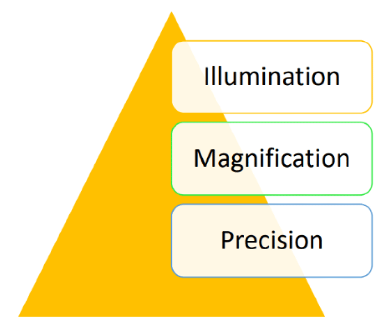
An operating microscope offers three distinct advantages: illumination, magnification, and enhanced precision in surgical technique delivery, collectively referred to as the microsurgical triad [15] (Figure 1).
The use of fiberoptic technology has enhanced illumination methods by allowing focused light to be directed onto specific areas, which has become a standard feature of surgical operating microscopes [17]. The second element of the microsurgical triad, magnification can be attained through either loupes or an operating microscope [6]. The enhanced precision in surgical technique delivery, which constitutes the third component of the microsurgical triad, arises synergistically from the combined effects of illumination and magnification. Improved visual acuity allows for the utilization of smaller instruments with greater precision [16].
Types of magnification
Loupes: Simple loupes are rudimentary magnifying devices with restricted functionalities, comprising a pair of single, positive, side-by-side meniscus lenses. Compound lenses, which are achromatic and significantly enhanced in optical design, mitigate the effects of chromatic and spherical aberrations while bringing two wavelengths into focus in the same plane. Prism loupes offer superior magnification capabilities as they incorporate Schmidt or rooftop prisms [18].
Surgical operating microscope: Surgical microscopes employ Galilean optical principles. In periodontal surgery, an ideal magnification range of 5x to 12x is recommended. These microscopes feature a sophisticated lens configuration that facilitates stereoscopic vision at magnifications ranging approximately from 4x to 40x [18].
Microsurgical instruments
A fundamental microsurgical instrument set includes a needle holder, micro scissors, micro scalpel holder, anatomical and surgical forceps, and a variety of elevators. Enhanced visual acuity allows for the utilization of smaller instruments with greater precision. Microsurgical instruments should be slightly top-heavy, have a circular cross-section, and be approximately 18 cm in length to facilitate proper handling and high-precision movements. To prevent fatigue of hand and arm muscles, the weight of microsurgical instruments should be kept below 15 g – 20 g. Among the variety of suture materials accessible, monofilament non-absorbable sutures are favoured; however, they should be removed at the earliest biologically acceptable time [19].
Utilizing small-sized needles and sutures under magnification enables wound closure with adequate tension and minimizes dead space. Suturing techniques in microsurgery deviate from those in conventional surgery. In microsurgical procedures, needles should penetrate tissues perpendicularly and exit at uniform distances. The suture bite size should be around 1.5 times the tissue thickness to ensure proper wound approximation. Knot tying under the microscope is performed using instrument ties, with a microsurgical needle holder in the dominant hand and a microsurgical tissue pickup in the non-dominant hand [19].
Clinical applications
Application in scaling and root planing: The microscope, with its enhanced magnification and illumination capabilities, facilitates the completion of fundamental periodontal treatments such as scaling and root planing, particularly in challenging areas like furcations. The effectiveness of any periodontal therapy hinges on the meticulous debridement of the root surface [20]. This procedure constitutes a vital aspect of periodontal therapy, exhibiting effectiveness when performed under illumination and with the aid of micro ultrasonic instruments [21]. These instruments, with diameters ranging from 0.2 to 0.6 mm and variable power settings ranging from 25,000 to over 40,000 cycles per second, enable subgingival treatment in deep pockets [22]. Additionally, they feature active working sides on all surfaces, facilitate ultrasonically activated lavage in the treatment area, and can be utilized with minimal water spray, contributing to an improved early healing index and reduced postoperative pain [23].
Application in flap surgery: In Periodontics, flap reflection serves the purpose of exposing the underlying tissues, including bone and the root surface. Employing microsurgical techniques allows for the elevation of periodontal flap margins with consistent thickness, resulting in a scalloped butt joint. This enables precise adaptation of the tissue to the teeth or opposing flap in an edentulous area, effectively eliminating gaps and dead spaces. Consequently, the necessity for new tissue formation is minimized, thereby enhancing periodontal regeneration. The integration of a surgical microscope enhances surgical precision, rendering it an indispensable component of periodontal surgical practice [24].
Application in periodontal regeneration: The benefits of employing a microsurgical approach in regenerative therapy are associated with enhanced illumination and magnification of the surgical field. This facilitates precise access to and debridement of the intrabony defect, resulting in increased accuracy and reduced trauma [25]. Additionally, the capability to achieve and sustain primary wound closure minimizes bacterial contamination, creating more favorable conditions for periodontal regeneration [26].
Application in papilla reconstruction procedures: The microsurgical procedure is a minimally invasive technique utilized to position donor tissue beneath a deficient interdental papilla. Due to the compact size of the interdental papilla and restricted access, surgical magnification and microsurgical instruments are advised. These aids enhance visibility, eliminate the need for unnecessary or unintentional incisions, and facilitate access, thereby enhancing the predictability of the procedure [27].
Application in mucogingival surgery: In contrast to the traditional macrosurgical method for treating gingival recession, the microsurgical approach has demonstrated several advantages. These include enhanced vascularization of grafts, higher rates of root coverage, substantial increases in the width and thickness of keratinized tissue, improved aesthetic outcomes, and reduced patient morbidity [17].
Application in subepithelial connective tissue graft harvesting: By employing magnification and precise instruments, microsurgical techniques facilitate the accurate identification of anatomical landmarks, enabling the harvesting of Subepithelial Connective Tissue Grafts (SCTGs) from the suitable portion of the lamina propria with uniform thickness and minimal risk of injury to the greater palatine artery and surrounding tissues. Additionally, microsurgery preserves a minimal amount of connective tissue in the donor site flap, even in borderline cases, ensuring wound nutrition and enhancing the healing process and postoperative comfort for the patient [28].
Esthetic crown lengthening microsurgery: The crown-lengthening procedure serves two purposes: aesthetic and functional. In both scenarios, the surgical objective is to restore the biological width apically while exposing additional tooth structure [29]. In regions requiring increased aesthetic precision, where preserving papilla and soft tissue is crucial, employing a microsurgical technique is advised. This approach involves smaller incisions that avoid impacting the papillae. Flap reflection is kept minimal, and suturing allows for precise adaptation of the flaps, minimizing inflammation, scarring, and patient discomfort. With procedures being minimally invasive and offering superior wound adaptation, rapid healing, and improved aesthetics can be anticipated [29].
Application in implant therapy: The surgical microscope holds significant value in implant dentistry. Various stages of implant treatment, spanning from implant placement to implant recovery and peri-implantitis management, can be executed with enhanced precision when performed under magnification [13]. Implant microsurgery offers an opportunity for implant therapy that can enhance the esthetic results. Its benefits include rapid healing, minimal discomfort, and improved patient acceptance [30]. The SMILE technique introduces a systematic microsurgical approach for the immediate replacement and restoration of teeth in the esthetic zone [13]. It reduces surgical trauma, shortens the postoperative healing period, and yields improved aesthetic and functional outcomes. Leveraging a transmission operating microscope offers unparalleled advantages for capturing authentic photo and video documentation [31]. The microscope proves advantageous in visualizing the final sutures of the implant for subcrestal placement, ensuring implant recovery with minimal trauma to adjacent tissues, managing peri-implantitis, visualizing the sinus membrane during sinus lift procedures, and reducing the risk of perforations or tears [32].
Application in sinus lift procedures: A modern and innovative application of microsurgery is evident in sinus lift procedures. The use of a surgical microscope reduces the risk of perforations and enables indirect visualization of the sinus membrane, improving accuracy and safety during the procedure. With sinus lift treatments performed under a microscope, success rates reach 97%, enabling indirect observation of the sinus membrane and decreasing the risk of perforation [33].
Wound healing after microsurgery
The recovery process following periodontal/peri-implant microsurgical procedures presents challenges due to the surgical wound’s placement on a rigid, avascular surface of the tooth (or implant). This location leads to reduced local immune defenses and nutrient supply to the involved tissues [34]. Microsutures promote the primary closure of the surgical wound, resulting in edge-to-edge alignment and facilitating primary intention healing [34]. Aside from the minimal scarring risk, microsurgical techniques can expedite graft revascularization [35].
Limitations of microsurgery
Although microsurgical principles and techniques offer numerous advantages, the limited integration of microsurgery into periodontal surgical practice may stem from its inherent limitations. The establishment of a microsurgical setup is costly, time-consuming, and requires specialized training, making it significantly more demanding. Achieving clinical proficiency entails a steep learning curve and mastering physiological tremor control to execute finer movements during surgery [36].
Contemporary advances in microsurgery
Recent innovations in microsurgery encompass the adoption of three-dimensional microsurgery systems, which have not only simplified procedures but also improved precision. Robotic microsurgery stands as a pioneering supplementary instrument, offering heightened magnification and unparalleled accuracy. The incorporation of robotic microsurgery represents an innovative frontier in periodontal therapy that is yet to be fully explored [37].
Conclusion
In conclusion, the review highlights the significant advancements and transformative potential of periodontal and peri-implant microsurgery in contemporary dental practice. From the precision afforded by magnification and specialized instruments to the enhanced healing and aesthetics resulting from minimally invasive approaches, microsurgery emerges as a cornerstone of modern periodontal care. As the field continues to evolve, periodontal surgeons must stay acquainted with the latest developments and incorporate microsurgical principles into their practice.
- Zucchelli G, Mounssif I. Periodontal plastic surgery. Periodontol 2000. 2015 Jun; 68(1):333-68. doi: 10.1111/prd.12059. PMID: 25867992.
- Ramanauskaite A, Obreja K, Schwarz F. Surgical Management of Peri-implantitis. Curr. Oral Health Rep. Springer Science and Business Media B.V. 2020; 7: 283– 303. doi: 10.1111/prd.12417.
- Tibbetts LS, Shanelec D. Periodontal microsurgery. Dent Clin North Am. 1998 Apr; 42(2):339-59. PMID: 9597340.
- Serafin D. Microsurgery: past, present, and future. Plast Reconstr Surg. 1980 Nov; 66(5):781-5. PMID: 7001524.
- Kawaldeep K, Deepak G, Viniti G, Sumit K, Gurpreet K. Periodontal Microsurgery and Microsurgical Instrumentation: A Review. Dental Journal of Advance Studies. 2016; 04:074-080. doi:10.1055/s-0038-1672050.
- Leeuwenhoek AV. The select works of Antony van Leeuwenhoek: containing his microscopical discoveries in many of the works of nature. Arno Press. 1800; 1: 1632-1723.doi: 10.5962/bhl.title.5700
- Daniel RK. Microsurgery: through the looking glass. N Engl J Med. 1979 May 31; 300(22):1251-7. doi: 10.1056/NEJM197905313002205. PMID: 372810.
- Barraquer JI. The history of the microscope in ocular surgery. J Microsurg. 1980 Jan-Feb; 1(4):288-99. doi: 10.1002/micr.1920010407. PMID: 7000963.
- Apotheker H, Jako GJ. A microscope for use in dentistry. J Microsurg. 1981 Fall; 3(1):7-10. doi: 10.1002/micr.1920030104. PMID: 7341737.
- Tibbetts LS, Shanelec DA. An overview of periodontal microsurgery. Curr Opin Periodontol. 1994:187-93. PMID: 8032459.
- Belcher JM. A perspective on periodontal microsurgery. Int J Periodontics Restorative Dent. 2001 Apr; 21(2):191-6. PMID: 11829393.
- Cortellini P, Tonetti MS. Microsurgical approach to periodontal regeneration. Initial evaluation in a case cohort. J Periodontol. 2001 Apr; 72(4):559-69. doi: 10.1902/jop.2001.72.4.559. PMID: 11338311.
- Shanelec DA. Anterior esthetic implants: microsurgical placement in extraction sockets with immediate plovisionals. J Calif Dent Assoc. 2005 Mar; 33(3):233-40. PMID: 15918405.
- Cortellini P, Tonetti MS. A minimally invasive surgical technique with an enamel matrix derivative in the regenerative treatment of intra-bony defects: a novel approach to limit morbidity. J Clin Periodontol. 2007 Jan;34(1):87-93. doi: 10.1111/j.1600-051X.2006.01020.x. PMID: 17243998.
- Mamoun J, Wilkinson ME, Feinbloom R. Surgical and Dental Ergonomic Loupes Magnification and Microscope Design Principles Technical Aspects and Clinical Usage of Keplerian and Galilean Binocular Surgical Loupe Telescopes used in Dentistry or Medicine. Res Sq. 2013; 17:11-22
- Marron-Tarrazzi I. Fundamentals of the operating microscope. Microsurgery in Periodontal and Implant Dentistry: Concepts and Applications. Springer Nat. 2022;1: 47-68.doi:10.1007/978-3-030-96874-8_3
- Chinthakunta V, Sambashivaiah S. Periodontal Microsurgery-A Review. J. adv. res. dent. oral health. 2023;8(2):1–5.doi:10.24321/2456.141X.202301
- Tripathi S, Gupta S, Ahmad Khan M, Gowrav P, Jalali V, Sharda India A. Periodontal Microsurgery-The growing wave of magnification. Tripathi World J. Pharm. Res. 2019; 8(7):375. doi:10.22271/oral.2022.v8.i1h.1474
- Yadav VS, Salaria SK, Bhatia A, Yadav R. Periodontal microsurgery: Reaching new heights of precision. J Indian Soc Periodontol. 2018 Jan-Feb; 22(1):5-11. doi: 10.4103/jisp.jisp_364_17. PMID: 29568165; PMCID: PMC5855270.
- Lindhe J, Westfelt E, Nyman S, Socransky SS, Haffajee AD. Long-term effect of surgical/non-surgical treatment of periodontal disease. J Clin Periodontol. 1984 Aug; 11(7):448-58. doi: 10.1111/j.1600-051x.1984.tb01344.x. PMID: 6378986.
- Pihlstrom BL, Ortiz-Campos C, McHugh RB. A randomized four-years study of periodontal therapy. J Periodontol. 1981 May; 52(5):227-42. doi: 10.1902/jop.1981.52.5.227. PMID: 7017103.
- Reinhardt RA, Johnson GK, Tussing GJ. Root planing with interdental papilla reflection and fiber optic illumination. J Periodontol. 1985 Dec; 56(12):721-6. doi: 10.1902/jop.1985.56.12.721. PMID: 3908643.
- Kwan JY. Enhanced periodontal debridement with the use of micro ultrasonic, periodontal endoscopy. J Calif Dent Assoc. 2005 Mar; 33(3):241-8. Erratum in: J Calif Dent Assoc. 2005 Apr; 33(4):282. PMID: 15918406.
- Rathore P, Manjunath S, Singh R. Evaluating and comparing the efficacy of the microsurgical approach and the conventional approach for the periodontal flap surgical procedure: A randomized controlled trial. Dent Med Probl. 2024 Jan-Feb; 61(1):23-28. doi: 10.17219/dmp/147183. PMID: 35904770.
- Cortellini P, Tonetti MS. Minimally invasive surgical technique and enamel matrix derivative in intra-bony defects. I: Clinical outcomes and morbidity. J Clin Periodontol. 2007 Dec; 34(12):1082-8. doi: 10.1111/j.1600-051X.2007.01144.x. Epub 2007 Oct 22. PMID: 17953696.
- Cortellini P, Tonetti MS. Improved wound stability with a modified minimally invasive surgical technique in the regenerative treatment of isolated interdental intrabony defects. J Clin Periodontol. 2009 Feb; 36(2):157-63. doi: 10.1111/j.1600-051X.2008.01352.x. PMID: 19207892.
- Suryavanshi PP, Bhongade ML. Periodontal Microsurgery: A New Approach to Periodontal Surgery. Int. J. Sci. Res. 2015; 6(3):785-89.
- Kumar MP, Jaswitha V, Gautami SP, Ramesh KSV. Applications of microscope in periodontal therapy- Role in magnification really matters! IP Int J Periodontology Implantology. 2019; 4(1):1–5. https://doi.org/10.18231/j.ijpi.2019.001
- Cortellini P, Cortellini S, Bonaccini D, Stalpers G, Mollo A. Treatment of Teeth with Insufficient Clinical Crown. Part 1: One-Year Clinical Outcomes of a Minimally Invasive Crown-Lengthening Approach. Int J Periodontics Restorative Dent. 2021 Jul-Aug; 41(4):487-496. doi: 10.11607/prd.5660. PMID: 34328465.
- Shanelec DA, Tibbetts LS, Ishikawa I, Butler B, Aoki A, McGregor A, et al. Recent Advances in Surgical Technology. Elsevier sci. 2012; 601–7. doi:10.1016/B978-1-4377-0416-7.00064-0
- Shakibaie-M B. Uses of the operating microscope in minimally invasive implantology. Quintessence Int. 2010; 61(3):293-308
- Awasthi R, Jalaluddin M, Agrawal U, Singh D. Conceptual approach to periodontal microsurgery: An insight. J Prim Care Dent O Hlth. 2022; 3(2):29. doi:10.4103/jpcdoh.jpcdoh_35_21
- Steiner GG, Steiner DM, Herbias MP, Steiner R. Minimally invasive sinus augmentation. J Oral Implantol. 2010; 36(4):295-304. doi: 10.1563/AAID-JOI-D-09-00010. PMID: 20735266.
- Jain R, Kudva P, Kumar R. Periodontal microsurgerymagnifying facts, maximizing results. J Adv Med Dent Sci Res. 2014; 2:24-34.
- de Sanctis M, Clementini M. Flap approaches in plastic periodontal and implant surgery: critical elements in design and execution. J Clin Periodontol. 2014 Apr; 41 Suppl 15:S108-22. doi: 10.1111/jcpe.12189. PMID: 24640996.
- Avhad R, Laddha R, Sewane S, Agrawal S, Sharma D, Upadhye K. Microsurgery in Periodontics: A Review. J Adv Med Dent Scie Res. 2019; 7(6):41-47. doi: 10.21276/jamdsr
- Brahmbhatt JV, Gudeloglu A, Liverneaux P, Parekattil SJ. Robotic microsurgery optimization. Arch Plast Surg. 2014 May; 41(3):225-30. doi: 10.5999/aps.2014.41.3.225. Epub 2014 May 12. PMID: 24883272; PMCID: PMC4037767.
Article Alerts
Subscribe to our articles alerts and stay tuned.
 This work is licensed under a Creative Commons Attribution 4.0 International License.


 Save to Mendeley
