Journal of Surgery and Surgical Research
Gastrointestinal Stromal Tumor (GIST) presenting as acute abdomen: Image in surgery
Med Dheker Touati1,2*, Marwa Idani1,2, Mohamed Raouf Ben Othmane1,2, Fahd Khefacha1,2, Nadhem Khelifi1,2, Anis Belhadj1,2, Ahmed Saidani1,2 and Faouzi Chebbi1,2
2Faculty of Medicine of Tunis, University of Tunis El Manar, R534+F9H, Tunis, Tunisia
Cite this as
Touati MD, Idani M, Ben Othmane MR, Khefacha F, Khelifi N, et al. (2023) Gastrointestinal Stromal Tumor (GIST) presenting as acute abdomen: Image in surgery. J Surg Surgical Res 9(2): 013-014. DOI: 10.17352/2455-2968.000157Copyright
© 2023 Touati MD, et al. This is an open-access article distributed under the terms of the Creative Commons Attribution License, which permits unrestricted use, distribution, and reproduction in any medium, provided the original author and source are credited.Gastrointestinal stromal tumors originating from the small bowel are uncommon. We present the case of a 45-year-old man with no prior medical or surgical history who presented to our hospital's emergency department with abdominal pain, vomiting, inability to pass gas, and no bowel movements. Physical examination revealed abdominal distention and tenderness. His white blood cell count was 10.9 x 10^3/µl, and hemoglobin levels were 13.3 g/dl.
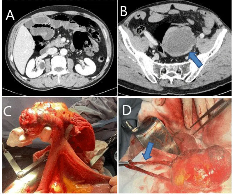
A thoraco-abdomino-pelvic CT scan showed air-fluid levels in the ileo-jejunal loops, a soft tissue density mass measuring approximately 77 mm x 35 mm, and liver metastasis (Figure 1A,B).
The patient underwent surgery due to an acute abdomen from mechanical ileus. Proximal small bowel dilatation and a 9 cm x 4 cm soft-tissue mass protruding from the serosa (Figure 1C) were found without luminal obstruction, approximately 70 cm proximal to the ileocecal valve. Additionally, a flange (Figure 1D) between the tumor and sigmoid caused ileum loop obstruction. Tumor resection with a linear surgical stapler was performed, and pathology confirmed a gastrointestinal stromal tumor.
The patient was discharged on the fourth postoperative day without complications and an Imatinib treatment plan was initiated.
This case underscores the exceptional clinical and diagnostic aspects of small bowel gastrointestinal stromal tumors, contributing valuable insights to the existing medical literature.
Images in surgery
Gastrointestinal Stromal Tumors (GISTs) are rare mesenchymal tumors believed to originate from the interstitial cells of Cajal (ICC), recognized as the pacemaker cells in the Gastrointestinal (GI) tract, bridging autonomic innervation and smooth muscle function [1]. Despite their infrequency, GISTs are the most common mesenchymal tumors affecting the GI tract, accounting for 1% - 3% of all GI malignancies. The annual incidence is estimated at approximately 7 - 20 cases per million population; however, recent data suggest a higher prevalence of microscopic, subclinical lesions [2]. Notably, subcentimetric gastric GISTs may be more common than previously assumed. The median age of presentation is typically 60 years - 65 years, although GISTs can manifest at any age, with a similar occurrence in both men and women [3].
In a specific case, a 45-year-old man, with no prior medical or surgical history, presented to our hospital's emergency department with complaints of abdominal pain, vomiting, inability to pass gas, and absence of bowel movement. Physical examination revealed abdominal distention and tenderness. The white blood cell count was 10.9 x 10^3/µl, and hemoglobin levels were 13.3 g/dl.
A thoraco-abdomino-pelvic Computed Tomography (CT) scan demonstrated air-fluid levels in the ileo-jejunal loops, along with a soft tissue density mass measuring approximately 77 mm x 35 mm at its widest dimension and liver metastasis (Figures 1A,B).
Based on the examination and diagnostic findings, the patient underwent surgery with a diagnosis of acute abdomen due to mechanical ileus. Proximal small bowel dilatation was observed. A tumoral mass of approximately 9 cm x 4 cm, with a soft consistency, protruding from the serosa (Figure 1C), without luminal obstruction, was detected approximately 70 cm proximal to the ileocecal valve. Additionally, a flange (Figure 1D) between the tumor and the sigmoid caused an ileum loop obstruction. Tumor resection was performed using a linear surgical stapler. Pathological examination confirmed the presence of a gastrointestinal stromal tumor.
The patient was discharged on the fourth postoperative day without any complications, and an Imatinib treatment plan was initiated.
Patient consent
Written informed consent was obtained from the patient for the publication of this case report and its accompanying images. A copy of the written consent is available for the Editor-in-Chief of this journal to review upon request.
- Fletcher CD, Berman JJ, Corless C, Gorstein F, Lasota J, Longley BJ, Miettinen M, O'Leary TJ, Remotti H, Rubin BP, Shmookler B, Sobin LH, Weiss SW. Diagnosis of gastrointestinal stromal tumors: A consensus approach. Hum Pathol. 2002 May;33(5):459-65. doi: 10.1053/hupa.2002.123545. PMID: 12094370.
- Kawanowa K, Sakuma Y, Sakurai S, Hishima T, Iwasaki Y, Saito K, Hosoya Y, Nakajima T, Funata N. High incidence of microscopic gastrointestinal stromal tumors in the stomach. Hum Pathol. 2006 Dec;37(12):1527-35. doi: 10.1016/j.humpath.2006.07.002. Epub 2006 Sep 25. PMID: 16996566.
- Besana-Ciani I, Boni L, Dionigi G, Benevento A, Dionigi R. Outcome and long term results of surgical resection for gastrointestinal stromal tumors (GIST). Scand J Surg. 2003;92(3):195-9. doi: 10.1177/145749690309200304. PMID: 14582540.
Article Alerts
Subscribe to our articles alerts and stay tuned.
 This work is licensed under a Creative Commons Attribution 4.0 International License.


 Save to Mendeley
