Journal of Surgery and Surgical Research
Detection of potentially clinically relevant lung nodules with breath-hold CT compared to free-breathing CT in PET-CT in oncology patients and the value of MIP reconstructions
Alemany Montserrat1, Sörensen Jens2, Trampal Carlos1 and Hansen Tomas1*
2Section of Nuclear Medicine and PET, Department of Surgical Sciences, Uppsala University Hospital, SE-75185 Uppsala, Sweden
Cite this as
Montserrat A, Jens S, Carlos T, Tomas H (2020) Detection of potentially clinically relevant lung nodules with breath-hold CT compared to free-breathing CT in PET-CT in oncology patients and the value of MIP reconstructions. J Surg Surgical Res 6(2): 173-177. DOI: 10.17352/2455-2968.000126Background: The value of an additional thoracic breath-hold (BH) CT in PET-CT for detecting small nodules is controversial.
Purpose: To determine the value of a BH-CT compared to the routinely used free-breathing (FB) CT, and the value of maximum intensity projection (MIP) technique in these two CT series for detection and its potential clinical relevance in staging of small lung nodules in oncology patients.
Material and methods: This retrospective study included 200 patients from February to September 2016 who were referred for oncological FDG PET-CT. Thin slice (1.25 mm) and MIP (10 mm) images were analyzed for detection of small lung nodules (1-10 mm). Binominal test and descriptive statistics were analyzed.
Results: It was possible to evaluate 186 patients, with a mean age of 70.0 yrs ± 11.1 yrs, range 27-96 yrs, consisting of 84 females (mean age 68.5 yrs ± 12.2, 27-90 yrs) and 102 men (71.3 yrs ± 10.1, 27-96 yrs). In FB-CT, 393 nodules were detected in thin slice CT images, with MIP in 578. In BH-CT, 534 nodules were detected and 905 with MIP (p< 0,001). The extra detected nodules were considered as having a potential clinical relevance in 4.8% (9 of 186) of the patients. The total radiation dose from the routine PET-CT, including FB-CT, was 26 mSv and an additional 4 mSv from the BH-CT.
Conclusion: An additional BH-CT in deep inspiration compared with FB-CT detects more small lung nodules, which potentially could alter the TNM stage.
Abbreviations
BH: Breath Hold; FB: Free Breathing; MIP: Maximum Intensity Projection
Introduction
With the introduction of a national Swedish healthcare program in 2015 called “Standardized Care Pathway” (SCP), the clinical work up for cancer changed [1]. Previously, a full radiation dose contrast enhanced Computed Tomography (CECT) often existed for staging and treatment planning. The value of Positron Emission Tomography (PET) CT was primarily to detect metabolic active lesions, increasing the likelihood of malignant tumor or spread. In practice, SCP means that the patient should be examined within one week after referral. It is quite common that a PET-CT is performed directly after detection of a suspected lung lesion found on a plain chest X-ray. PET-CT then becomes the baseline examination if the suspected lesion proves to be a cancer.
Due to the lack of a previous diagnostic CT in these patients, the protocol for the CT performed in conjunction with PET has been changed in most PET facilities in Sweden. A full radiation dose CECT is performed albeit in Free-Breathing (FB) due to the need for morphological correlation to the PET data. Images of the lungs in FB often contain moving artifacts, which could hide small nodules crucial for cancer staging and management. The follow-up of cancer patients is most frequently done with CT. Later, when therapy response is assessed with the CT from the previous PET-CT, comparison could be problematic especially for the basal parts of the lungs. Changes in size and number of small nodules are difficult to assess.
Therefore, some institutions add an additional thoracic Breath-Hold (BH) CT in deep inspiration to the routine PET-CT examination. This practice has created controversy based on concerns regarding the increased radiation dose. The clinical value of adding an additional thoracic BH-CT has not been proven and is also controversial in literature [2,3].
Another strategy for detection of nodules could be the addition of 10 mm Maximum Intensity Projection (MIP) reconstructions to the thin slice CT images of the lungs, based on the knowledge that MIP increases the detection of small nodules [4].
The aim of this study was to determine the value of an additional BH-CT in deep inspiration compared to FB-CT in PET-CT and the value of MIP reconstructions in these two CT series for the detection of potentially clinically relevant small lung nodules in oncology patients.
Material and methods
Patient population
This retrospective study included 200 oncology patients from February to September 2016 who were referred for fluorodeoxyglucose (FDG) PET-CT. The most common indication was characterization of a newly detected lung lesion (Table 1).
Image acquisition
The examinations were performed in a Discovery VCT PET-CT scanner (GE Healthcare, Waukesha, WI, U.S.A.). The system was equipped with a 64 slice CT detector. The patients rested while 4 MBq/kg dose of FDG was injected intravenously. After 60 minutes, the patients were placed on the tabletop with arms over the head. After an initial CT scout image, a low radiation dose CT (100 kVp, 40-110 mAs, noise index 20, 3.75 mm slice collimation) images in free-breathing from the base of the skull to the mid-thigh were acquired and later used for the attenuation correction (AC) of PET data. These images were not transferred to the Picture Archive and Communication System (PACS) due to their low image quality. Thereafter, the PET emission scans were collected for three minutes for each bed position; thus, 6 to 8 bed positions were acquired depending on the patient’s height. Iodine contrast agent was injected intravenously, and a breath-hold thoracic CT during deep inspiration was performed after 35 sec (120 kV, 70-780 mAs, noise index 48, 0.625 mm slice collimation). After 70 seconds from the start of the injection, the routine shallow free-breathing CT in venous phase was scanned (120 kV, 70-780 mAs, noise index 48, 0.625 mm slice collimation). The FB-CT images were used for the PET-CT fusion images. The whole-body CT scan with free-breathing was positioned as the range for PET acquisition from the base of the skull to the mid-thigh and included the neck, thorax, and abdomen. The CT scan with deep inspiration only covered the thorax.
CT images of the lung parenchyma were reconstructed with a sharp lung algorithm, with 1.25 mm slice thickness and no overlap. The CT images were analyzed in a PACS workstation (Carestream Health, Rochester, NY, U.S.A.). MIPs were reconstructed with 10 mm slice thickness.
A “small lung nodule” was defined as a non-calcified rounded opacity, smaller than 10 mm surrounded by lung parenchyma. A “potentially clinically relevant nodule” was defined as a nodule that could potentially influence the patient’s management by altering the stage based on the TNM classification system.
2.3 Image analysis: Two radiologists with 15 years (MA), respectively, 12 years (TH) of experience in oncology staging analyzed the 4 sets of CT images for 100 patients each. The series were reviewed in the following order to minimize any bias from pre-existing knowledge of the presence of a nodule: First, the FB scans, then the BH series. Afterwards, the MIP reconstructions of the FB series and finally, the MIP of the BH series were reviewed. The original report for the PET-CT examination was blinded for the radiologists. The number of small lung nodules and the location of the nodules considered as potentially clinically relevant was registered. Consensus readings were performed only for the potentially clinically relevant nodules.
Data were analyzed with institutional radiation review board approval and a waiver of the requirement for informed consent.
Statistical analysis
Binominal test for proportions were used for test of significance of the number of nodules detected on BH MIP versus the other series. Significance level were defined as p< 0.05. Descriptive statistics were calculated for the sample. Mean, ± standard deviation, and range are given. Graphpad (San Diego, CA, USA) were used for statistical analysis.
Results
Fourteen patients were excluded due to missing data set for the evaluation (No. =9), scan excluding parts of the lungs (No. =2) and inability of the patient to hold their breath (No. =3). The remaining sample consisted of 186 patients (70.0 yrs ± 11.1 yrs, 27-96 yrs) with 84 women (68.5 yrs ± 12.2, 27-90 yrs) and 102 men (71.3 yrs ± 10.1, 27-96 yrs).
Small lung nodules were detected in 64.5% (120 of 186) of the patients that were possible to evaluate. With the additional BH-CT, it was possible to detect more small nodules than FB-CT in 42 patients. This figure increased to 51 patients when the MIP reconstructions of the two series of images were analyzed.
The number of the detected nodules for the thin slice FB-CT and the BH-CT series with 1.25 mm slice thickness and MIP reconstructions are given in Table 2. An additional 512 (130.3%, 905/393) small nodules were detected upon reviewing the BH-CT series with the MIP reconstructions as compared with the thin slice 1.25 mm FB-CT series without the MIP reconstructions (p< 0.001).
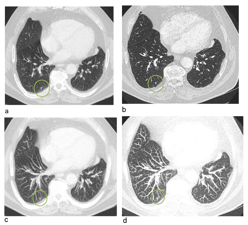
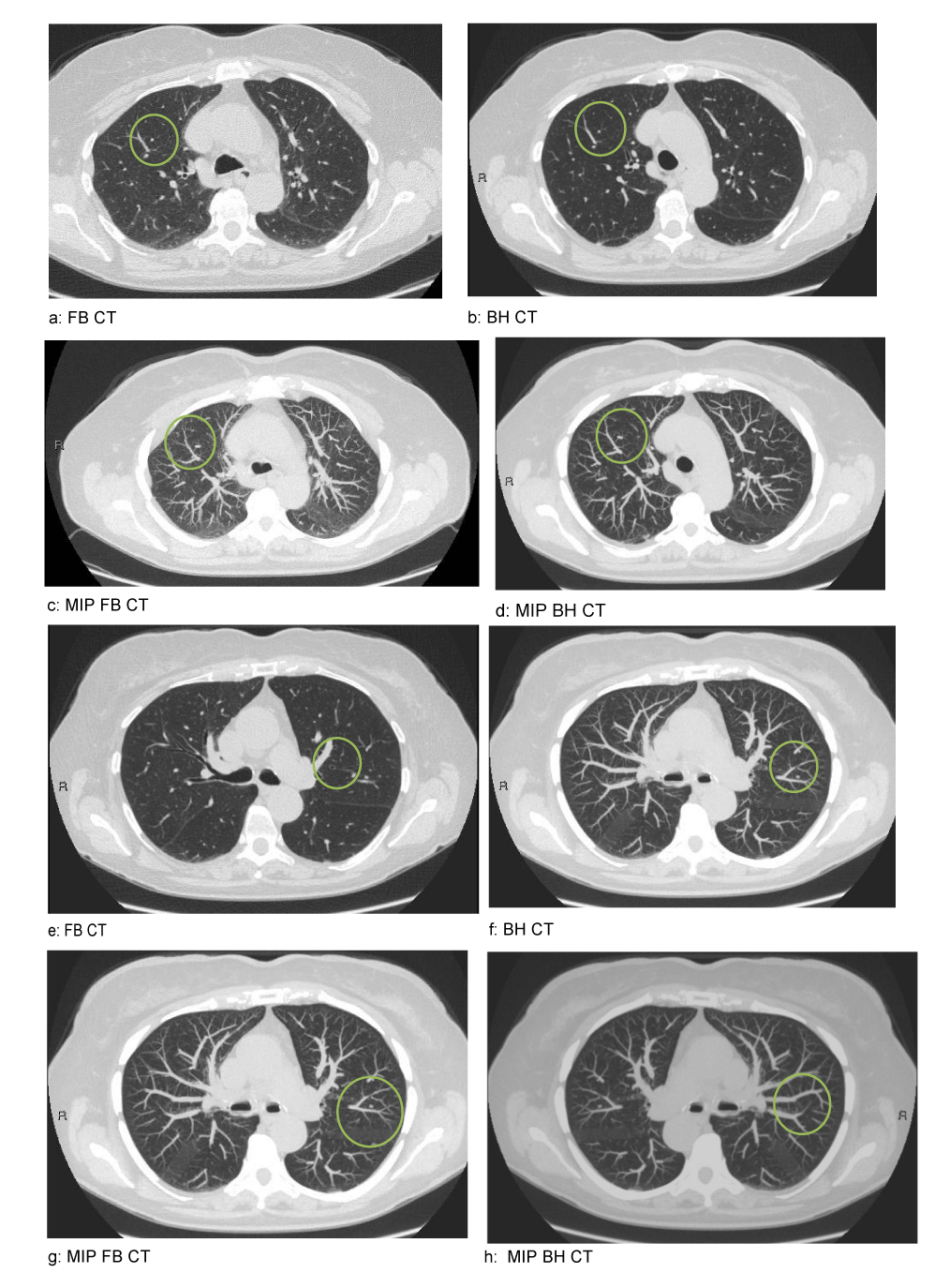
Most of the additionally detected nodules were considered as having no potential clinical relevance as their location could not have an impact on a change in the TNM staging. The thin slice FB-CT missed nodules that potentially could have changed the TNM stage in 4.8% (9 of 186) of the patients as compared with the thin slice BH-CT and additional use of the MIP reconstructions of the FB-CT and BH-CT series. When reviewing the thin slice FB-CT, potentially clinically relevant nodules were not detected in 3.2% (6 of 186) of the patients in comparison with the thin slice BH-CT series. In two patients, two potentially clinically relevant nodules were detected in each patient with a total of eleven nodules in nine patients (Table 3). Examples of potentially clinically relevant nodules are given in Figures 1,2.
The total radiation dose from the routine PET-CT was 26 mSv consisting of 6 mSv from the CT for AC, 6 mSv from the Whole-Body (WB) PET, and 14 mSv from the WB FB-CT from the base of the skull to the mid-thigh in a person weighing 75 kg. The additional thoracic BH-CT in deep inspiration gave an estimated dose of 4 mSv.
Discussion
The value of an additional thoracic BH-CT when performing an FDG PET-CT in oncology patients to detect small lung nodules is controversial. In this study, an additional BH-CT detected more small lung nodules than the FB-CT performed in a conventional PET-CT examination (393 vs. 539). Even more nodules were depicted (578 vs. 905) when MIP reconstructions were used on these two CT series. The nodules not detected on the thin slice FB-CT series were considered as having a potential clinical relevance in 4.8% (9 of 186) of the patients. When reviewing the thin slice FB-CT, comparing with the thin slice BH-CT, potentially clinically relevant nodules were not detected in 3.2% (6 of 186) of the patients.
This finding supports a previous report which stated that BH-CT detected more nodules than FB-CT [2]. In contrast, in a study reporting on the use of thin 2 mm slices instead of thicker 3-5 mm slices for the FB-CT, the 2 mm slices were found sufficient to detect nodules, and an additional BH-CT was considered to be unnecessary [3]. In the present study, thin 1.25 mm slices were used on both FB-CT and BH-CT, and our results support the finding that BH-CT detects more nodules than FB-CT.
When comparing the MIP reconstructions for the thin slice FB-CT scan, potentially clinically relevant nodules were detected in four more patient’s, increasing the percentage of detected nodules to 4.3% (8 of 186) and decreasing the percentage of missed nodules to 0.5% (1 of 186). The last nodule was found only on MIP reconstructions from the BH-CT series. A previous report also stated that more nodules are detected with the use of MIP reconstructions as compared with thin slice images, and the result from the present study supports that statement [5].
This study reports on the ability of the radiologist to detect nodules. Perception is multi-factorial, including components such as image quality, experience, and reader fatigue [6]. Even though a nodule could be clearly depicted on an image, it could still be overlooked by the radiologist and be undetected. There are high expectations regarding the concept of artificial intelligence to support the radiologist in this detection process [7]. The findings in this study support the theory that better image quality, i.e., images of the lungs in deep inspiration and breath-hold for reducing motion artifacts and post processing such as MIP reconstructions, helps the radiologist to detect more nodules [5].
The concept of “potentially clinically relevant nodule” was used in the present study because merely detecting a nodule could be of no importance for the clinical decision. No attempt was made to evaluate if a detected nodule was proven to be benign or malignant, simply considering if the location of such a detected nodule could potentially require further work up to stage the patient in a correct manner. In most of the cases, the nodules would have been too small to be able to biopsy or unlikely to be FDG avid due to the small size. For example, in a lung cancer patient with a tumor size between 5-7 cm, the detection of an additional nodule in a different lobe in the ipsilateral lung would not have the potential to alter the T stage as it already is T3 due to the tumor size according to TNM 8th edition [8]. A nodule detected in another lobe in the ipsilateral lung could change the stage to T4. The finding of a nodule in the contralateral lung could change the M stage from M0 to M1a. Most of the additionally detected nodules in the present study had no potential clinical relevance as their location could not change the TNM stage.
The potential benefit of detecting more nodules is associated with a risk for the patient, in terms of increased radiation dose. If the patient would be under-staged due to nodules not perceived, the likelihood of a more aggressive treatment plan with potential hazards for the patient increases. The use of health care resources would also increase, possibly without benefit for the patient.
The BH-CT added 4 mSv to the patient’s total radiation dose of 26 mSv. In literature, PET dose ranges from 3.5-10.5 mSv, WB CT 11-20 mSv, and CT for AC 3-6 mSv [9]. As technical development occurs, the CT incorporated in the PET-CT could be able to achieve similar image quality for both the FB-CT and the BH-CT series with a reduced radiation dose [10]. In this context and in oncology patients, it seems to possible to justify the additional 4 mSv in most cases.
Motion artefacts are primarily located in the lower lung and upper abdomen. Recently introduced applications for motion correction could increase the spatial correlation between a focal increased uptake in PET and the morphological correlate on CT [11]. Despite these technical advances, an additional CT in deep inspiration would still be needed because small nodules usually do not have enough metabolic activity to exhibit visible FDG uptake, and nodules still need to be detected morphologically on the CT.
Several limitations of the study exist; the CT images were only reviewed by a single radiologist, but this reflects the workflow of clinical reporting in our institution. These results are based on a sample of oncology patients with a skewness toward characterization of lung lesions; thus, the findings of this study could be inappropriate for non-oncology patients and oncology patients without referral for lung findings. No follow-up has been performed on these patients because the aim of the study was to find out if more nodules could be detected with BH-CT as compared with FB-CT, and not whether a detected potentially clinically relevant nodule was benign or malignant.
Conclusion
The addition of BH-CT in deep inspiration and in conjunction with MIP reconstructions of FB-CT and BH-CT can detect more potentially clinically relevant nodules in oncology patients referred for PET-CT.
Funding
This research did not receive any specific grant from funding agencies in the public, commercial, or not-for-profit sectors.
- Wilkens J, Thulesius H, Schmidt I, Carlsson C (2016) The 2015 National Cancer Program in Sweden: Introducing standardized care pathways in a decentralized system. Health Policy 120: 1378-1382. Link: http://bit.ly/2IT3Uv1
- Allen-Auerbach M, Yeom K, Park J, Phelps M, Czernin J (2006) Standard PET/CT of the chest during shallow breathing is inadequate for comprehensive staging of lung cancer. J Nucl Med 47: 298-301. Link: http://bit.ly/2LztbLK
- Flavell RR, Behr SC, Mabray MC, Hernandez-Pampaloni M, Naeger DM (2016) Detecting Pulmonary Nodules in Lung Cancer Patients Using Whole Body FDG PET/CT, High-resolution Lung Reformat of FDG PET/CT, or Diagnostic Breath Hold Chest CT. Acad Radiol 23: 1123-1129. Link: http://bit.ly/3moREQS
- Valencia R, Denecke T, Lehmkuhl L, Fischbach F, Felix R, et al. (2006) Value of axial and coronal maximum intensity projection (MIP) images in the detection of pulmonary nodules by multislice spiral CT: comparison with axial 1-mm and 5-mm slices. Eur Radiol 16: 325-332. Link: http://bit.ly/37tAEVr
- Gruden JF, Ouanounou S, Tigges S, Norris SD, Klausner TS (2002) Incremental benefit of maximum-intensity-projection images on observer detection of small pulmonary nodules revealed by multidetector CT. AJR Am J Roentgenol 179: 149-157. Link: http://bit.ly/3adFR5n
- Krupinski EA (2010) Current perspectives in medical image perception. Atten Percept Psychophys 72: 1205-1217. Link: http://bit.ly/2WkZq3x
- Pehrson LM, Nielsen MB, Lauridsen CA (2019) Automatic Pulmonary Nodule Detection Applying Deep Learning or Machine Learning Algorithms to the LIDC-IDRI Database: A Systematic Review. Diagnostics 9: 29. Link: http://bit.ly/2LLkZIF
- TNM classification of malignat tumors, 8:th edition, John Wiley and Sons Ltd2017.
- Akin EA, Torigian DA, Colletti PM, Yoo DC (2020) Optimizing Oncologic FDG-PET/CT Scans to Decrease Radiation Exposure. https://www.imagewisely.org/Imaging-Modalities/Nuclear-Medicine/Optimizing-Oncologic-FDG-PETCT-Scans. Link: http://bit.ly/3r0WCGX
- Kubo T, Ohno Y, Seo JB, Yamashiro T, Kalender WA, et al. (2017) Securing safe and informative thoracic CT examinations-Progress of radiation dose reduction techniques. Eur J Radiol 86: 313-319. Link: http://bit.ly/34fbAiz
- De Ponti E, Morzenti S, Crivellaro C, Elisei F, Crespi A, et al. (2018) Motion Management in PET/CT: Technological Solutions. Curr Radiopharm 11: 79-85. Link: http://bit.ly/38aluDp
Article Alerts
Subscribe to our articles alerts and stay tuned.
 This work is licensed under a Creative Commons Attribution 4.0 International License.


 Save to Mendeley
