Journal of Surgery and Surgical Research
Adrenal extramedullary hematopoiesis, an entity to consider in the diagnosis of adrenal mass
Garazi Elorza-Echaniz*, Nerea Borda-Aguirrezabalaga, Ignacio Aguirre-Allende, Ainhoa Andres-Imaz, Elisabet Bollo-Arocena and Jose Maria Enriquez-Navascues
Cite this as
Radjenović MR, Radjenović B (2020) Echaniz GE, Aguirrezabalaga NB, Allende IA, Imaz AA, Arocena EB, et al. (2020) Adrenal extramedullary hematopoiesis, an entity to consider in the diagnosis of adrenal mass. J Surg Surgical Res 6(1): 030-032. DOI: 10.17352/2455-2968.000092Extramedullary Hematopoiesis (EMH) is frequently seen in the liver, spleen or lymph nodes in patients with hematologic disorders such as beta-thallasemia or hereditary spherocytosis. We report the first case of adrenal hematopoiesis in a patient with Chronic Myelomonocytic Leukemia.
The patient was a 63 year-old man who was in the study of a thrombocytopenia. A CT and MRI demonstrated a unilateral adrenal mass with multiple splenic lesions.
The patient underwent right adrenalectomy and splenectomy. Microscopic examination showed extramedullary hematopoiesis in the adrenal gland and spleen.
The diagnosis of extramedullary hematopoiesis must be considered in any patient with hematologic disorders and thoracic or abdominal mass. An unnecessary procedure could be avoided.
Palabras clave: Extramedullary hematopoiesis; adrenal mass; adrenal incidentaloma; Chronic Myelomonocytic Leukemia.
Introduction
EMH is defined as a proliferation of the hematopoietic cells outside of the bone marrow, generally due to a failure of blood cells production or to a increased destruction of red blood cells. It is relatively frequent in the spleen, liver and lymph nodes. EMH is associated with hematologic disorders including beta-thalassemia, primary myelofibrosis and hereditary spherocytosis. Hematopoiesis in the adrenals is rare, this is the first case of adrenal hematopoiesis in a patient with Chronic Myelomonocytic Leukemia.
Case report
A 63-year-old man, without previous medical record of interest was incidentally diagnosed of an adrenal mass with splenic lesions. The asymptomatic patient was being studied for thrombocytopenia (75×109/L platelets; Haemoglobin 15.10 g/dL; Mean Corpuscular Volume 92.60 fL, Leukocytes 19×109/L), an alcoholic chronic liver disease was suspected. An ultrasound and a contrast enhanced-CT of the abdomen and pelvis showed an enhancing right adrenal mass, solid, with high uptake, sized 6x6 cm and splenic lesions with contrast enhancement in the arterial phase and portal washout (Figure 1). The finding was confirmed in the MRI. Based on the images the differential diagnosis included a malignant adrenal tumor with splenic metastases or splenic lymphoma. Positron emission tomography revealed hypermetabolic right adrenal mass. Serum biochemical endocrine tests were normal.
The patient underwent right adrenalectomy and splenectomy. In the postoperative course the patient showed intense leucocytosis, monocytosis and improvement of the thrombocytopenia. Considering the blood test Discussion, a peripheral blood smear and a subsequent bone marrow biopsy were performed, with the diagnosis of Chronic Myelomonocytic Leukemia.
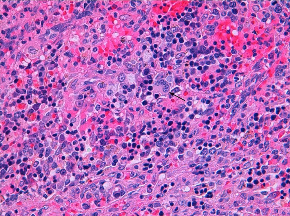
Histopathological examination of adrenal mass was reported as encapsulated hematoma with foci of hematopoiesis (Figure 2) and the splenic lesions also proved massive hematopoiesis.
The postoperative outcome was uneventful and 12 months after surgery he has a stable disease with Hydroxyurea.
Discussion
EMH is relatively common in haematological diseases such as β-thalassemia or myeloproliferative disorders [1], usually located in the spleen, liver or lymph nodes. The adrenal localization is exceptional and only 10 cases have been reported in the literature [1-12]. This is the first published case of adrenal EMH in a patient with chronic myelomonocytic leukemia.
EMH is a compensatory mechanism that occurs in an inappropriate bone marrow function or insufficient to meet circulatory demands. The physiopathological mechanism is unknown; it is believed that the adrenal gland has hematopoietic capacity in the fetus and EMH may arise from embryological rests. There is another hypothesis that hold it may happen by embolization of primitive hematopoietic cells [3].
EMH is usually asymptomatic and is incidentally diagnosed. In symptomatic patients, symptoms are related to the mass effect upon the affected organ. Furthermore, adrenal gland may be involved uni or bilaterally [4].
The difficulty in this case lies in its differential diagnosis. Radiological assessment of a big adrenal mass along with splenic lesions, ignoring the mild haematological disorder, brought us to an adrenal carcinoma with splenic metastasis as the most probable diagnosis. EMH appears on ultrasound examination as a homogeneous, well defined, hypoechoic mass [5] and shows low density in CT [4]. A high index of suspicion, an adrenal biopsy and the previous diagnosis of Chronic Myelomonocytic Leukemia would have been the key for an absolute certainty in diagnosis.
One of the most severe complications after splenectomy in patients with EMH is the massive hepatic hematopoiesis, which may result in fulminant hepatic failure.
Conclusion
In patients with known haematological disease, the presence of hepatic and splenic lesions is relatively common and EMH should be considered. The differential diagnosis in patients with hematologic disorders along with a mass in any location should include EMH, in order to avoid unnecessary procedures and morbidity.
- Banerji JS, Kumar RM, Devasia A (2013) Extramedullary hematopoiesis in the adrenal: Case report and review of literature. Can Urol Assoc J 7: e436-e437. Link: https://bit.ly/3bd83C2
- Martinez-Losada C, Alhambra-Exposito M, Sanchez-Sanchez R, Casaño J, Tenorio-Jimenez C, et al. (2013) Dysplastic extramedullary hematopoiesis with ringed sideroblasts mimicking adrenal adenoma. Histopathology 63: 738-739. Link: https://bit.ly/2SGIvH8
- Keikhaei B, Shirazi AS, Pour MM (2012) Adrenal extramedullary hematopoiesis associated with β-thalassemia major. Hematol Rep 4: e7. Link: https://bit.ly/3doVIME
- King BF, Kopecky KK, Baker MK, Clark SA (1987) Extramedullary hematopoiesis in the adrenal glands: CT characteristics. J Comput Assist Tomogr 11: 342-343. Link: https://bit.ly/2W86LnM
- Lau HY, Lui DC, Ma JK, Wong RW (2011) Sonographic Features of Adrenal Extramedullary Hematopoiesis. J Ultrasound Med 30: 706-7013. Link: https://bit.ly/2W8pu2t
- Porcaro AB, Novella G, Antoniolli SZ, Martignoni G, Brunelli M, et al. (2001) Adrenal extramedullary hematopoiesis: report on a pediatric case and update of the literature. Int Urol Nephrol 33: 601-603. Link: https://bit.ly/3dpc2x3
- Calhoun SK, Murphy RC, Shariati N, Jacir N, Bergman K (2001) Extramedullary hematopoiesis in a child with hereditary spherocytosis: an uncommon cause of an adrenal mass. Pediatr Radiol 31: 879-881. Link: https://bit.ly/3b8Xj80
- Chuang CK, Chu SH, Fang JT, Wu JH (1998) Adrenal extramedullary hematopoietic tumor in a patient with beta-thalassemia. J Formos Med Assoc 97: 431-433. Link: https://bit.ly/2SJro7F
- Wat NM, Tse KK, Chan FL, Lam KS (1998) Adrenal extramedullary haemopoiesis: diagnosis by a non-invasive method. Br J Haematol 100: 725-727. Link: https://bit.ly/3b6fECw
- Karami H, Kosaryan M, Taghipour M, Sharifian R, Aliasgharian, et al. (2014) Extramedullary Hematopoiesis presenting as a right adrenal mass in patient with Beta thalassemia. Nephrourol Mon 6: e19465. Link: https://bit.ly/2W8ADjW
- Kolev NH, Genov PP, Dunev VR, Stoykov BA (2020) A rare case of extramedullary hematopoiesis in adrenal mass. Urol Case Rep 30: 101120. Link: https://bit.ly/2L841QV
- Kannan S, Kulkarni P, Lakshmikantha A, Gadabanahalli K (2017) Extramedullary Haematopoiesis presenting as an adrenal mass. J Clin Diagn Res 11:TJ01
Article Alerts
Subscribe to our articles alerts and stay tuned.
 This work is licensed under a Creative Commons Attribution 4.0 International License.


 Save to Mendeley
