Journal of Surgery and Surgical Research
Closed removable thread vascular anastomosis stent (Lasheen Vascular Stent)
Ahmed E Lasheen*
Cite this as
Lasheen AE (2019) Closed removable thread vascular anastomosis stent (Lasheen Vascular Stent). J Surg Surgical Res 5(2): 071-073. DOI: 10.17352/2455-2968.000076Background: Vascular anastomosis is a most common and important part of many reconstructive transplant procedures. Venous anastomosis can be done by using coupling devices. In arterial anastomosis still the suture method is the main method, which is time consuming, need much experience, and associated by many complications specially with small arteries (microanastomosis). This study offers new type of stent (Lasheen vascular stent) for vascular anastomosis.
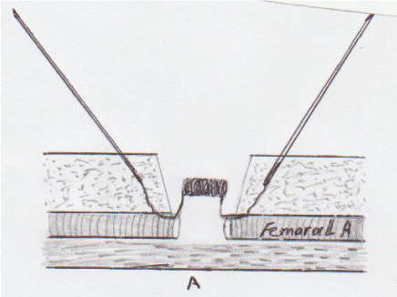
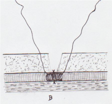
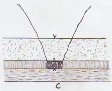
Methods: This experimental study included 7 Egyptian native dogs. Under general anesthesia, the femoral artery was exposed and cut, then re-anastomosis by using thread vascular stent of suitable caliber. Where one end of stent passed from the lumen of distal end of femoral artery and passed through the tissue to appear on skin. Other end of stent passed from the lumen of proximal end of femoral artery through the tissue to appear on skin. Four sutures were taken to approximate two femoral ends on stent. Then, the wound closed in its layers with stent in its position and both stent ends were fixed on skin. After, two weeks the stent was removed by traction on both stent ends to change the stent to thread and come out of body.
Results: The mean time to put the stent in right position was 10 minutes. The prophylactic dose of antithrombotic drug given for three days and antiplatelet aggregation drug for 15 days. All anastomosis were clearly patent by clinical observation and duplex scanning if needed during period of follow up (3 months). No leakage during procedure or hematoma after stent removal were observed.
Conclusion: Lasheen vascular stent for vascular anastomosis is rapid , less time consumed, has short learning curve and good results, and with no special complications.
Introduction
Many surgical procedures as organ and tissue transplantation of re-implantation and reconstruction the vascular anastomosis represent the most important part [1]. The main cause of these surgical procedures failure is problems in vascular anastomosis technique as intima laceration, vessels distortion, unsuitable sutures. This accidents well leading to thrombosis and tissue ischemia [2]. Many researches were done to improved the vascular anastomosis techniques steps and outcome results as using clips, coupling devices, adhesive and gel substances, leaser, and stents [3-7]. The ideal method for vascular anastomosis must be easy (has short learning cure), short time consuming, maintain normal blood flow, and not leading to thrombosis and ischemia immediately or later on.
Methods
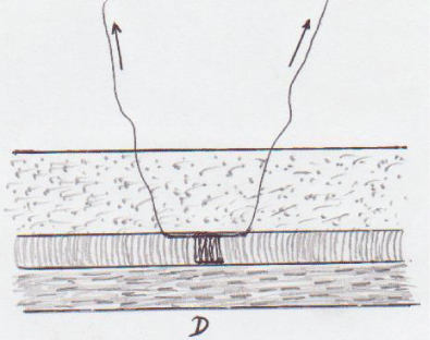
The General Surgery Department, Zagazig University Ethical Committee approved this study before its initiation at September 2018. The thread Vascular stent was prepared by corresponding author for this study. The stent formed from nylon or Prolene of No. 0 or 2/0 twisted in equal multiple circles to form tube (about 1.5 to 2 cm) and these circles adherent together by fibrin glue. The both stent ends are thread in length 10 to 15 cm and each of them fixed inside long fine needle (spinal needle No. 20 or 22) Figure 1. Seven Egyptian native dogs were included in this study and their weight ranged from 15 to 20 kg. For the surgery, in each animal an antecubital vein was cannulated sodium pentothal injected, then intubated and anesthetized with 2% isoflurane in oxygen. With the animal in supine position the medial thigh and groin were shaved and prepared with povidone iodine. Tourniquet was applied on thigh proximal to site of femoral artery exposure. The femoral artery was exposed and transected. The suitable thread stent for femoral artery caliber was prepared. The needle with one stent end passed from lumen of one femoral artery end, then transected artery wall about 1 cm and through tissue to bring stent end on skin. Then do same steps with other stent end and other femoral artery end. The both stent ends were fixed in skin of thigh. Few stitches were taken to approximate two femoral ends. The wound closed in layers with stent in position inside the artery, then removed tourniquet and notice flow, and leakage. Prophylactic dose of anticoagulant (heparin )for three days and antiplatelet aggregation (aspirin) for 2 weeks were given. Broad spectrum antibiotics were given for all dogs for one week. After 2 weeks the stent was removed by application of traction on both stent ends until the stent change to thread, then cut one end on skin and continuous traction on other stent end until all stent come out in form of thread. The patency of each anastomosis was assessed clinically and confirm by duplex ultrasound scanning if needed daily at first 2 weeks, every week for next 2 weeks, every one month for next 2 months.
Results
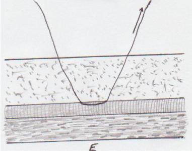
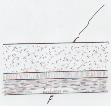
The mean time to put Lasheen vascular stent in right position and finish of anastomosis was 10 minutes. The stent length was varied from 1 to1.5 cm. All anastomosis were clearly patent by palpitation, vitality of dog limb, and confirm by duplex scanning during period of follow up (3 months). No leakage during procedure or hematoma after removal of stent were observed. Stent removal was easy and not associated by any complications (Figures A-E).
Discussion
The tissue transplantation and many reconstructive procedures depends on successful of vascular anastomosis. The characters of ideal vascular anastomosis procedure are simple and has short learning curve, rapid (decrease tissue ischemia time), less vessel wall trauma, and provides good short and long-term patency rates. Many researches were done to find this technique [8,9]. Clips and staples were used as alternative to suture anastomosis technique which suitable to big vessels and with less advantages [10,11]. Glue and adhesives substances as cyanoacrylates and thrombin-based (fibrin-glues) with some success and complications as thrombus formation [12]. Laser assisted vascular anastomosis is largely confined to experimental studies [8,13]. Stents for vascular anastomosis divided to temporary and permanent. Temporary stent may be dissolve, which lysis immediately after finishing of anastomosis procedure as gels [14]. Non dissolved temporary stent, which put during anastomosis technique and removed before closure of line of anastomosis, with some complications as time consuming, rupture of line of anastomosis, and leakage 9. Stent temporary for time of healing and the lysis spontaneously inside the vessel my be leading to vascular obstruction by stent remnants or thrombus formation [6]. Permanent stent as metallic type which suitable for big artery, need for anticoagulant drugs for life, and my be associated by tissue overgrowth [15]. Lasheen vascular stent stay at anastomosis line until complete healing (staying time is complete controlled), when removed not producing any trauma for vessel wall and tissue, and not leaving any remnants. Anticoagulant and antiplatelet agents were given for stent staying time only. In spite our stent was used in dogs (experimental study), no limitation to use lasheen vascular stent in human being by same technique.
Conclusion
Vascular anastomosis with thread stent is quick, simple, has short learning curve, associated with good results on short- and long-term time, and removed without any trauma for vessel wall and tissue (closed removable).
- Byvaltsev VA, Akshulakov Sk, Polkin RA, Ochkal SV, Stepanov IA, et al. (2018) Microvascular anastomosis training in neurosurgery A review. Minim Invasive Surg 2018: 9. Link: http://bit.ly/33ivvLz
- Lebowitz C, Matzon JL (2018) Arterial injury in the upper extremity: Evaluation, strategies, and anticoagulation management. Hand Clin 34: 85-95. Link: http://bit.ly/2MMfFD1
- Kim JT, Kim YH, Kim SW (2017) Effect of fibrin sealant in positioning and stabilizing microvascular pedicle : A comparative study. Microsurgery 37: 206-209. Link: http://bit.ly/2YSMYqD
- Knapp JF, Maxwell M, Briggs C, Shaw ET, Marshall MV, et al. (2017) A sutureless vascular closure device for emergent bovine xenograft implantation. Mil Med 182: 59-65. Link: http://bit.ly/2GVkUws
- Ritschi LM, Ficher AM, Von During M, Mitchell DA, Wolff KD, et al. (2015) Risk of thromboembolus after application of different tissue glues during microvascular anastomosis. Plast Reconstr Surg 136: 1216-1225. Link: http://bit.ly/2MLQHUw
- Farzin A, Miri AK, Sharifi F, Faramarzi N, Jaberi A, et al. (2018) 3D-Printed sugar-based stents facilitating vascular anastomosis. Adv Health Mater 7: e1800702. Link: http://bit.ly/2ZCOZIy
- Wang HH, Ma J, Wang SP, Ma F, Lu JW, et al. (2018) Magnetic anastomosis rings to create portacaval shunt in a canine model of portal hypertension. J Gastrointest Surg Link: http://bit.ly/2YOnbnh
- Leclere FMP, Schoofs M, Buys B, Mordon SR (2010) Outcomes after 1.9-u m diode laser-assisted anastomosis in reconstructive microsurgery: results in 27 patients. Plast Reconstr Surg J 125: 1167-1175. Link: http://bit.ly/2TdqXS9
- Bauer F, Fichter AM, Loeffelbein DJ, Wolff KD, Schutz K, et al. (2015) Microvascular anastomosis using modified micro-stents: A pilot in vivo study. J of Cranio-Maxillofacial Surgery 43: 204-207. Link: http://bit.ly/2ThA6sT
- Reddy C, Pennington D, Stern H (2012) Microvascular anastomosis using the vascular closure device in free flap reconstructive surgery : A13-year experience. J Plast Reconstr Surg 65: 195-200. Link: http://bit.ly/2YKp4BH
- Rozen WM, Whitaker LS, Acosta R (2010) Venous coupler for free flap anastomosis: Outcomes of 1,000 cases. Anticancer Res 30: 1293-1294. Link: http://bit.ly/2yJaKuj
- Cho AB, Wei TH, Torres LR, Junior RM, Rugiero GM, et al. (2009) Fibrin glu application in microvascular anastomosis: Comparative study of two free flaps series. Microsurgery 29: 24-28. Link: http://bit.ly/2KnejgH
- Puca A, Esposito G, Albanese A, Maira G, Rossi F, et al. (2009) Minimally occlusive laser vascular anastomosis (MOLVA); Experimental study. Acta Neurochir 151: 363-368. Link: http://bit.ly/2OMvKLH
- Qassemyar Q, Michel G (2015) A new method of sutureless microvascular anastomosis using a thermosensitive poloxamer and cyanoacrylate: An experimental study. Microsurgery. 35: 315-319. Link: http://bit.ly/2M72tZY
- Prabhu IS, Homer-Vanniasinkam S (2013) A proof-of-principle study on animals for a new method of anastomosing vessels using intraluminal stents. J Craniomaxillofac Surg 41: 327-330. Link: http://bit.ly/2ZFqTNi
Article Alerts
Subscribe to our articles alerts and stay tuned.
 This work is licensed under a Creative Commons Attribution 4.0 International License.


 Save to Mendeley
