Journal of Surgery and Surgical Research
Scrotal emergencies-Two case reports on scrotal exploration scenarios
Abhijit Shah*
Cite this as
Shah A (2018) Scrotal emergencies-Two case reports on scrotal exploration scenarios. J Surg Surgical Res 4(2): 019-022. DOI: 10.17352/2455-2968.000054This case report evaluates the management of acute scrotal emergency in two similar cases with presenting complaints and different outcomes post-scrotal exploration. One case of young male discusses the eventual outcome of testicular torsion and importance of urgent exploration combined with careful cord examination, whilst second case was simply a hematocele requiring excision of sac. There is overall discussion of importance of scrotal exploration, even if at times it may not yield any positive findings.
Introduction
An acute scrotum is defined as an acute painful swelling of the scrotum or its contents, accompanied by local signs or general symptoms. Early identification and skillful management of testicular torsion is critical, as it may threaten testicular viability and future fertility if not managed expediently and appropriately. The cremasteric reflex and testicular sonography are frequently used, yet imperfect, diagnostic tools in assessing for testicular torsion. Other emergent conditions include incarcerated inguinal hernia, Fournier's gangrene, and any form of genitourinary trauma until proven otherwise [1]. Hence the option of scrotal exploration. The aim of these case descriptions is to emphasize the urgency of scrotal exploration and mistakes that occur during the same, and not to be deterred by negative exploration during the same.
Case Scenarios
Case 1
A 19 year old male, presented to GT Hospital Emergency Dept, with acute onset scrotal pain, fever and multiple episodes of vomiting. Patient complains of mild pain 24 hours before the episode during night time whilst the patient was sleeping and with no apparent h/o trauma. Patient was immediately admitted.
On admission patient was tachycardiac (102/min), normotensive (124/70mmHg) and appeared in distress with no evidence of palor or signs of dehydration.
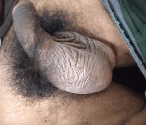
Clinical Examination further revealed no abdominal tenderness, palpable organomegaly or signs of trauma/ previous surgeries. Local Scrotal examination revealed tenderness in the Right Hemiscrotal region with no relief experienced on lifting the testis to relax the cord on the affected side, with no obvious signs of tenderness on the contralateral side. (Figure 1) Chest Xray, Abdominal Xray depicted no significant abnormality. On an emergency ultrasound revealed Right sided Torsion testis with reduced vascularity and emergency urological consult advised emergency Orchidectomy with contralateral Orchidopexy.
Patient was shifted to the Emergency OT within the hour. After due consents and pre-operative preparations, patient was induced under Spinal Anesthesia with careful monitoring.
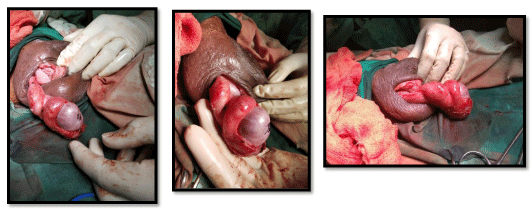
Scrotal exploration, however, revealed healthy viable testis with certain amount of cord edema at the head of epididymis without evidence of any blackening over the testes and without evidence of torsion along the length of cord or the testis itself. Careful re-placement of the testes done with fixation at 3 points with 00-Silk sutures done. Similar steps carried out on the contralateral side. (Figures 2-4).
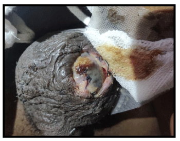
Post-op Day 6 revealed gape in suture line with evidence of serous discharge from side. Careful wound management demonstrated expansion of this gape over next few days and exposure of testes with underlying suture visible. Day 10 revealed blackening over exposed aspect of testes which increased spread over the next 24 hours (Figure 5).
Urgent repeat Doppler performed, demonstrating similar reports of torsion as mentioned previously. Patient shifted to the OT on emergency basis, and scrotal re-exploration done to reveal infracted testes at the local site without evidence of Torsion along the length of cord.
Non-viability of testes prompted Orchidectomy of the affected side. Patient tolerated the procedure well, and shifted back to ward with stable vitals.
Suture site healed well and patient discharged after 7 days of observation.
Case 2
A 63 year old male, visited during OPD hours at GT Hospital Surgery OPD with complaints of Left sided scrotal swelling, pain, fever and cough. Scrotal symptoms were noticed almost 24 hours before presentation. Patient gave no apparent h/o trauma. An outside Ultrasound report was s/o a scrotal Hematoma with Normal echotexture of testes.
Patient was posted for OT under spinal anesthesia with careful monitoring done throughout the procedure. Scrotal exploration revealed almost 50-75cc of tobacco-brown colored fluid with thick wall of the sac (Figures 6,7). Excision of margins performed to reveal a chronic brown fibrosed layer over the testes.Careful separation of this layer revealed healthy testes underneath with viable albuginea surface (Figure 8). Portex Corrugated rubber drain inserted (Figure 9).
Closure performed in layers. Patient shifted back to ward with stable vitals.
Drain removal done on day 3 followed by suture removal on Day 7. Patient discharged on POD 8 with no further complications witnessed.
Case discussion and review of literature
The aim of this study was to detect rate of nosocomial pathogens in burned patients with their susceptibility pattern, and to determine the environmental contamination in Burn and plastic surgery department, aljallahospital, Benghazi.
Testicular torsion refers to the torsion of the spermatic cord structures and subsequent loss of the blood supply to the ipsilateral testicle. This is a urological emergency; early diagnosis and treatment are vital to saving the testicle and preserving future fertility. The rate of testicular viability decreases significantly after 6 hours from onset of symptoms.
A bimodal peak in the incidence of TT is observed beginning in the neonatal period and early adolescence. Most boys who present with an acute scrotum (∼50%) will have torsion of the appendix testis (TAT). However, around 13–20% will have TT, with epididymo-orchitis the most common of the other contributing conditions [2]. However, torsion may occasionally occur in men 40-50 years old.
Testicular torsion is caused by twisting of the spermatic cord and the blood supply to the testicle (see the image below). With mature attachments, the tunica vaginalis is attached securely to the posterior lateral aspect of the testicle, and, within it, the spermatic cord is not very mobile. If the attachment of the tunica vaginalis to the testicle is inappropriately high, the spermatic cord can rotate within it, which can lead to intravaginal torsion. This defect is referred to as the bell clapper deformity. This occurs in about 17% of males and is bilateral in 40% [3].
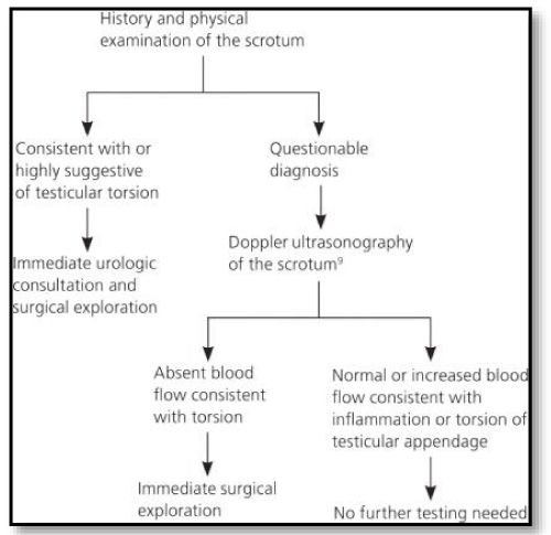
Surgical treatment may prevent further ischemic damage to the testis [4]. Rarely, observation is appropriate, depending on the pathology. Diagnosis of testicular torsion is clinical, and diagnostic testing should not delay treatment.
Testicular rupture, extra-testicular haematoma and a haematocele are the most common sequelae of testicular trauma. An uncommon finding is an isolated intra-testicular haematoma, which has a variable appearance on ultrasound with a temporal change in characteristics on repeat ultrasound. A history of trauma to the scrotum should prompt the diagnosis. Acutely the haematoma appears as patchy increased reflectivity that becomes darker and decreases in size as the haematoma retracts, to eventually resolve completely. The haematoma demonstrates no colour Doppler flow, a low reflective rim thought to represent surrounding oedema, and internal echoes. The most important differential is that of an intra-testicular tumour but a lack of lesion vascularity and the clinical history should lead to the correct diagnosis. The addition of normal tumour markers and a decrease in lesion size on sequential scans are useful [5,6].
Scrotal Doppler can also detect Segmetal ischaemia, which is a rare scenario usually idiopathic or seen in rheumatologic or hematological cases [7].
In the first case of confirmed torsion, scrotal exploration performed had revealed a viable testis. However over period of days, testis eventually underwent gangrenous changes. Cause being a suspected high lying torsion along the cord structures or a possible bell clapper deformity.
Aggressive exploration is a potential possibility of salvaging such cases on an emergency basis.
In the second case presenting with similar complaints, it proved to be a clear-cut case of fluid filled hematocele with a fibrotic layer lining the testis, on removal of which revealed a viable testis. Kept under observation, to reveal a healthy suture line without any complaints. However like the preceding case even this case failed to present a traumatic event predisposing to this condition.
This report is in agreement to the findings of BARADA, WEINGARTEN AND CROMIE [8] about role of salvage surgery prior to the 10 hour mark after which the resultant outcome is less than 20% of salvaging the testis. As per the study, in the first case permanent fixation was performed with non absorbable suture, as there have been reports of repeated torsion with absorbale sutures [9]. However Rodriguez and Kaplan document similar results in absorbable vs non absorbable suture fixation via experimental animal studies [10].
Why should a trauma surgeon/emergency doctor be aware of such a scenario?
Scrotal exploration is a must in any patient presenting with such complaints. However one should be prepared for a negative exploration as well, as torsion of a testicular or epididymal appendage can present similar to various other conditions like-Orchitis, Testis neoplasm, with or without hemorrhage, Testicular abscess, Traumatic hydrocele or hematocele, Henoch-Schönlein purpura, Idiopathic scrotal edema, Scrotal fat necrosis, Scrotal skin infection or inflammation: cellulitis, infected sebaceous cyst, Incarcerated scrotal hernia, Other intraperitoneal process manifesting in scrotum (e.g., meconium peritonitis) [11].
In the event it does turn out to be torsion of testis, there is a significant chance of salvaging the same on a simple de-rotation and orchiopexy procedure. Total ischemia time is the single most important factor in testicular salvage after torsion. Any diagnostic studies beyond those performed at the bedside further delay definitive treatment [8].
The role of ancillary studies should not be used to confirm a diagnosis of torsion preoperatively.
If treated either manually or surgically within six hours, there is a high chance (approx. 90%) of saving the testicle. At 12 hours the rate decreases to 50%; at 24 hours it drops to 10%, and after 24 hours the ability to save the testicle approaches 0 [12]. About 40% of cases result in loss of the testicle [13].
“Comparative study of the fertility potential of men with only one testis.” By Ferreira U and team, concluded that the fertility potential was definitely lower in men with one testis, and hence the role of early diagnosis and management of scrotal emergency in these young males has tremendous value, with scrotal exploration taking precedence over Doppler Ultrasound studies.
Conclusion
These two cases give a very ideal comparative scenario in a case of acute scrotum and the similar line of managements however different outcomes for the same. It ascertains the fact, on lines of proven medical dictum, that scrotal exploration within the first six hours gives the best chance for survival of the affected testis along with the need for aggressive, higher up exploration of the scrotal and cord contents, with a risk of negative exploration at the same time in these patients. This can salvage the testicular function and also have an improved psychological outcome in the young male patients.
- Davis JE, Silverman M (2011) Scrotal Emergencies. Emerg Med Clin North Am. 29:469-484.
- Ta A, D’Arcy FT, Hoag N, D’Arcy JP, Lawrentschuk N (2016) Testicular torsion and the acute scrotum: Current emergency management. Eur J Emerg Med. 23:160-165. Link: https://goo.gl/5FcQwB
- Testicular Torsion (2018) Practice Essentials, Anatomy, Pathophysiology. Link: https://goo.gl/aPHh6y
- Molokwu CN, Somani BK, Goodman CM (2011) Outcomes of scrotal exploration for acute scrotal pain suspicious of testicular torsion: a consecutive case series of 173 patients. BJU Int. 107:990-993. Link: https://goo.gl/NtG39b
- Hematocele - an overview Science Direct Topics (2018). Link: https://goo.gl/gJQe8W
- Cass AS, Luxenberg M (1988) Value of early operation in blunt testicular contusion with hematocele. J Urol. 5347. Link: https://goo.gl/Mj1Tsz
- White WM, Brewer ME, Kim ED (2006) Segmental ischemia of testis secondary to intermittent testicular torsion. Urology. 68: 670-671. Link: https://goo.gl/jQNmf5
- Barada JH, Weingarten JL, Cromie WJ (1989) Testicular salvage and age-related delay in the presentation of testicular torsion. J Urol. 142:746-748. Link: https://goo.gl/Nixjqk
- Vorstman B, Rothwell D (1982) Spermatic cord torsion following previous surgical fixation. J Urol. Link: https://goo.gl/Zc6NxD
- Rodriguez LE, Kaplan GW (1988) An experimental study of methods to produce intrascrotal testicular fixation. J Urol. Link: https://goo.gl/FEQzBX
- Hulbert WC, Rabinowitz R, Mevorach RA (2007) Testicular Torsion. Pediatr Clin Advis. 554-555.
- Wampler SM, Llanes M (2010) Common scrotal and testicular problems. Prim Care. 37: 613-26. Link: https://goo.gl/UapveS
- Sharp VJ, Kieran K, Arlen AM (2013) Testicular torsion: diagnosis, evaluation, and management. Am Fam Physician. 88:835-840. Link: https://goo.gl/wyLMVo
- Liang T, Metcalfe P, Sevcik W, Noga M (2013) Retrospective review of diagnosis and treatment in children presenting to the pediatric department with acute scrotum. Am J Roentgenol. 200: 444-449. Link: https://goo.gl/pWFyDh
- American Academy of Family Physicians. VJ, Kieran K, Arlen AM (2013) American Family Physician. Vol 88. American Academy of Family Physicians; 1970. Link: https://goo.gl/r8aB7j
Article Alerts
Subscribe to our articles alerts and stay tuned.
 This work is licensed under a Creative Commons Attribution 4.0 International License.


 Save to Mendeley
