Journal of Surgery and Surgical Research
Arthroscopic Morphology of Labrum Tear in Recurrent Anterior Dislocation of Shoulder
Dung Tran Trung1,2, Manh Nguyen Huu2, Tuyen Nguyen Trung1,2, Hieu Pham Trung1,2, Nam Vu Tu1,2, Phuong Nguyen Huy1,2, Lam Tran Quoc2, Thai Nguyen Hoang1 and Sang Nguyen Tran Quang1
2Department of Orthopaedic Surgery, Saint Paul Hospital, Ha Noi, Vietnam
Cite this as
Trung DT, Huu MN, Trung TN, Trung HP, Tu NV, et al. (2017) Arthroscopic Morphology of Labrum Tear in Recurrent Anterior Dislocation of Shoulder. J Surg Surgical Res 3(2): 057-060. DOI: 10.17352/2455-2968.000048Objective: Evaluate the characters of labrum tear morphology in recurrent anterior dislocation of shoulder via arthroscopy.
Material and method: Retrospective demographic study 55 recurrent shoulder dislocation patients underwent arthroscopic labrum repair in Saint Paul University Hospital and Hanoi Medical University Hospital from 2009 to 2013.
Results: 65.5% patients with grade 2 tear with 2-3 cm length; The location of tear is anterior and anterior inferior occupied 81.9%; mobile torn labrum is 50.9% and bone healed labrum is 34.6%.
Conclusion: The morphology of labrum tear in recurrent shoulder dislocation is various.
Objective
Recurrent anterior dislocation of shoulder is a popular lesion in rheumatic musculoskeletal as well as orthopaedics and trauma clinical practice. Recurrent anterior dislocation of shoulder accounts for more than 80% of shoulder dislocation cases [1-5]. If first aid for the lesion is not properly given, or if the lesion is too serious, the recurrence is highly likely [1]. At early stage, when the lesion occurs mostly in the labrum, and the joint capsule is not much stressed, the dislocation happens in complicated sport actions. When dislocation is recurrent for a number of times, it becomes easier to happen; patients can even reduce the joint by themselves. This often involves the lesion of glenoid bone and possibly a part of the humerus (Hill Sachs Lesion) [1-3].
Arthroscopic labrum repair has been agreed by most authors to be used as a treatment for simple labrum tear, without the lesion in glenoid bone and humerus. Clinically, most of these cases have a small number of recurrence, under 10 times, and dislocation reduction is still difficult to perform [2-7].
However, labrum tear morphology in recurrent anterior dislocation varies in sizes, grade and combination. Morphology of labrum tear is quite important because it is directly related to the possibility of labrum repair with suture, possibility of recovery of shoulder muscle and especially the possibility of preventing dislocation, possibility of the labrum to heal and continue to develop after the repairing arthoscopy [1,3].
During 5 years, from 2009 to 2013, in Saint Paul University Hospital and Hanoi Medical University Hospital, we performed arthroscopy labrum repair with suture for 55 patients with recurrent anterior dislocation of the shoulder.
This report aims to discuss the morphology of labrum tear in recurrent anterior dislocation of shoulder via arthroscopy.
Patients and Research Method
Patients
55 patients with labrum tear in recurrent anterior dislocation of the shoulder underwent labrum tear arthroscopy surgery in Saint Paul University Hospital and Hanoi Medical University Hospital from 2009 to 2013.
Research methodology
Applying the description method.
Surgery method
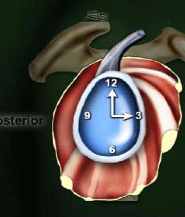
The patients were given endotracheal anesthesia and underwent the surgery in 1 of the 2 position: the “beach chair” position and Decubitus position. The joints were entered in three portals: posterior portal, anterior superior portal and anterior inferior portal, in which 2 portals (anterior and anterior inferior portal used plastic trocar to facilitate the arthroscopy labrum repair with suture. Water pressure pump was used to prevent joint stretching, all anterior, superior and posterior labrum was assessed. The area edge and location of the anterior labrum tear was identified and the labrum was partitioned following clock map to facilitate labrum tear morphology.
- Labrum tear was categorized based on location, following the clock map.
The size of labrum tear was identified based on the number of anchors used to repair the labrum with the average distance between the anchors of 1-1.5cm, with 3 grades:
+ Grade 1: repair with 1 anchor only
+ Grade 2: repair with 2 anchors
+ Grade 3: repair with 3 anchors
The labrum tear morphology was categorized into 3 types:
+ Type 1: Labrum tear separated from the glenoid fossa, mobile torn labrum
+ Type 2: Labrum tear with the labrum tear healed to the glenoid bone
+ Type 3: Labrum tear, the tear has been digested or degenerated
- Data were processed using SPSS 16.0 with bio-medical statistical algorithms
- Research ethics: All patients agreed to give permission for surgery, allowing their medical information to be used for research purpose with personal information unrevealed.
Research Results
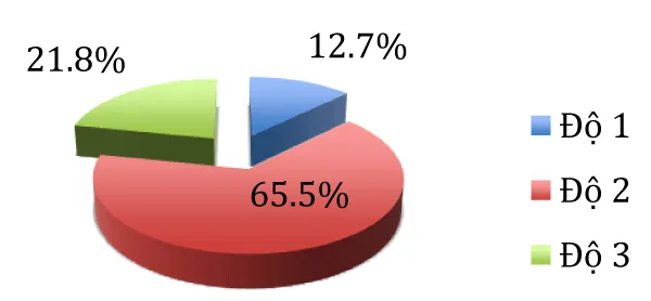
Lesion was found on the right hands of all patients, no patient had the lesion on the left hand. A majority of patients were male, accounting for 98.2% (54/55 patients); the only one female patient was a volleyball player who encountered lesion during competition. The average age of patients was 24.6 ±3.34 years old, in which the youngest was 20 years old and the oldest was 30 years old (Chart 1).
Evaluation
Most of the lesion felt in to grade 2, with 2 anchors – equivalent to the tear length of 2-3 cm (Table 1).
Evaluation
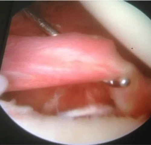
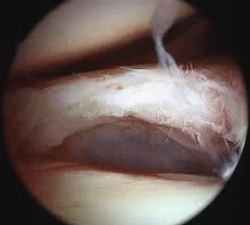
Most of the cases encountered the anterior labrum tear (2-4h on clock map) and anterior inferior labrum tear (3-5h on clock map). There were 3 cases with complicated lesion on the whole anterior labrum (from 1 to 6 h). There were 7 cases with anterior superior labrum tear (1-3h on clock map) (Figures 1,2) (Table 2).
Evaluation
In most of the cases, the labrum tear was mobile out of glenoid bone, accounting for 50.9%. There were 7 cases (12.7%) with degeneration signs and digestion for a part of the labrum.
Discussion
Labrum tear occurred mostly among young people in sports activities, therefore more popular in male than female. The average age was 24.6 ±3.34 years old. The characteristics of patients in our research quite resembled that of researches by other authors of the same topic [4,8,9].
Most of the lesions felt into grade 2, with the labrum tear length from 2-3cm. Among the rest, the proportion of grade 1 and grade 3 was approximate. We encountered mostly grade-two lesion because of our regulation that arthroscopy surgery was given as treatment for cases with labrum tear without lesion in glenoid bones and humerus.
In most of the dislocation cases in our research, the number of dislocation times was low, under 10 times; therefore labrum lesion was of average grade, the arthroscopy performance was quite quick and easy to perform. Most of other authors also stated that a majority of the cases they encountered were of the grade-two lesion, undergoing two-anchor arthroscopy [4,9]. Whereas, 21.8% of cases were complicated grade-three labrum tear with a length greater than 3 cm, with 3 anchors. In such cases, the patients encountered a number of dislocations while treatment at the first time of dislocation was not appropriate, which caused the tear unable to be stablized. The percentage of grade-three tear that we encountered was higher than that in the researches of some authors [4,7] but lower than some of the others [5,9]. This might be explained that the grade of lesion depended on the sample group of patients; and other authors also explained the difference in the proportion of grade-three lesion in their reseaches. As much as 12.7% of cases were grade-one lesion, with a small number of dislocation, under 5 times, however the patients wanted to receive the surgery treatment so that they could get back to playing sports. This group of patient is not large because in actual clinical practices, some patients who have grade-one lesion are not willing to undergo surgery and choose not to play sports to prevent future dislocation. In grade-one lesion, the joint capsule is not stretched and the labrum is not degenerated yet, which is the ideal condition for positive result of arthroscopy surgery. Other authors also reported almost perfect result of arthrocopy surgery among grade-one patients [4,5,7,9].
Table 2 shows that most of the cases encountered anterior labrum tear and anterior inferior labrum tear (Figure 2), accounting for 81.9%. This result quite resembled the results reported by other authors [2]. Most of the authors believed that the mechanic of the lesion was related to the movement of the shoulder joint that let the humerus move to the front, which was the popular movement in hand-dominant sports such as throwing balls, using rackets,.. [1,3]. As much as 12,7% of cases encountered anterior superior labrum tear, in which the tear reached nearly the long head attaches to the top of glenoid, but with no damage to the long head attaches. There were 3 patients, or 5.4%, having complicated lesion with damage in almost whole of the anterior labrum, from the long head attaches to 6h on clock map (Figure 3). This lesion was rarer compared to other types of lesion, mostly encountered by patients with heavy and lengthy first time dislocation of the shoulder joint, the difficulties in reducing the dislocation caused more and more damages in the following dislocation reduction. Among these patients, the frequency of dislocation was not high, however the possibility of dislocation was high, which affected their daily activities, so the patients desired to undergo surgery. Other authors also reported the low ratio of this lesion [2,8,9].
Post-surgery check-up showed labrum involution among 12.7% of patients undergoing the surgery, even loss of a fragment in one patient, possibly because a part of the mobile labrum was torn mobile and then digested. We had to repair the remained labrum using suture, and the digested fragment was supplemented by capsular plication into place labrum lesions. As much as 50.9% of cases had mobile labrum tear, and 36.4% cases had torn labrum healed to the glenoid bone. Some authors described the similar lesion morphology and argued that the bone healed tear might have had happened from the first time of dislocation, therefore the chance of recurrence was high, however the labrum lesion was less likely to get more serious. In cases with mobile labrum tear, the involution was more highly likely due to low blood supply [1,2].
Conclusion
Based on researches on 55 cases of labrum tear in recurrent anterior dislocation of shoulder undergoing arthroscopy, we found that:
- 65.5% of cases had grade-two lesion, with length from 2-3 cm.
- 81.9% of cases were anterior and inferior anterior labrum tear
- 50.9% of cases were mobile labrum tear and 36.4% were bone healed labrum tear
- Dumont GD, Russell RD, Robertson WJ (2011) Anterior shoulder instability: a review of pathoanatomy, diagnosis and treatment. Curr Rev Musculoskelet Med 4: 200-207. Link: https://goo.gl/2XAuZP
- Godin J, Sekiya JK (2011) Systematic review of arthroscopic versus open repair for recurrent anterior shoulder dislocations. Sports Health 3: 396-404. Link: https://goo.gl/QHwJcN
- Anakwenze OA, Huffman GR (2011) Evaluation and treatment of shoulder instability. Phys Sportsmed 39: 149-157. Link: https://goo.gl/gXYfeT
- Privitera DM, Bisson LJ, Marzo JM (2012) Minimum 10-year follow-up of arthroscopic intra-articular Bankart repair using bioabsorbable tacks. Am J Sports Med 40: 100-107. Link: https://goo.gl/ioZQB8
- Lenart BA, Sherman SL, Mall NA, Gochanour E, Twigg SL, et al. (2012) Arthroscopic repair for posterior shoulder instability”. Arthroscopy 28: 1337-1343. Link: https://goo.gl/gZH1pd
- Martetschläger F, Kraus TM, Hardy P, Millett PJ (2013) Arthroscopic management of anterior shoulder instability with glenoid bone defects”. Knee Surg Sports Traumatol Arthrosc 21: 2867-2876. Link: https://goo.gl/ZyQmrc
- Ricchetti ET, Ciccotti MC, O'Brien DF, DiPaola MJ, DeLuca PF, et al. (2012) Outcomes of arthroscopic repair of panlabral tears of the glenohumeral joint. Am J Sports Med 40: 2561-2568. Link: https://goo.gl/BGnoiJ
- Park MJ, Tjoumakaris FP, Garcia G, Patel A, Kelly JD 4th (2012) Arthroscopic remplissage with Bankart repair for the treatment of glenohumeral instability with Hill-Sachs defects. Arthroscopy 27: 1187-1194. Link: https://goo.gl/QEaFwr
- van der Linde JA, van Kampen DA, Terwee CB, Dijksman LM, Kleinjan G, et al. (2011) Long-term results after arthroscopic shoulder stabilization using suture anchors: an 8- to 10-year follow-up. Am J Sports Med 39: 2396-2403. Link: https://goo.gl/NNDZ8U
Article Alerts
Subscribe to our articles alerts and stay tuned.
 This work is licensed under a Creative Commons Attribution 4.0 International License.

 Save to Mendeley
