Journal of Surgery and Surgical Research
An Exceptional Accident: Penetrating Craniofacial and Head Injury with a Double Harpoon – A Case Report
Dokuz Eylül University Faculty of Medicine, Department of Anesthesiology and Reanimation, Turkey
Author and article information
Cite this as
Şahin O, et al. An Exceptional Accident: Penetrating Craniofacial and Head Injury with a Double Harpoon – A Case Report. J Surg Surgical Res. 2026; 12(2): 17-20. Available from: 10.17352/2455-2968.000172
Copyright License
© 2026 Şahin O, et al. This is an open-access article distributed under the terms of the Creative Commons Attribution License, which permits unrestricted use, distribution, and reproduction in any medium, provided the original author and source are credited.Background: Penetrating craniofacial and head injuries caused by underwater harpoon guns are exceedingly rare, particularly when occurring outside an aquatic environment. Simultaneous injury with a double-pronged harpoon presents exceptional biomechanical, surgical, and anesthetic challenges due to dual trajectories traversing critical neurovascular and upper airway structures.
Case Presentation: A 24-year-old male presented to the emergency department following an accidental submental penetrating injury caused by a double harpoon. Despite significant oral and nasal hemorrhage and severely restricted mouth opening (<2 cm), the patient was conscious with a Glasgow Coma Scale (GCS) score of 15 and no focal neurological deficits. Computed tomography (CT) revealed that both metallic shafts traversed the tongue, hard palate, and nasal cavity, terminating against the inner table of the frontal sinus without dural violation or major vascular injury. Prior to surgical extraction, firefighters shortened the externally protruding shafts to facilitate positioning and airway access. Due to impossible orotracheal and nasotracheal intubation, a surgical tracheostomy was performed under deep sedation using a multimodal regimen centered on dexmedetomidine, preserving spontaneous respiration and protective laryngeal reflexes. Following airway securement, general anesthesia was induced, and both harpoons were extracted smoothly without hemorrhage or cerebrospinal fluid (CSF) leakage. The patient recovered uneventfully and remained neurologically and cognitively intact at the six-month follow-up.
Conclusion: Managing double-harpoon craniofacial injuries requires a coordinated multidisciplinary approach. Preoperative shaft shortening, targeted cranial and vascular imaging, and spontaneous-breathing tracheostomy under dexmedetomidine sedation are decisive strategies to prevent catastrophic airway loss and secondary neurovascular trauma.
Penetrating craniofacial and traumatic brain injuries (TBIs) resulting from spearguns or harpoon guns are rare clinical entities, especially when discharged out of water. Underwater harpoon guns store substantial static mechanical energy designed to overcome water resistance; consequently, accidental discharge in open air imparts high kinetic energy to the projectile, resulting in deep penetration and severe tissue disruption. While isolated single-spear injuries to the head and neck have been sporadically documented in the literature, penetrating trauma involving a simultaneous double-pronged harpoon is exceptionally uncommon [1-4].
The clinical significance of a double-harpoon injury lies in its complex dual-trajectory biomechanics, which threaten multiple anatomical compartments simultaneously, including the submental space, floor of the mouth, upper airway, paranasal sinuses, skull base, and intracranial vault. Furthermore, the presence of long, rigid metallic foreign bodies protruding externally creates immediate mechanical obstacles to patient positioning, neuroimaging, and conventional endotracheal intubation. Although previous reports have primarily focused on surgical extraction techniques, comprehensive descriptions of emergency airway securement and perioperative anesthesiological management remain scarce [3,5]. This report highlights the unique clinical presentation, multidisciplinary coordination, and spontaneous-breathing anesthetic strategy utilized to safely manage a life-threatening double-harpoon craniofacial injury.
Case presentation
A 24-year-old male was admitted to the emergency department via ambulance following an accidental discharge of a double-pronged harpoon gun beneath his chin. Upon arrival, the patient was conscious, alert, and fully oriented, with a Glasgow Coma Scale (GCS) score of 15 and bilaterally equal, reactive pupils. His initial vital signs were stable, with an arterial blood pressure of 130/80 mmHg and a respiratory rate of 20 breaths/min. Comprehensive neurological and ophthalmological examinations revealed intact cranial nerve function, normal visual acuity, and no motor or sensory deficits.
Physical examination demonstrated two parallel metallic harpoon shafts entering through the submental region, accompanied by active bleeding from the oral and nasal cavities (Figures 1 and 2). Inspection of the nasal cavity revealed no clear rhinorrhea suggestive of cerebrospinal fluid (CSF) leakage. Interincisal mouth opening was severely restricted to less than 2 cm due to mechanical fixation of the tongue and floor of the mouth by the metallic shafts.
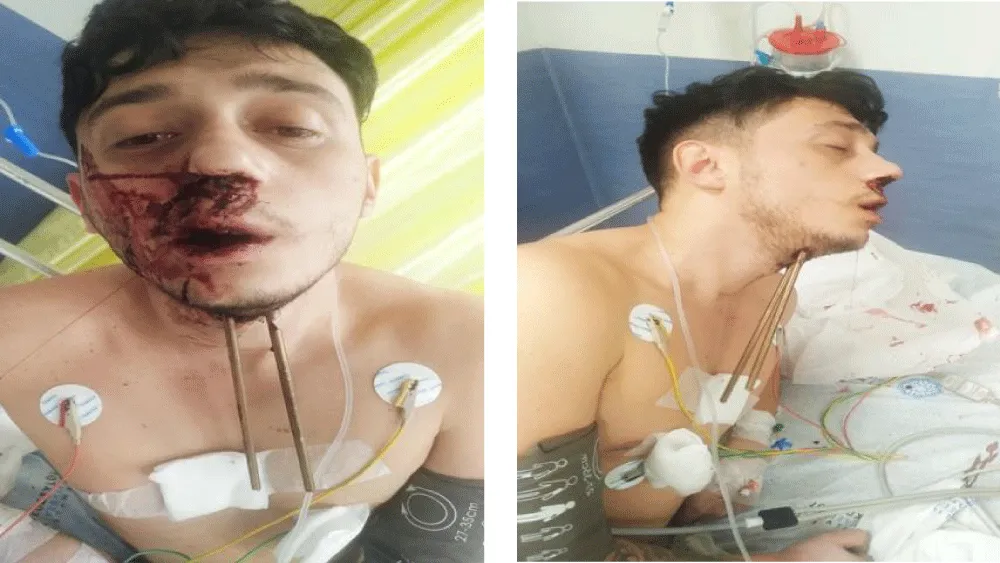
Clinical photographs of the patient upon admission to the emergency department, showing two parallel harpoon shafts entering the submental region with active oral and nasal bleeding.
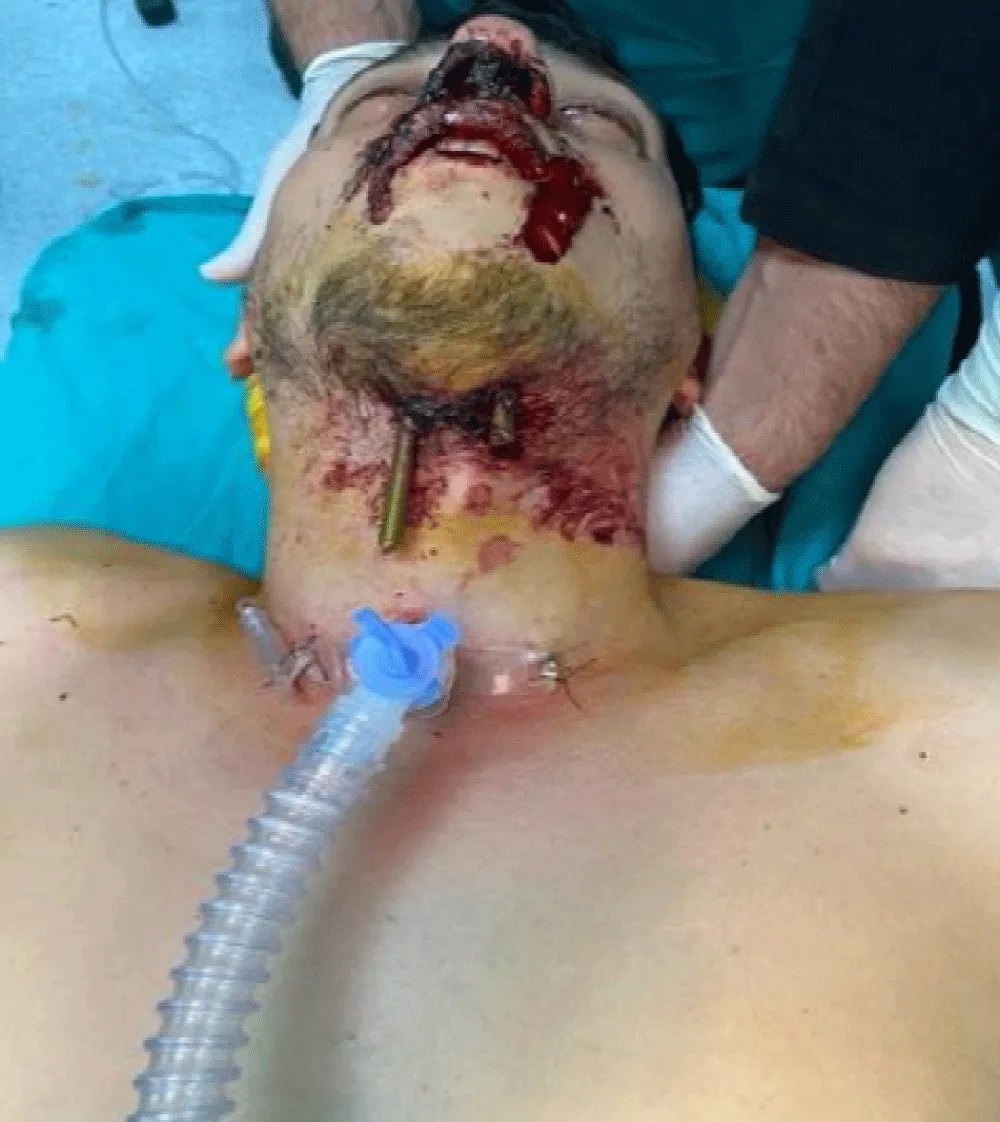
Intraoperative photograph following successful surgical tracheostomy and shortening of the externally protruding harpoon shafts prior to extraction.
Non-contrast craniofacial and brain computed tomography (CT) scans demonstrated that both harpoons penetrated the submental soft tissues, pierced the base of the tongue and hard palate, traversed the nasal cavity adjacent to the septum, and impacted the inner table of the frontal sinus (Figures 4 and 5). Crucially, the tips did not breach the posterior wall of the frontal sinus into the brain parenchyma, and no intracranial hemorrhage, pneumocephalus, or major cervical and intracranial vascular injury was identified.
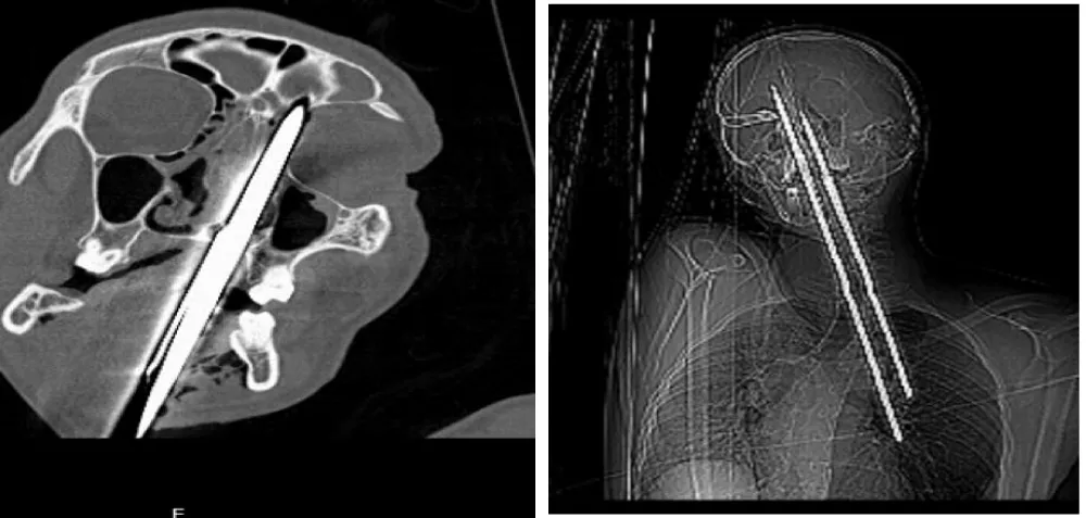
Preoperative computed tomography (CT) scans demonstrating the upward trajectory of both metallic shafts piercing the base of the tongue, hard palate, and nasal cavity, terminating against the inner table of the frontal sinus without intracranial breach.
A multidisciplinary team comprising anesthesiologists, otolaryngologists, and neurosurgeons was immediately assembled. Because the externally protruding harpoon shafts were excessively long and obstructed access to the anterior neck and airway, firefighters were summoned to the emergency unit to carefully shorten the external metal rods using specialized cutting tools while the surgical team stabilized the shafts to prevent micro-movements and secondary tissue trauma.
In the operating room, standard monitoring (electrocardiography, non-invasive blood pressure, peripheral oxygen saturation [SpO₂], and end-tidal carbon dioxide [EtCO₂]) was established, and supplemental oxygen was delivered at 4–6 L/min. Given the trismus (<2 cm), intraoral bleeding, and anatomical distortion, both direct laryngoscopy and nasal fiberoptic intubation were deemed unsafe. A surgical tracheostomy under deep sedation with preserved spontaneous ventilation and protective airway reflexes was planned. Premedication consisted of intravenous midazolam (2 mg), ketamine (30 mg), and fentanyl (50 µg). Subsequently, a dexmedetomidine loading dose of 1 µg/kg was administered over 10 minutes, followed by a continuous maintenance infusion at 0.7 µg/kg/h.
Following antiseptic preparation and local anesthetic infiltration of the anterior neck, a surgical tracheostomy was performed uneventfully while the patient maintained spontaneous respiration. Once tracheal cannula placement was confirmed via capnography and auscultation, general anesthesia was induced with intravenous rocuronium (50 mg) and maintained with sevoflurane. Under direct visualization and controlled surgical exposure, both harpoons—including their articulated wishbone mechanism—were carefully extracted along their entry trajectories without resistance or significant bleeding (Figures 3 and 6). Hemostasis was achieved, and broad-spectrum intravenous antibiotic prophylaxis and tetanus immunization were administered.
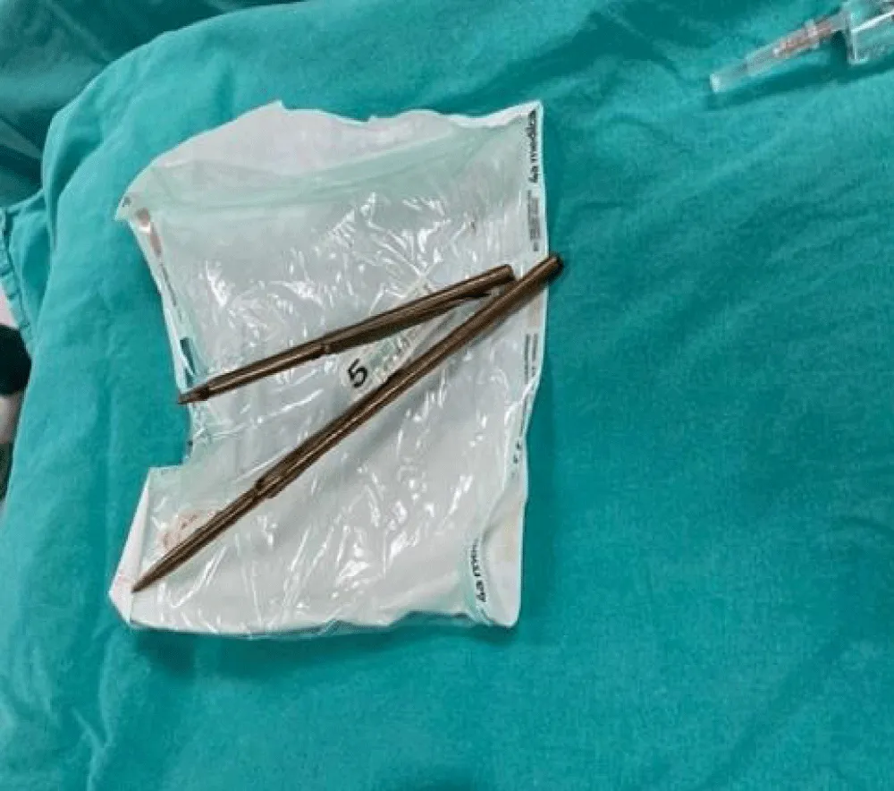
The two extracted harpoon shafts along with their articulated wishbone mechanism following controlled surgical removal.
Postoperative analgesia was provided using intravenous paracetamol (1 g), dexketoprofen (50 mg), and morphine (2 mg). The patient was monitored in the post-anesthesia care unit (PACU) for 24 hours before being transferred to the otolaryngology ward, where he was closely followed for seven days. His postoperative course was uncomplicated, with no signs of infection, secondary hemorrhage, or CSF fistula. Successful decannulation was performed on postoperative day 7, and he was discharged home on postoperative day 8 in good clinical condition. At the six-month follow-up, physical, neurological, and cognitive evaluations—including memory and executive function—were completely normal, and control CT imaging confirmed satisfactory bone and soft tissue healing without structural defects.
Discussion
Penetrating craniofacial and brain injuries represent a complex neurosurgical and anesthesiological emergency characterized by high morbidity and mortality rates. Unlike high-velocity ballistic trauma, harpoon guns operate as low-to-medium velocity projectiles driven by elastic or pneumatic static energy. When fired out of water, the absence of hydrodynamic drag allows the spear to retain maximum kinetic energy, enabling penetration through dense facial bones and the cranial vault. In the present case, the simultaneous penetration of two parallel shafts compounded the anatomical risk, creating a rigid mechanical lock across the submental, oral, and sinonasal compartments [1-6].
Airway and anesthesiological challenges
Securing the airway is the foremost priority in penetrating maxillofacial trauma. Even in patients presenting with initial airway patency and a GCS score of 15, rapid deterioration can occur due to progressive soft tissue edema, hematoma expansion, or aspiration of blood. Standard rapid-sequence induction and neuromuscular blockade are hazardous in such scenarios, as loss of pharyngeal muscle tone can precipitate a "cannot intubate, cannot oxygenate" crisis. In our patient, severe mechanical trismus (<2 cm) and intraoral foreign bodies precluded both oral and nasal intubation. Preoperative shortening of the external harpoon shafts by emergency rescue personnel was a critical preparatory step that eliminated leverage forces and provided sufficient anatomical clearance for anterior neck access.
Dexmedetomidine, a selective α₂-adrenergic agonist, played a pivotal role in facilitating safe sedated tracheostomy. Unlike propofol or high-dose opioids, dexmedetomidine provides cooperative sedation and analgesia without causing significant respiratory depression, thereby preserving spontaneous ventilation and laryngeal reflexes during airway manipulation. Combining low-dose ketamine and fentanyl with dexmedetomidine further optimized analgesia and hemodynamic stability while minimizing adverse respiratory effects [7].
Medical, neurosurgical, and long-term considerations
From a neurosurgical and otolaryngological perspective, blind removal of penetrating cranial foreign bodies outside the operating room is strictly contraindicated, as the object may exert a tamponade effect on lacerated vessels. Comprehensive preoperative neuroimaging using multidetector CT and vascular assessment is essential to delineate the projectile trajectory, assess skull base integrity, and exclude traumatic intracranial aneurysms, arteriovenous fistulas, or major cervical vascular injury. In our case, CT confirmed that the harpoon tips arrested precisely at the inner table of the frontal sinus without dural penetration or parenchymal laceration. Had the projectiles breached the posterior frontal sinus wall or cranial vault, extensive craniotomy, watertight dural repair, and aggressive debridement would have been mandatory to prevent life-threatening meningitis, brain abscess, or CSF rhinorrhea [8-10].
Furthermore, severe penetrating TBIs frequently necessitate decompressive craniectomy to control refractory intracranial hypertension, subsequently requiring delayed cranioplasty. Literature indicates that post-traumatic cranioplasty carries substantial complication risks—including surgical site infection, bone flap resorption, subdural hygroma, and hydrocephalus—which are strongly influenced by surgical timing and underlying cerebrospinal fluid dynamics. In patients with parenchymal involvement, maintaining optimal cerebral perfusion pressure (CPP) is equally critical during intensive care management to prevent secondary ischemic brain injury or hyperperfusion-induced cerebral edema, as cerebrovascular autoregulation is often impaired following trauma [11,12].
Finally, survivors of penetrating craniofacial and frontal-region trauma require structured long-term neurocognitive follow-up. Even when gross motor function recovers, injuries affecting frontal and temporal networks can result in persistent cognitive deficits, particularly involving attention, executive functioning, and episodic memory—the ability to encode and retrieve personal experiences. Fortunately, because the frontal sinus inner table absorbed the terminal kinetic energy in our patient without parenchymal contusion, he experienced neither acute intracranial hypertension nor long-term episodic memory impairment at six months [8-12].
Conclusion
Double-harpoon penetrating craniofacial injury is an extraordinary trauma mechanism that demands rapid, coordinated intervention by anesthesiologists, otolaryngologists, neurosurgeons, and emergency rescue teams. Key clinical takeaways from this case include: (1) stabilizing and shortening externally protruding metallic shafts prior to surgery to prevent secondary leverage trauma and facilitate airway access; (2) utilizing targeted CT and vascular imaging to map projectile trajectories and rule out occult neurovascular or dural breaches prior to extraction; and (3) employing a dexmedetomidine-based multimodal sedation protocol to safely perform a spontaneous-breathing tracheostomy when conventional intubation is anatomically impossible. Adherence to these principles ensures airway security, prevents catastrophic hemorrhage, and promotes full neurological and cognitive recovery.
Informed consent
This case report was written with informed consent obtained from the patient for the publication of this case report and all accompanying clinical photographs and radiographic images.
- World Health Organization. WHO global air quality guidelines: particulate matter (PM2.5 and PM10), ozone, nitrogen dioxide, sulfur dioxide and carbon monoxide. Geneva: World Health Organization; 2021. Available from: https://www.who.int/publications/i/item/9789240034228
- Kampa M, Castanas E. Human health effects of air pollution. Environ Pollut. 2008;151(2):362-7. Available from:https://doi.org/10.1016/j.envpol.2007.06.012
- Monks PS, Granier C, Fuzzi S, Stohl A, Williams ML, Akimoto H, et al. Atmospheric composition change—global and regional air quality. Atmos Environ. 2009;43(33):5268-5350. Available from:https://doi.org/10.1016/j.atmosenv.2009.03.004
- Seinfeld JH, Pandis SN. Atmospheric chemistry and physics: from air pollution to climate change. 3rd ed. Hoboken (NJ): Wiley; 2016.
- Pope CA 3rd, Dockery DW. Health effects of fine particulate air pollution: lines that connect. J Air Waste Manag Assoc. 2006;56(6):709-42. Available from: https://doi.org/10.1080/10473289.2006.10464485
- Brook RD, Rajagopalan S, Pope CA 3rd, Brook JR, Bhatnagar A, Diez-Roux AV, et al. Particulate matter air pollution and cardiovascular disease: an update to the scientific statement from the American Heart Association. Circulation. 2010;121(21):2331-78. Available from:https://doi.org/10.1161/CIR.0b013e3181dbece1
- Loomis D, Grosse Y, Lauby-Secretan B, El Ghissassi F, Bouvard V, Benbrahim-Tallaa L, et al. The carcinogenicity of outdoor air pollution. Lancet Oncol. 2013;14(13):1262-3. Available from:https://doi.org/10.1016/S1470-2045(13)70487-X
- Herting MM, et al. Outdoor air pollution and brain development in childhood and adolescence. Trends Neurosci. 2024;47(8):593-607. Available from: https://doi.org/10.1016/j.tins.2024.06.008
- Lee KK, et al. Adverse health effects associated with household air pollution: a systematic review, meta-analysis, and burden estimation study. Lancet Glob Health. 2020;8(11):e1427-34. Available from:https://doi.org/10.1016/S2214-109X(20)30343-0
- World Health Organization. WHO guidelines for indoor air quality: household fuel combustion. Geneva: World Health Organization; 2014. Available from: Available from: https://www.who.int/publications/i/item/9789241548885
- India State-Level Disease Burden Initiative Air Pollution Collaborators. Health and economic impact of air pollution in the states of India: the Global Burden of Disease Study 2019. Lancet Planet Health. 2021;5(1):e25-38. Available from:https://doi.org/10.1016/S2542-5196(20)30298-9
- Bond TC, Doherty SJ, Fahey DW, Forster PM, Berntsen T, DeAngelo BJ, et al. Bounding the role of black carbon in the climate system: a scientific assessment. J Geophys Res Atmos. 2013;118(11):5380-5552. Available from:https://doi.org/10.1002/jgrd.50171
- Shindell D, Kuylenstierna JCI, Vignati E, van Dingenen R, Amann M, Klimont Z, et al. Simultaneously mitigating near-term climate change and improving human health and food security. Science. 2012;335(6065):183-9. Available from:https://doi.org/10.1126/science.1210026
- Intergovernmental Panel on Climate Change. Transport. In: Climate change 2022: mitigation of climate change. Cambridge: Cambridge University Press; 2022. Available from: https://doi.org/10.1017/9781009157926.012
- Lelieveld J, Evans JS, Fnais M, Giannadaki D, Pozzer A. The contribution of outdoor air pollution sources to premature mortality on a global scale. Nature. 2015;525:367-71. Available from: https://doi.org/10.1038/nature15371
- Pöschl U. Atmospheric aerosols: composition, transformation, climate and health effects. Chem Rev. 2005;105(2):731-8. Available from: https://doi.org/10.1021/cr0500785
- World Bank, Institute for Health Metrics and Evaluation. The cost of air pollution: strengthening the economic case for action. Washington (DC): World Bank; 2016. Available from: https://doi.org/10.1596/25013
- International Agency for Research on Cancer. Outdoor air pollution. IARC Monogr Eval Carcinog Risks Hum. 2016;109.
- Grigoratos T, Martini G. Brake wear particle emissions: a review. Environ Sci Pollut Res Int. 2015;22:2491-504. Available from:https://doi.org/10.1007/s11356-014-3696-8
- World Health Organization. Guidelines for indoor air quality: household fuel combustion. Geneva: World Health Organization; 2014.
- Nyadanu SD, Dunne J, Tessema GA, Mullins B, Kumi-Boateng B, Bell ML, et al. Prenatal exposure to ambient air pollution and adverse birth outcomes: an umbrella review of 36 systematic reviews and meta-analyses. Environ Pollut. 2022;306:119465. Available from: https://doi.org/10.1016/j.envpol.2022.119465
- Likens GE, Wright RF, Galloway JN, Butler TJ. Acid rain. Sci Am. 1979;241(4):43-51.
- Fowler D, et al. Atmospheric deposition of nutrients to land and ocean. Atmos Environ. 2009. Available from:https://doi.org/10.1016/j.atmosenv.2008.09.067
- Ainsworth EA, Yendrek CR, Sitch S, Collins WJ, Emberson LD. The effects of tropospheric ozone on net primary productivity and implications for climate change. Annu Rev Plant Biol. 2012;63:637-61. Available from: https://doi.org/10.1146/annurev-arplant-042110-103829
- Ministry of Environment, Forest and Climate Change, Government of India. National Clean Air Programme (NCAP). New Delhi: Government of India; 2019.
- Coelho S, Ferreira J, Lopes M. Source apportionment of air pollution in urban areas: a review of the most suitable source-oriented models. Air Qual Atmos Health. 2023;16:1185-94. Available from:https://doi.org/10.1007/s11869-023-01334-z
- Idrees Z, Zheng L. Low-cost air pollution monitoring systems: a review of protocols and enabling technologies. J Ind Inf Integr. 2020;17:100123. Available from:https://doi.org/10.1016/j.jii.2019.100123
- van Donkelaar A, Martin RV, Brauer M, Kahn R, Levy R, Verduzco C, et al. Global estimates of ambient fine particulate matter concentrations from satellite-based aerosol optical depth: development and application. Environ Health Perspect. 2010;118(6):847-55. Available from: https://doi.org/10.1289/ehp.0901623



 Save to Mendeley
Article Alerts
Subscribe to our articles alerts and stay tuned.
 This work is licensed under a Creative Commons Attribution 4.0 International License.