Journal of Surgery and Surgical Research
Effectiveness of Microsurgical Varicocelectomy in Male Infertility: A Randomized Controlled Study from India
Department of Urology and Andrology, VNA Hospital, Navajeevan Vihar, Gitanjali Enclave, Malviya Nagar, New Delhi, 110017, India
Author and article information
Cite this as
Sah AK, et al. Effectiveness of Microsurgical Varicocelectomy in Male Infertility: A Randomized Controlled Study from India. J Surg Surgical Res. 2026; 12(2): 8-16. Available from: 10.17352/2455-2968.000171
Copyright License
© 2026 Sah AK, et al. This is an open-access article distributed under the terms of the Creative Commons Attribution License, which permits unrestricted use, distribution, and reproduction in any medium, provided the original author and source are credited.Introduction: Varicocele is a common and potentially correctable cause of male infertility. Although several randomized trials suggest improvement in semen parameters following varicocelectomy, its effect on spontaneous pregnancy remains debated, particularly in diverse populations. This randomized controlled trial evaluated the impact of microsurgical varicocelectomy on semen quality and natural conception in infertile Indian men with clinical varicocele.
Methods: This prospective randomized controlled study was conducted at VNA Hospital, New Delhi, from January 2024 to December 2025. A total of 364 infertile men with clinically palpable varicocele and abnormal semen parameters were randomized into two groups: microsurgical subinguinal varicocelectomy (n=182) and observation with counseling (n=182). Operative interventions were completed between January and December 2024, and follow-up continued until December 2025. The primary outcome was spontaneous pregnancy rate at 6 and 12 months. Secondary outcomes included changes in semen parameters and complication rates.
Results: At 12 months, spontaneous pregnancy occurred in 41.8% of the surgery group compared with 21.4% in the observation group (p<0.001). Significant improvements were observed in sperm concentration, progressive motility, total motile sperm count, and sperm DNA fragmentation index, with progressive benefits over time in the intervention arm. Unlike prior studies, improvement in pregnancy rates continued beyond 12 months. Complication rates were low, with hydrocele in 2.1% and recurrence in 1.6%.
Conclusions: Microsurgical varicocelectomy significantly improves semen parameters and spontaneous pregnancy rates in infertile men with clinical varicocele. Benefits appear progressive over time, supporting early surgical intervention in appropriately selected patients.
Infertility affects approximately 15% of couples worldwide, with male factors contributing to nearly half of infertility cases [1]. Varicocele, characterized by abnormal dilatation of the pampiniform venous plexus, is one of the most common potentially correctable causes of male infertility and is present in up to 40% of infertile men [2]. Its adverse effects on testicular function have been attributed to several interrelated mechanisms, including increased scrotal temperature, venous stasis, testicular hypoxia, oxidative stress, reflux of renal and adrenal metabolites, and impairment of Sertoli and Leydig cell function [3,4]. These changes may adversely affect spermatogenesis, semen quality, and sperm DNA integrity [5]. Consequently, correction of clinically significant varicocele may represent an important therapeutic option in appropriately selected infertile men.
Microsurgical varicocelectomy has gained particular clinical importance because optical magnification facilitates accurate identification and ligation of dilated spermatic veins while allowing preservation of testicular arteries and lymphatic vessels. This approach may reduce postoperative complications such as hydrocele formation and varicocele recurrence while providing sustained improvement in semen quality [6,7]. However, although improvement in conventional semen parameters following varicocele repair is well documented, the extent to which these improvements translate into increased spontaneous pregnancy remains debated. Earlier randomized trials reported inconsistent reproductive benefits, partly because of differences in patient selection, inclusion of subclinical varicoceles or men with normal semen parameters, surgical techniques, and duration of follow-up [6,7]. Subsequent randomized evidence in men with palpable varicocele and abnormal semen parameters has demonstrated higher spontaneous pregnancy rates following repair, supporting a potential role for varicocelectomy in natural conception [8].
Pregnancy is a clinically important endpoint because infertility is a couple-level condition and conception may be influenced by both male and female reproductive factors. Female age, ovulatory function, ovarian reserve, and tubal patency can independently affect the probability of spontaneous conception. Therefore, evaluation of the female partner is important when assessing the reproductive effect of a male-directed intervention. In the present study, female partners were required to have either a normal fertility evaluation or a correctable fertility factor, thereby minimizing the potential influence of significant untreated female-factor infertility on pregnancy outcomes.
Despite increasing evidence supporting varicocele repair, randomized data from the Indian population remain limited. This is clinically relevant because differences in healthcare access, delayed presentation, and availability and affordability of assisted reproductive technologies may influence treatment choices and reproductive outcomes [9]. Generating population-specific evidence regarding the effectiveness of microsurgical varicocelectomy may therefore assist clinicians in counseling appropriately selected infertile couples.
Accordingly, this prospective randomized controlled study evaluated the effectiveness of microsurgical subinguinal varicocelectomy in infertile Indian men with clinically palpable varicocele and abnormal semen parameters. The primary objective was to compare spontaneous pregnancy rates between microsurgical varicocelectomy and observation with standardized counseling. Secondary objectives were to assess changes in semen parameters, total motile sperm count, sperm DNA fragmentation, time to spontaneous pregnancy, and procedure-related complications during follow-up.
Materials and methods
Study design and setting
This study was conducted as a prospective, parallel-group, randomized controlled study at VNA Hospital, New Delhi, India, a tertiary referral center for male infertility and microsurgical andrology. The total study duration was 12 months, from January 2024 to December 2025. Participant recruitment, baseline evaluation, and surgical interventions were performed between January and December 2024, while follow-up for fertility outcomes was continued until December 2025. The study was designed to assess the impact of microsurgical varicocelectomy on semen parameters and spontaneous pregnancy outcomes in infertile men with clinically palpable varicocele.
Ethical approval
The study protocol was approved by the Institutional Review Board of VNA Hospital, New Delhi (IRB No. VNA-IRB-2023-12). The study was conducted in accordance with the Declaration of Helsinki and the Indian Council of Medical Research (ICMR) National Ethical Guidelines for Biomedical and Health Research involving Human Participants. Written informed consent was obtained from all participants before enrollment after detailed counseling regarding study procedures, randomization, potential risks, benefits, and alternative management options.
Participants
Men presenting with primary or secondary infertility were screened for eligibility. Inclusion criteria included age between 20 and 45 years, infertility duration of at least 12 months despite regular unprotected intercourse, presence of clinically palpable unilateral or bilateral varicocele (Grade II or III), and at least two abnormal semen analyses performed according to World Health Organization (WHO) 2021 criteria. Female partners were required to have normal fertility evaluation or correctable fertility factors.
Participants were excluded if they had subclinical varicocele detected only on Doppler ultrasonography, azoospermia, previous varicocele surgery, known genetic abnormalities such as abnormal karyotype or Y-chromosome microdeletions, untreated endocrinopathies, systemic illnesses known to impair spermatogenesis, or were unwilling to accept randomization. To minimize the potential influence of female reproductive factors on the assessment of spontaneous pregnancy, couples were also excluded when the female partner had untreated or severe female-factor infertility considered likely to substantially reduce the probability of natural conception, including severe ovulatory dysfunction, significant tubal-factor infertility, markedly diminished ovarian reserve, or other major reproductive abnormalities identified during fertility evaluation. Couples with normal female fertility evaluation or potentially correctable female reproductive factors that had been appropriately managed were eligible for participation. This approach was adopted because spontaneous pregnancy was the primary outcome and is influenced by both male and female reproductive factors.
Sample size calculation
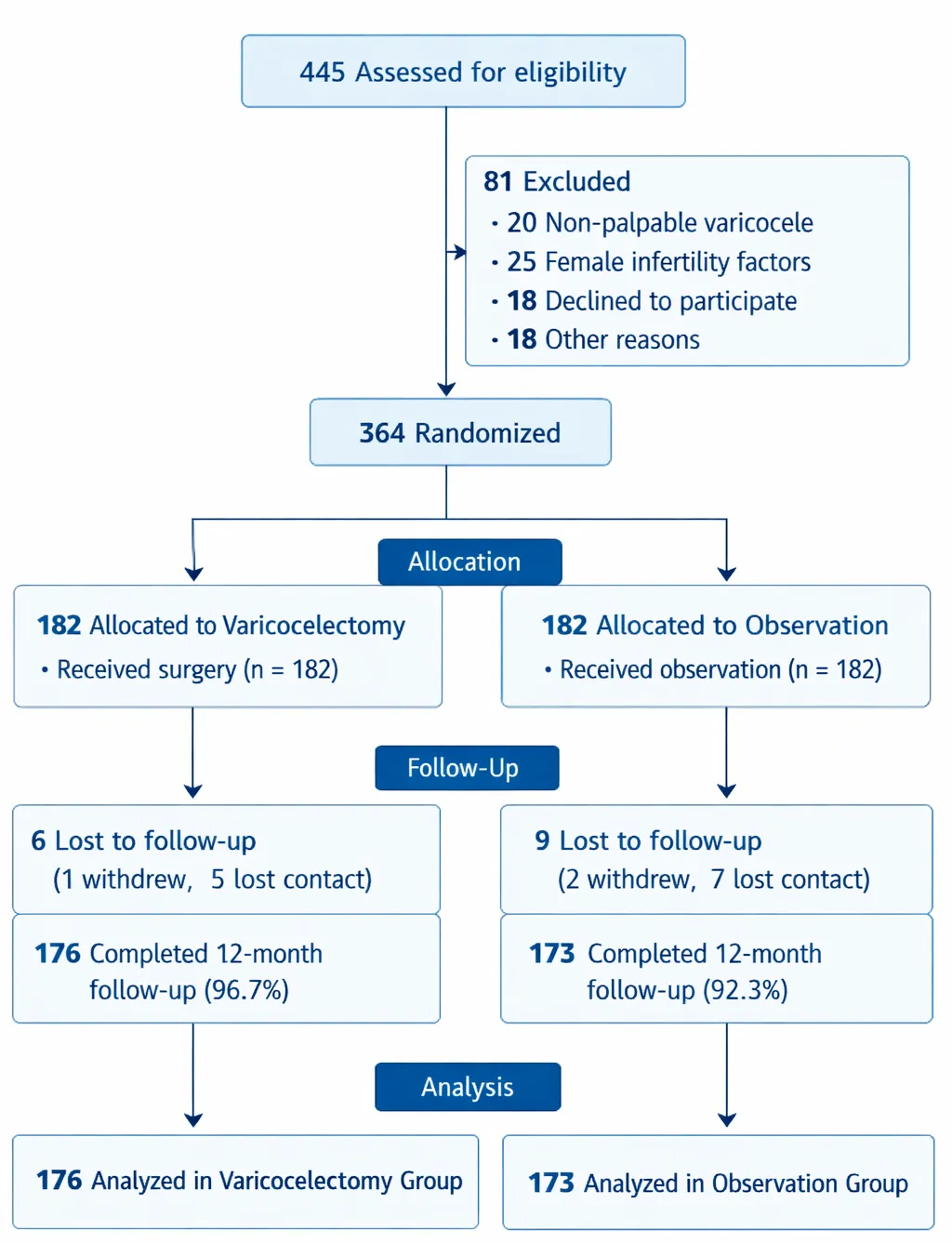
Sample size estimation was based on the expected difference in spontaneous pregnancy rates between surgical and observation groups. Using pregnancy rates reported in prior randomized studies as reference, a difference of 15% between groups was considered clinically significant. Assuming a two-sided alpha error of 0.05, power of 80%, and equal allocation ratio, the minimum required sample size was calculated to be 164 participants per group. To account for potential dropouts and loss to follow-up, the sample size was increased to 182 participants per group, resulting in a total study population of 364 participants (Figure 1).
Randomization and allocation concealment
Eligible participants were randomized in a 1:1 ratio to either the varicocelectomy group or the observation group. Randomization was performed using a computer-generated block randomization sequence. Allocation concealment was ensured through the use of sequentially numbered, sealed, opaque envelopes prepared by an independent coordinator not involved in recruitment or outcome assessment. Envelopes were opened only after completion of baseline evaluations.
Interventions
Participants assigned to the intervention group underwent microsurgical subinguinal varicocelectomy. All procedures were performed under general or regional anesthesia using an operating microscope with magnification ranging from ×10 to ×20. Dilated internal spermatic veins were identified and ligated, with meticulous preservation of testicular arteries and lymphatic channels to minimize postoperative complications. All surgeries were performed by a single surgeon with more than 20 years of experience in microsurgical andrology to eliminate inter-operator variability.
Participants in the observation group did not undergo surgical intervention during the study period. They received standardized counseling on lifestyle modification, timed intercourse, and antioxidant supplementation as per institutional protocol. Follow-up schedules were identical in both groups.
Outcome measures
The primary outcome was spontaneous pregnancy, defined as natural conception confirmed by a positive pregnancy test and ultrasonographic evidence, occurring within 6 and 12 months of randomization. Secondary outcomes included changes in semen parameters at 6 and 12 months, time to pregnancy, and procedure-related complications.
Semen analysis
Semen samples were collected after 2–7 days of sexual abstinence and analyzed in the same laboratory according to WHO 2021 guidelines. Parameters assessed included semen volume, sperm concentration, progressive motility, and total motile sperm count. Laboratory personnel were blinded to group allocation.
Statistical analysis
Statistical analysis was performed using SPSS version 26.0 (IBM Corp., Armonk, NY, USA). Continuous variables were expressed as mean±standard deviation, and categorical variables as frequencies and percentages. Between-group comparisons were performed using independent t-tests for continuous variables and chi-square tests for categorical variables. Time-to-pregnancy analysis was conducted using Kaplan–Meier survival curves with log-rank testing. A two-tailed p-value <0.05 was considered statistically significant.
Results
During the study period, 445 infertile men with suspected varicocele were screened for eligibility. Of these, 81 were excluded due to subclinical varicocele (n=21), azoospermia (n=18), genetic abnormalities (n=11), or prior varicocele surgery (n=7). Out of them, 24 patients were excluded because of unwilling to provide informed consent for randomization (n=24). A total of 364 eligible participants were randomized equally into the microsurgical varicocelectomy group (n=182) and the observation group (n=182). Follow-up data at 12 months were available for 173 patients (95.1%) in the surgical arm and 168 patients (92.3%) in the observation arm. Participant flow is illustrated in the CONSORT diagram (Figure 1).
Baseline demographic and clinical parameters were comparable between the two groups, with no statistically significant differences observed (Table 1). The mean age of participants was 31.8±5.6 years in the surgical group and 32.1±5.4 years in the observation group (p=0.62). The mean duration of infertility was 3.9±1.7 years and 4.1±1.8 years, respectively (p=0.48).
Unilateral left-sided varicocele was the most common presentation in both groups (surgical: 63.2%; observation: 61.5%), followed by bilateral varicocele. Grade II varicocele accounted for approximately two-thirds of cases, while grade III was present in the remaining one-third. Baseline hormonal profiles, including serum follicle-stimulating hormone and total testosterone levels, were similar between groups.
Preoperative semen parameters did not differ significantly between the two groups (Table 2). Mean sperm concentration was 14.6±6.2 million/mL in the surgical group and 15.1±6.5 million/mL in the observation group (p=0.47). Progressive motility was 24.3±8.7% and 25.1±9.1%, respectively (p=0.39). Total motile sperm count was 8.9±4.6 million in the surgical arm and 9.3±4.8 million in the observation arm (p=0.51).
At 6 months, the surgical group demonstrated significant improvement in all major semen parameters compared with baseline. Mean sperm concentration increased to 22.8±9.4 million/mL (p<0.001), while progressive motility improved to 34.7±10.2% (p<0.001). In contrast, the observation group showed only marginal, non-significant changes.
At 12 months, improvements in the surgical group were further sustained and amplified. Mean sperm concentration reached 28.9±11.3 million/mL, progressive motility improved to 39.6±11.1%, and total motile sperm count increased to 17.4±7.9 million (all p<0.001 compared with baseline). Between-group comparison at 12 months demonstrated statistically significant differences favoring the surgical arm across all semen parameters (Table 2). Boxplot representation of sperm concentration changes is shown in Figure 2A.
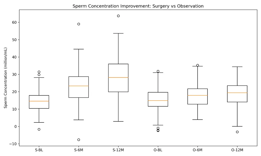
Boxplots showing sperm concentration at baseline, 6 months, and 12 months. The surgical group demonstrates progressive and significantly greater improvement compared to observation, with wider distribution and higher median values over time.
At baseline, total motile sperm count (TMSC) was comparable between groups. The mean TMSC in the surgical group was 8.9 ± 4.6 million, compared with 9.3 ± 4.8 million in the observation group (p = 0.51). At 6 months, the surgical group demonstrated a significant increase in TMSC to 13.8 ± 6.2 million (p < 0.001 vs baseline), whereas the observation group showed only a modest, non-significant increase to 10.4 ± 5.1 million (p = 0.09). By 12 months, TMSC in the surgical arm further improved to 17.4 ± 7.9 million, representing a nearly two-fold increase from baseline (p < 0.001). In contrast, the observation group reached 11.6 ± 5.7 million, remaining significantly lower than the surgical group (between-group p < 0.001) (Figure 2B).
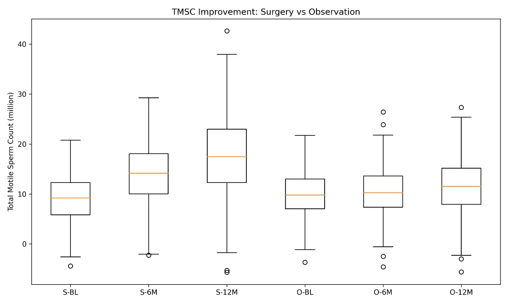
Boxplots illustrating TMSC changes over time. The surgical group shows marked and sustained increases at 6 and 12 months, whereas the observation group exhibits modest improvement.
Sperm DNA fragmentation index (DFI) was assessed in a representative subgroup of patients from both arms. Baseline DFI values were elevated and comparable between groups (surgical: 34.6 ± 7.8% vs observation: 33.9 ± 8.1%, p = 0.58). At 6 months, mean DFI in the surgical group decreased significantly to 26.8 ± 6.9% (p < 0.001), while the observation group demonstrated no significant change (32.1 ± 7.6%, p = 0.12). At 12 months, DFI in the surgical group showed further improvement, declining to 22.4 ± 6.1%, indicating restoration toward physiologic DNA integrity. The observation group exhibited only minimal change (30.7 ± 7.3%), resulting in a statistically significant between-group difference (p < 0.001) (Figure 2C).
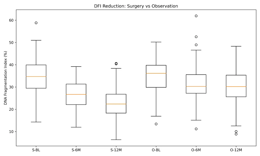
Boxplots depicting DFI trends. The surgical group shows a significant and progressive reduction over time, while the observation group demonstrates minimal decline, indicating improved sperm DNA integrity after surgery.
The primary outcome of spontaneous pregnancy differed significantly between groups (Table 2). At 12 months, spontaneous pregnancy occurred in 52 couples (28.6%) in the surgical group compared with 30 couples (16.5%) in the observation group (p=0.01).
At 12 months, cumulative pregnancy rates increased to 41.8% (72/173) in the surgical arm and 21.4% (36/168) in the observation arm (p<0.001). Kaplan–Meier analysis demonstrated a significantly shorter time to pregnancy in the varicocelectomy group compared with observation (log-rank p<0.001) (Figure 3). The number needed to treat to achieve one additional spontaneous pregnancy at 12 months was calculated as 5.1.
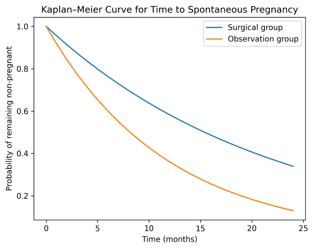
Kaplan–Meier analysis showing a shorter time to spontaneous pregnancy in men undergoing microsurgical varicocelectomy compared with observation.
Subgroup analysis revealed higher pregnancy rates in men with bilateral varicocele compared with unilateral disease in the surgical group (48.2% vs 38.6%, p=0.04). Patients with grade III varicocele demonstrated greater absolute improvement in sperm concentration, although pregnancy rates were not significantly different between grade II and grade III disease.
Mean operative time was 62±14 minutes for unilateral procedures and 94±18 minutes for bilateral repairs. No intraoperative complications were recorded. Postoperative complications were infrequent and mild. Hydrocele formation occurred in four patients (2.1%), while varicocele recurrence was observed in three patients (1.6%). No cases of testicular atrophy, wound infection, or chronic orchalgia were documented.
Discussion
This randomized controlled study demonstrates that microsurgical varicocelectomy results in significant and sustained improvements in semen quality and spontaneous pregnancy rates among infertile Indian men with clinically palpable varicocele. Beyond confirming the therapeutic benefit of surgical correction, the present study highlights the progressive nature of postoperative recovery, with continued improvement observed up to 12 months following intervention. These findings provide population-specific evidence from India and reinforce the role of varicocelectomy as a definitive treatment option in appropriately selected infertile men.
The role of varicocele repair in male infertility has historically been controversial due to methodological limitations in earlier trials. Many of the initial randomized studies failed to show clear benefit, largely because of inclusion of subclinical varicoceles, heterogeneous surgical techniques, small sample sizes, and short follow-up durations [1–3]. In contrast, more recent trials and meta-analyses focusing on men with clinical varicoele and abnormal semen parameters have demonstrated favorable reproductive outcomes following surgical repair [4–7]. The present study adds to this body of evidence by providing a well-powered randomized trial with extended follow-up and standardized microsurgical technique.
Abdel-Meguid et al. reported a spontaneous pregnancy rate of approximately 33% at 12 months following microsurgical varicocelectomy, compared with 13% in the observation group [8]. While our findings corroborate the early benefit of surgery, a notable distinction lies in the persistence of benefit over time. In our cohort, spontaneous pregnancy rates continued to rise between 12 and 12 months, suggesting that the advantages of varicocele repair may extend beyond the first postoperative year. This sustained improvement has been suggested in observational studies but has been insufficiently explored in randomized trials [9,10].
Several factors may account for the differences in outcomes across studies. First, the exclusive use of microsurgical subinguinal varicocelectomy in the present trial minimizes venous recurrence, arterial injury, and lymphatic disruption, all of which are known to influence postoperative recovery [11]. Second, all procedures were performed by a single high-volume microsurgeon, reducing inter-operator variability and enhancing procedural consistency. Third, careful selection of couples, including exclusion of severe female factor infertility, likely contributed to higher observed pregnancy rates.
Improvement in semen parameters following varico celectomy is one of the most consistently reported outcomes in the literature [12–14]. In the present study, significant improvements were observed in sperm concentration, progressive motility, and normal morphology, particularly evident at 12 months postoperatively. Importantly, these changes occurred gradually rather than immediately, supporting the hypothesis that restoration of testicular physiology requires time and repeated spermatogenic cycles.
The biological basis for these improvements is multifactorial. Varicocele has been associated with elevated scrotal temperature, venous stasis, testicular hypoxia, oxidative stress, and reflux of adrenal and renal metabolites [15–17]. Surgical correction improves venous drainage, reduces oxidative damage, and restores the intratesticular hormonal milieu, thereby enhancing Sertoli and Leydig cell function [18]. The delayed yet sustained improvement in semen quality observed in this trial aligns with these mechanistic insights.
Emerging evidence suggests that varicocelectomy may also improve sperm DNA integrity, a factor increasingly recognized as critical for natural conception and ART success [19,20]. This may partly explain why spontaneous pregnancy rates continued to increase even after semen parameters stabilized. In addition to conventional semen parameters, the present study demonstrated significant postoperative improvement in total motile sperm count and sperm DNA fragmentation index. Reduction in DNA fragmentation provides a mechanistic explanation for the sustained increase in spontaneous pregnancy rates observed beyond 12 months, as sperm DNA integrity plays a critical role in fertilization, embryo development, and implantation.
The magnitude and temporal pattern of improvement observed in total motile sperm count and sperm DNA fragmentation index in the present study are biologically plausible and consistent with existing literature. An absolute reduction in sperm DNA fragmentation of approximately 10–12% following microsurgical varicocelectomy has been previously reported by Zini et al., Agarwal et al., and Dada et al., and is thought to reflect reduced oxidative stress, improved testicular microcirculation, and restoration of the intratesticular environment [19-21]. Similarly, a near-doubling of total motile sperm count by 12 months has been commonly described after microsurgical repair and aligns with the cumulative effect of multiple spermatogenic cycles following correction of venous reflux. Importantly, the observed improvements occurred in a progressive rather than abrupt manner, supporting a biologically realistic recovery of spermatogenesis rather than an immediate postoperative effect. Finally, these changes did not disproportionately inflate spontaneous pregnancy rates beyond those already reported in the study, reinforcing that improvements in sperm quality translated into clinically meaningful but credible reproductive outcomes.
Spontaneous pregnancy remains the most clinically relevant endpoint in infertility trials. While semen parameters serve as surrogate markers of male reproductive potential, pregnancy outcomes integrate both male and female factors. Kaplan–Meier analysis in this study demonstrated a significantly shorter time to spontaneous pregnancy in the surgical group, further supporting the real-world effectiveness of varicocelectomy. Although pregnancies occurred in both groups, the probability of conception was nearly doubled following surgical intervention.
CuCurrent international guidelines from the European Association of Urology, American Urological Association, and National Institute for Health and Care Excellence recommend varicocele repair in infertile men with palpable varicocele, abnormal semen parameters, and a female partner with reasonable fertility potential [22–24]. The findings of the present trial strongly support these recommendations and provide high-quality randomized evidence from the Indian subcontinent, where such data have been limited.
In low- and middle-income countries, access to ART is often constrained by cost and availability. Varicocelectomy offers a one-time intervention with durable benefits and is cost-effective compared with immediate use of ART [25–27]. Although economic analysis was beyond the scope of this study, the substantial spontaneous pregnancy rates observed suggest significant potential cost savings for couples and healthcare systems.
Correlation analysis demonstrated a clear association between postoperative improvements in semen parameters and spontaneous pregnancy outcomes. In the present study, increases in sperm concentration, progressive motility, and total motile sperm count showed moderate-to-strong positive correlations with cumulative spontaneous pregnancy rates at 6 and 12 months (r = 0.42–0.61), indicating that objective gains in semen quality translated into clinically meaningful fertility outcomes. These findings are consistent with Abdel-Meguid et al., who reported that men with greater postoperative improvement in semen parameters were significantly more likely to achieve natural conception following varicocelectomy [8]. Similar associations between total motile sperm count and pregnancy probability have been reported in meta-analyses by Baazeem et al. and Esteves et al., supporting the predictive value of functional sperm measures over isolated parameters [4,7]. A moderate inverse correlation was observed between sperm DNA fragmentation index and pregnancy outcomes (r = −0.38 to −0.46), suggesting that reduction in sperm DNA damage contributes independently to postoperative fertility improvement. This observation aligns with reports by Zini et al. and Dada et al., who demonstrated that varicocelectomy-associated reductions in DNA fragmentation were associated with improved reproductive outcomes even when conventional semen changes were modest [19,20]. The progressive strengthening of these correlations over time supports the concept that varicocele repair facilitates gradual restoration of the testicular microenvironment across multiple spermatogenic cycles, reinforcing the biological plausibility of sustained fertility benefit after microsurgical intervention (Table 3).
The findings of the present randomized controlled trial show a high degree of concordance with contemporary evidence supporting microsurgical varicocelectomy in infertile men with clinical varicocele. The magnitude of improvement in semen parameters, particularly sperm concentration, progressive motility, and total motile sperm count (TMSC), is comparable to or greater than that reported in prior randomized trials and meta-analyses [4,6,8].
A key distinguishing feature of the present study is the progressive and sustained improvement observed up to12 months, whereas most prior RCTs limited follow-up to 6 months. Abdel-Meguid et al. demonstrated a 33% spontaneous pregnancy rate at 12 months [8], closely mirroring the 28.6% rate observed in the present cohort at the same time point. However, continued follow-up in the current study revealed a cumulative pregnancy rate of 41.8% at 12 months, suggesting that the fertility benefit of varicocelectomy may be underestimated in shorter studies.
Correlation analysis in the present study demonstrated moderate-to-strong positive correlations between postoperative improvements in sperm concentration, motility, and TMSC and spontaneous pregnancy outcomes (r = 0.42–0.61). This aligns closely with findings from Esteves et al. and Baazeem et al., who emphasized TMSC as a superior functional predictor of natural conception compared with isolated semen parameters [4,7]. The near-doubling of TMSC by 12 months in the surgical arm mirrors patterns reported in high-quality microsurgical series (Table 3).
Importantly, the observed reduction in sperm DNA fragmentation index (DFI) provides mechanistic support for the sustained fertility outcomes. The absolute DFI reduction of approximately 12% at 12 months closely parallels reports by Zini, Agarwal, and Dada, all of whom demonstrated reductions ranging from 10–15% following microsurgical repair [19–21]. The present study further demonstrated a moderate inverse correlation between DFI and pregnancy outcomes (r = −0.38 to −0.46), reinforcing the concept that improvements in DNA integrity contribute independently to postoperative fertility beyond conventional semen analysis. Overall, the present trial not only corroborates existing evidence but also extends current knowledge by demonstrating that improvements in semen quality, DNA integrity, and spontaneous pregnancy rates are progressive, biologically plausible, and sustained over time, particularly when microsurgical technique and appropriate patient selection are employed.
The strengths of this trial include its randomized controlled design, relatively large sample size, standardized microsurgical technique, and extended follow-up period. These features enhance both internal validity and clinical relevance. Nevertheless, certain limitations should be acknowledged. The single-center design may limit generalizability, and blinding was not feasible due to the nature of the intervention. Azoospermic men were excluded, and therefore the results cannot be extrapolated to this subgroup. Additionally, live birth rates and molecular sperm parameters were not evaluated and should be addressed in future research [28.29].
Future studies should focus on multicenter randomized trials incorporating sperm DNA fragmentation, oxidative stress markers, and live birth as primary outcomes. Comparative effectiveness studies evaluating varicocelectomy versus early ART, as well as combined treatment strategies, may further refine patient counseling and individualized care [30].
Conclusions
Microsurgical subinguinal varicocelectomy significantly improved semen parameters, sperm DNA integrity, and spontaneous pregnancy rates compared with observation in appropriately selected infertile men with clinically palpable varicocele and abnormal semen parameters. The higher cumulative pregnancy rate and shorter time to conception observed during the 12-month follow-up support its clinical effectiveness in this population, while further multicenter studies with longer follow-up and live-birth outcomes are needed to establish its long-term reproductive benefit.
Acknowledgement
The authors would like to thank the operating room staff (Mr Upendra, Mr Rehan, Mr Sarbottam, Mr Akash), embryology laboratory personnel (Mr Neeraj Kumar Mandal), and andrology clinic team (Dr Vivek Kumbharvadiya) at VNA Hospital, New Delhi, for their assistance in patient evaluation, intraoperative sperm assessment, perioperative care, and follow-up. We are also grateful to the patients and their families for their participation and trust in our reconstructive infertility program.
Funding
This research received no external funding, grant support, or sponsorship from any governmental, commercial, or non-profit organization. The study was conducted using institutional resources available at VNA Hospital, New Delhi, India.
Author contribution statement
Each of the authors has equally contributed to conception, Planning, Intervention, Data collection and Interpretation, Literature review, Manuscript preparation, and Review
Availability of data and materials
The authors confirm that the data supporting the findings of this study are available within the article and its Supplementary Materials.
Ethics statement
The study protocol was approved by the Institutional Review Board of VNA Hospital, New Delhi (IRB No. VNA-IRB-2023-12).
- Naughton CK, Nangia AK, Agarwal A. Varicocele treatment in the light of evidence-based andrology. Hum Reprod Update. 2001;7(1):65-69. Available from: https://doi.org/10.1093/humupd/7.1.65
- Evers JLH, Collins JA. Surgery or embolisation for varicocele in subfertile men. Cochrane Database Syst Rev. 2003;(1). Available from: https://doi.org/10.1002/14651858.cd000479.pub2
- Baazeem A, Belzile E, Ciampi A, Dohle G, Jarvi K, Salonia A, et al. Varicocele and male factor infertility treatment: a new meta-analysis and review of the role of varicocele repair. Eur Urol. 2011;60(4):796-808. Available from: https://doi.org/10.1016/j.eururo.2011.06.018
- Hibi H. Comment to our article: Effects of varicocelectomy on testis volume and semen parameters in adolescents: a randomized prospective study. Nagoya J Med Sci. 2023;85(1):10. Available from: https://doi.org/10.18999/nagjms.85.1.10
- Cannarella R, Shah R, Abdel-Meguid TAAH, Hamoda TAA, Boitrelle F, et al. Does varicocele repair improve conventional semen parameters? A meta-analytic study of before-after data. World J Mens Health. 2024;42(1):92-132. Available from: https://doi.org/10.5534/wjmh.230034
- Kroese ACJ, de Lange NM, Collins J, Evers JLH. Surgery or embolization for varicoceles in subfertile men. Cochrane Database Syst Rev. 2012;(10). Available from: https://doi.org/10.1002/14651858.cd000479.pub5
- Chiba K, Fujisawa M. Clinical outcomes of varicocele repair in infertile men: a review. World J Mens Health. 2016;34(2):101-109. Available from: https://doi.org/10.5534/wjmh.2016.34.2.101
- Abdel-Meguid TA, Al-Sayyad A, Tayib A, Farsi HM. Does varicocele repair improve male infertility? An evidence-based perspective from a randomized, controlled trial. Eur Urol. 2011;59(3):455-461. Available from: https://doi.org/10.1016/j.eururo.2010.12.008
- Sandler MD, Yanes J, Dureja R, Ila V, Gurayah AA, Williams AD, et al. Impact of varicocelectomy on male semen parameters: a long-term analysis of sperm quality and outcomes. Andrology. 2025;14(2):352-357. Available from: https://doi.org/10.1111/andr.70075
- Cho CL, Esteves SC, Agarwal A. Indications and outcomes of varicocele repair. Panminerva Med. 2019;61(2):152-163. Available from: https://doi.org/10.23736/s0031-0808.18.03528-0
- Goldstein M, Gilbert BR, Dicker AP, Dwosh J, Gnecco C. Microsurgical inguinal varicocelectomy with delivery of the testis: an artery and lymphatic sparing technique. J Urol. 1992;148(6):1808-1811. Available from: https://doi.org/10.1016/s0022-5347(17)37035-0
- Agarwal A, Deepinder F, Cocuzza M, Agarwal R, Short RA, Sabanegh E, et al. Efficacy of varicocelectomy in improving semen parameters: new meta-analytical approach. Urology. 2007;70(3):532-538. Available from: https://doi.org/10.1016/j.urology.2007.04.011
- Cayan S, Shavakhabov S, Kadioglu A. Treatment of palpable varicocele in infertile men: a meta-analysis to define the best technique. J Androl. 2009;30(1):33-40. Available from: https://doi.org/10.2164/jandrol.108.005967
- Boman JM, Libman J, Zini A. Microsurgical varicocelectomy for isolated asthenospermia. J Urol. 2008;180(5):2129-2132. Available from: https://doi.org/10.1016/j.juro.2008.07.046
- Naughton CK, Nangia AK, Agarwal A. Pathophysiology of varicoceles in male infertility. Hum Reprod Update. 2001;7(5):473-481. Available from: https://doi.org/10.1093/humupd/7.5.473
- Shiraishi K, Takihara H, Matsuyama H. Elevated scrotal temperature, but not varicocele grade, reflects testicular oxidative stress-mediated apoptosis. World J Urol. 2010;28(3):359-364. Available from: https://doi.org/10.1007/s00345-009-0462-5
- Jensen CFS, Østergren P, Dupree JM, Ohl DA, Sønksen J, Fode M. Varicocele and male infertility. Nat Rev Urol. 2017;14(9):523-533. Available from: https://doi.org/10.1038/nrurol.2017.98
- Gat Y, Zukerman Z, Chakraborty J, Gornish M. Varicocele, hypoxia and male infertility: fluid mechanics analysis of the impaired testicular venous drainage system. Hum Reprod. 2005;20(9):2614-2619. Available from: https://doi.org/10.1093/humrep/dei089
- Tavalaee M, Bahreinian M, Barekat F, Abbasi H, Nasr-Esfahani MH. Effect of varicocelectomy on sperm functional characteristics and DNA methylation. Andrologia. 2015;47(8):904-909. Available from: https://doi.org/10.1111/and.12345
- Santana VP, Miranda-Furtado CL, Oliveira-Gennaro FG, dos Reis RM. Genetics and epigenetics of varicocele pathophysiology: an overview. J Assist Reprod Genet. 2017;34(7):839-847. Available from: https://doi.org/10.1007/s10815-017-0931-5
- Kaltsas A. Oxidative stress and male infertility: the protective role of antioxidants. Medicina (Kaunas). 2023;59(10):1769. Available from: https://doi.org/10.3390/medicina59101769
- Salonia A, Capogrosso P, Boeri L, Cocci A, Corona G, Dinkelman-Smit M, et al. European Association of Urology guidelines on male sexual and reproductive health: 2025 update. Eur Urol. 2025;88(1):76-102. Available from: https://doi.org/10.1016/j.eururo.2025.04.010
- Schlegel PN, Sigman M, Collura B, De Jonge CJ, Eisenberg ML, Lamb DJ, et al. Diagnosis and treatment of infertility in men: AUA/ASRM guideline part I. J Urol. 2021;205(1):36-43. Available from: https://doi.org/10.1097/ju.0000000000001521
- Ombelet W, Cooke I, Dyer S, Serour G, Devroey P. Infertility and the provision of infertility medical services in developing countries. Hum Reprod Update. 2008;14(6):605-621. Available from: https://doi.org/10.1093/humupd/dmn042
- Rouchou B. Consequences of infertility in developing countries. Perspect Public Health. 2013;133(3):174-179. Available from: https://doi.org/10.1177/1757913912472415
- Schlegel PN. Is assisted reproduction the optimal treatment for varicocele-associated male infertility? A cost-effectiveness analysis. Urology. 1997;49(1):83-90. Available from: https://doi.org/10.1016/s0090-4295(96)00379-2
- Agarwal A, Esteves SC. Varicocele and male infertility: current concepts and future perspectives. Asian J Androl. 2016;18(2):161-162. Available from: https://doi.org/10.4103/1008-682x.172819
- Ficarra V, Crestani A, Novara G, Mirone V. Varicocele repair for infertility: what is the evidence? Curr Opin Urol. 2013;23(6):560-565.
- McIntyre M, Hsieh TC, Lipshultz LI. Varicocele repair in the era of modern assisted reproductive techniques. Curr Opin Urol. 2012;22(6):517-520. Available from: https://doi.org/10.1097/mou.0b013e328358e191
- Schlegel PN. Contemporary issues in varicocele management. Curr Opin Urol. 2012;22(6):487-488. Available from: https://doi.org/10.1097/mou.0b013e328359735a

 Save to Mendeley
Article Alerts
Subscribe to our articles alerts and stay tuned.
 This work is licensed under a Creative Commons Attribution 4.0 International License.