Journal of Surgery and Surgical Research
Uterine Perforation with Bowel Herniation and Gangrene following Manual Vacuum Aspiration: A Case Report
Department of Surgery, Abubakar Tafawa Balewa University Teaching Hospital, Bauchi, Nigeria
Author and article information
Cite this as
Ningi AB, Abdulkadir I. Uterine Perforation with Bowel Herniation and Gangrene following Manual Vacuum Aspiration: A Case Report. J Surg Surgical Res. 2025; 11(2): 005-008. Available from: 10.17352/2455-2968.000168
Copyright License
© 2025 Ningi AB, et al. This is an open-access article distributed under the terms of the Creative Commons Attribution License, which permits unrestricted use, distribution, and reproduction in any medium, provided the original author and source are credited.Uterine perforation is an uncommon but serious breach of the uterine wall, which may rarely progress to bowel herniation—a life-threatening condition where intestinal loops protrude into the uterine cavity or pelvis. It is primarily a gynecological problem that often requires the intervention of a general surgeon.
Most cases are iatrogenic occurring during Intrauterine Device (IUD) insertion (forty to fifty percent of perforations), Dilation and Curettage (D&C), hysteroscopy, surgical abortions or endometrial biopsies. Rarely, spontaneous perforation may occur in uterine malignancies or postpartum uterine necrosis.
Complications can include Bowel obstruction or ischemia (20% of cases), peritonitis, sepsis, fistula formation, hemorrhage or infertility from delayed diagnosis.
Management entails aggressive resuscitation and Immediate laparotomy for bowel reduction/resection and uterine repair (primary closure/hysterectomy if necrotic) as may be indicated by the specific presentation. Antibiotics and hemodynamic support for septic complications.
Report of uterine perforation with bowel herniation is rare especially in early pregnancy as in this index case. More so if the herniated bowel is gangrenous, hence the desire to present this rare occurrence.
Uterine perforation is a rare but serious complication of gynecological procedures, with incidence rates varying globally from 0.1 to 6.4 per 1,000 interventions [1]. The risk depends heavily on the type of procedure, ranging from low rates in diagnostic hysteroscopy (less than one percent) to higher rates in operative hysteroscopy, dilation and curettage, and manual vacuum aspiration, especially during second-trimester pregnancy terminations [2]. Patient-related factors such as previous uterine surgery, uterine anomalies, and advanced gestational age, alongside procedural factors like operator inexperience and blind techniques, significantly increase the risk [3]. Epidemiological data reveal marked disparities, with higher incidences and worse outcomes in low-resource settings due to delayed diagnosis, unsafe abortion practices, and limited access to skilled care [4]. Early recognition and prompt surgical intervention are crucial to preserving uterine integrity and reducing morbidity and mortality. This case report highlights the catastrophic potential of uterine perforation, emphasizing the importance of systematic evaluation and timely management to prevent life-threatening complications.
Case presentation
A 35-year old multigravida at 12 weeks gestation presented to us on referral with history of herniation of bowel through vagina, colicky abdominal pain and bilious vomiting of 10 hours duration.
Her problems started 2 days prior to presentation when she developed vaginal bleeding which was said to have soaked three pieces of wrapper which she improvised as sanitary pad, there is associated passage of clot, no associated of dizziness or loss of consciousness.
Lower abdominal pain started about the same time which was colicky in nature, mild to moderate in severe no known aggravating or relieving factor.
On account of the above symptoms she presented to a general hospital where she was told she had miscarriage and MVA was done. Soon after the procedure she noticed more bleeding and pain and feeling of protrusion through the vagina. She received a litre of normal saline and transfused one unit of blood before referral to this facility for expert management. She was initially received by the gynecologist who then invited the general surgery team.
All previous pregnancies were desired, carried to term and ended in vaginal deliveries. All are alive and doing well except second pregnancy child that died following febrile illness at two years of age.
She is not a known diabetic, hypertensive or sickle cell disease patient. No previous history of blood transfusion or prior history of surgery.
She is the third of 4 wives, not gainfully employed and has secondary School Certification. She doesn't smoke nor ingest alcohol.
On examination, we found a young woman in painful distress, not pale, anicteric, afebrile no pedal edema.
Vitals were stable (PR 98 bpm. BP 120/80 mmhg RR 26 cpm and SPO2 95% on room air.)
Abdomen was full, moves with respiration with suprapubic tenderness and no organ enlargement.
Vaginal examination reveals loops of bowel loops which were dark and lusterless.
Digital rectal examination was unremarkable.
A diagnosis of latrogenic uterine perforation secondary to MVA.
Baseline investigations were essentially normal. Nasogastric tube and urethral catheter were inserted.
Resuscitation was continued with Intravenous fluids, antibiotics and analgesics, while patient and relatives were counselled and consent obtained for exploratory laparotomy.
Procedure
Following general anesthesia and endotracheal intubation, she was positioned supine, cleaned and draped.
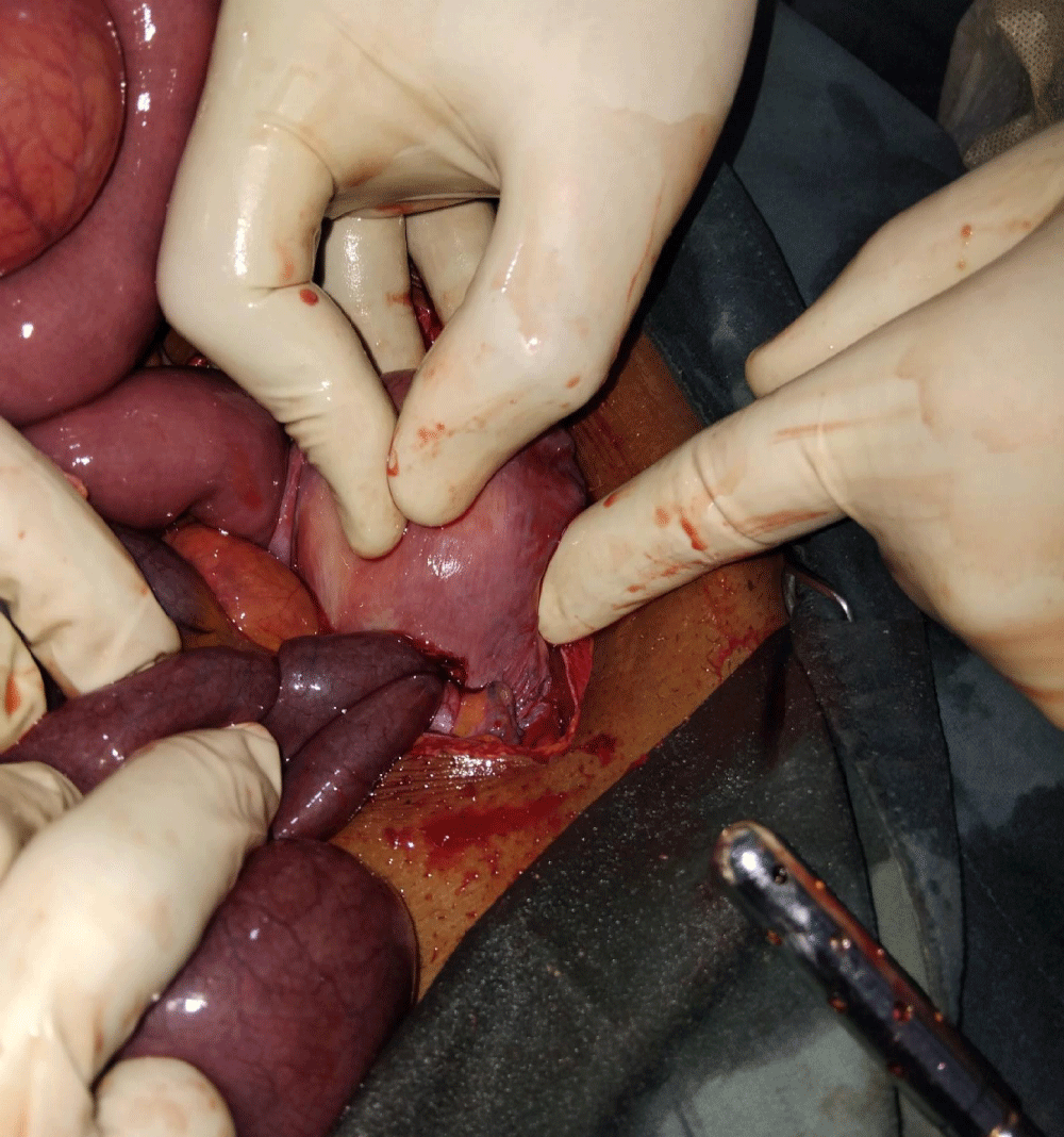
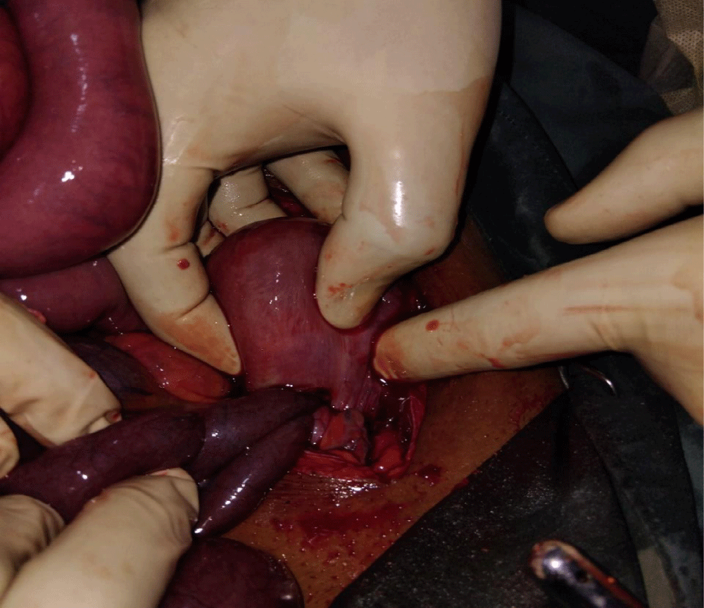
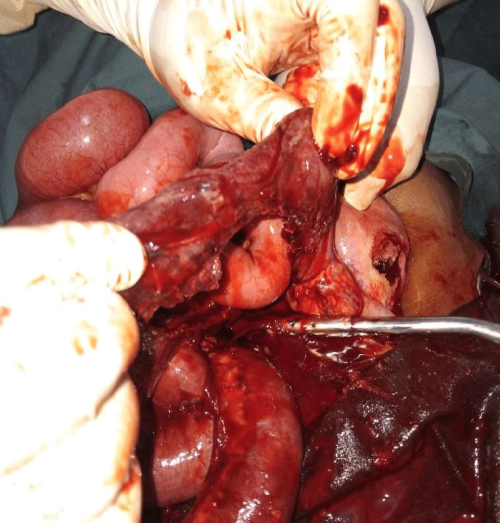
Access was via an extended lower midline incision. The proximal small bowel was distended (Figure 1). A loop of the distal ileum was seen herniating through a perforation on the anterior surface of the uterus (Figure 2). This segment was retracted back to the peritoneal cavity with some difficult and found to be gangrenous and measures eighty centimetres (80 cm) (Figure 3).
This segment was resected followed by ileo-colic anastomosis.
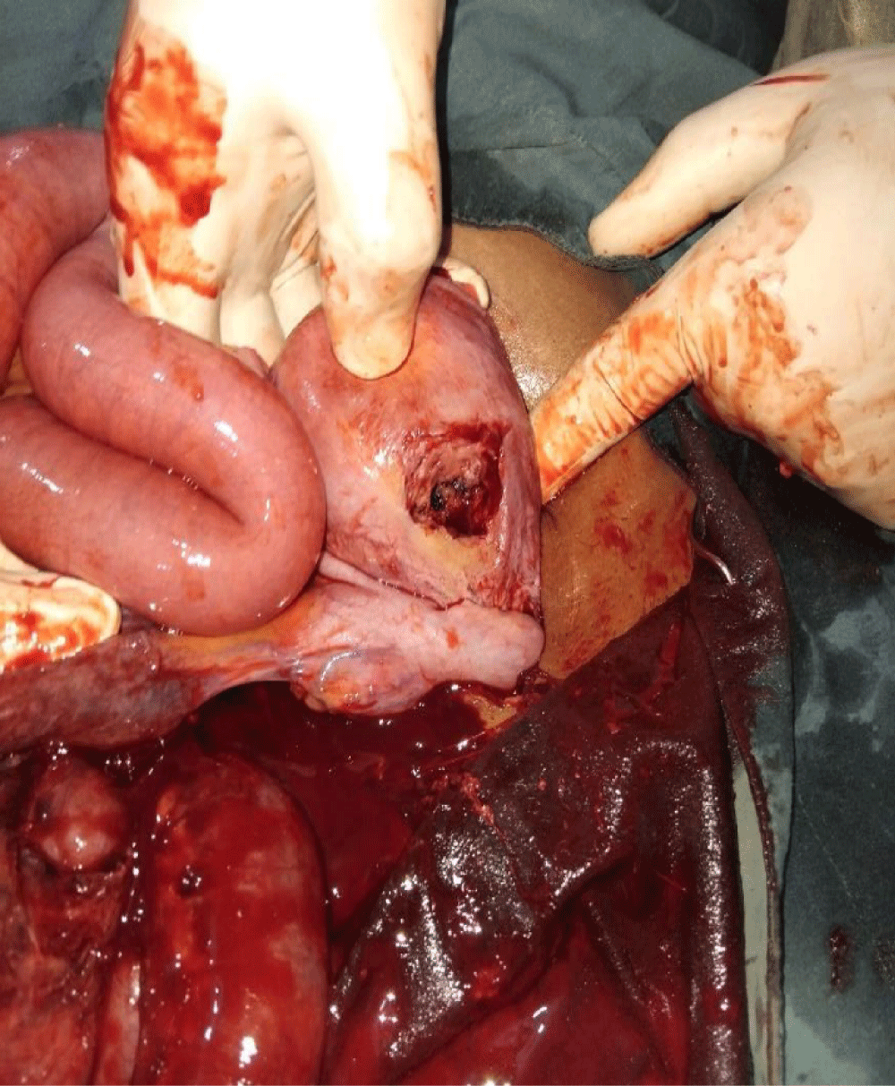
The uterine perforation of ten centimetres (10 cm) was closed in two layers (Figure 4).
Mass closure done for the laparotomy and skin tagged.
Postoperatively, she continued to receive Intravenous fluids, antibiotics and analgesics and. She moved her bowels 72 hours later and commenced oral intake. She was discharged 5 days post operatively to come for follow in a week.
Discussion
Uterine perforation represents one of the rarest and most serious complications of gynecological procedures, with reported incidence rates ranging from 0.1 to 6.4 per 1000 interventions, though actual numbers may be higher due to underreporting of asymptomatic cases [1]. The risk varies substantially by procedure type: while diagnostic hysteroscopy carries a 0.1% - 0.3% risk, operative hysteroscopy and Dilation and Curettage (D&C) demonstrate higher rates of 0.8% - 1.2%, increasing to six percent (6%) in cases of second-trimester pregnancy termination or Manual Vacuum Aspiration (MVA) [1,2].
Perforations may be simple (isolated uterine wall defect) or complex (associated with visceral injury, e.g., bowel, bladder or herniation of bowel through the defect) [5].
The perforation typically occurs through three primary mechanisms:
- Direct mechanical trauma from surgical instruments (curettes, forceps, or hysteroscopic equipment) [6]
- Excessive suction pressure during MVA (negative pressures up to 600 mmHg may draw adjacent bowel into the uterine cavity) [1]
- Uterine wall weakening from previous scarring (cesarean sections, myomectomies) or pathological conditions (adenomyosis, invasive molar pregnancy) [7].
We postulate that the first and second mechanisms above occurred in this index patient as the manual vacuum aspiration was performed by a medical officer in a general hospital.
Major risk factors can be categorized as:
A. Patient factors
- Uterine anomalies (retroversion, bicornuate uterus) [1].
- Myometrial thinning (multiparity, advanced gestational age > 12 weeks) [8].
- Previous uterine surgery (cesarean scars increase risk 3-fold) [7].
Except for the multiparity, none of the above factors were found in this patient.
B. Procedure factors
- Blind procedures without ultrasound guidance [1].
- Operator inexperience (novice surgeons have five times higher complication rates) [1].
- Emergency settings or unsafe abortion practices [1].
Again, all these three were present in this patient.
Clinical presentation varies dramatically based on time to diagnosis:
- Immediate recognition (intraprocedural): Sudden pain with instrument "pop-through" sensation [1]
- Early presentation (< 24 hours): Progressive abdominal distension, guarding, and rebound tenderness [1]
- Delayed presentation (days-weeks): Subacute bowel obstruction, sepsis, or incidentally discovered perforation [5].
The classic triad consists of acute abdominal pain (85 percent of cases), vaginal bleeding (60%), signs of peritonitis (40%) [1]. All these three were present in this index patient.
Untreated cases follow a predictable deterioration:
- 24–48 hours: Localized peritonitis develops as enteric contents leak through defect [1]
- 3–7 days: Bowel ischemia or necrosis occurs in 30% of cases with herniation [1]
- >1 week: Fistula formation (vesicouterine, enterouterine) or systemic sepsis develops [1].
Laparotomy within six (6) hours achieves > ninety-five percent (95%) uterine preservation; bowel resection rates drop from forty-five (45%) to less than ten percent (10%) with early diagnosis. Mortality decreases from eight to twelve percent (8% – 12%) to less than one percent (< 1%) with proper management [1].
The catastrophic potential of bowel herniation—occurring in 0.02% of perforations—necessitates heightened vigilance. This case exemplifies the diagnostic challenges and underscores the need for systematic evaluation of post-procedural abdominal pain to prevent morbidity and mortality [3,4,9].
Conclusion
Uterine perforation is a rare but life-threatening complication that can seldom lead to bowel strangulation and gangrene. Early recognition of post-procedural abdominal pain is critical to prevent these severe sequelae. Delayed diagnosis increases the risk of ischemia and necrosis, worsening patient outcomes. Prompt surgical intervention can preserve bowel viability and reduce mortality. This case emphasizes the importance of vigilance for bowel involvement in uterine perforation.
Ethical consideration
Consent was obtained from the patient after explaining the relevance of the publication and reassuring the patient that she would be anonymous. Ethical clearance was also obtained from the research and ethics committee of the hospital.
- Korkmazer E, Yildirim G, Korkmazer B. Uterine perforation as a complication of intrauterine procedures: A review. J Clin Med. 2023;11(15):4439.
- Coomarasamy A. Uterine perforation. In: Gynecologic and Obstetric Surgery. Wiley; 2016.
- Say L, Chou D, Gemmill A, Tunçalp Ö, Moller AB, Daniels J, et al. Global causes of maternal death: a WHO systematic analysis. Lancet Glob Health. 2014;2(6):e323-e333. Available from: https://doi.org/10.1016/s2214-109x(14)70227-x
- Miller CA, Lurie S. Uterine perforation: Clinical presentation and outcomes. J Minim Invasive Gynecol. 2014;21(5):740-745.
- Goyal LD, Singh K, Singh A. The frequency and management of uterine perforations during first-trimester abortions. Int J Gynaecol Obstet. 1989;30(4):311-314.
- American College of Obstetricians and Gynecologists. Practice Bulletin No. 212: Management of uterine perforation. Obstet Gynecol. 2020;135(3):e110-e117.
- Wildemeersch D, Hasskamp T, Pett A. Intrauterine device-related uterine perforation incidence and risk (APEX-IUD): a large multisite cohort study. Contraception. 2022;106:1-7.
- Schwarzman P, Baumfeld Y, Mastrolia SA, Yaniv-Salem S, Leron E, Silberstein T. Obstetric Outcomes after Perforation of Uterine Cavity. J Clin Med. 2022;11(15):4439. Available from: https://doi.org/10.3390/jcm11154439
- Kumar A, Singh N. Uterine perforation during intrauterine device insertion: Incidence, risk factors, and management. Contraception. 2018;97(3):221-225.
Article Alerts
Subscribe to our articles alerts and stay tuned.
 This work is licensed under a Creative Commons Attribution 4.0 International License.


 Save to Mendeley
