Journal of Clinical Research and Ophthalmology
Theory: Morphological and functional features of the structure of the Zonula Lens Fibers as a key executive link in the mechanism of the human eye accommodation
Ivan N Koshits1, Olga V Svetlova2*, Maksat B Egemberdiev3, Marina G Guseva4, Felix N Makarov5 and Nikolas M Roselo Kesada6
2Professor, Department of the Ophthalmology North-Western State Medical University named after I.I. Mechnikov, Saint-Petersburg, Russia
3Chief of the Department of Ophthalmology, Chuy Region United Hospital, Bishkek, Kyrgyzstan
4Ophthalmologist, Optometrist Medical Diagnostic Center of the Public company, Vodokanal of Saint-Petersburg, Saint-Petersburg, Russia
5Professor, Chief of the laboratory of Neuro Morphology of the Pavlov Institute of Physiology of the Russian Academy of Sciences, Saint-Petersburg, Russia
6Postgraduate student of the Department of ophthalmology, Pirogov Russian National Research Medical University, Moscow, Russia
Cite this as
Koshits IN, Svetlova OV, Egemberdiev MB, Guseva MG, Makarov FN, et al. (2020) Theory: Morphological and functional features of the structure of the Zonula Lens Fibers as a key executive link in the mechanism of the human eye accommodation. J Clin Res Ophthalmol. 7(2): 061-074. DOI: 10.17352/2455-1414.000075Purpose: Assess morphophysiological and functional features of the structure of the zonula lens fibers on the basis of an in-depth analysis consistent with the laws of mechanics. Define the purpose and scope of each portion of the zonula lens fibers. To analyze the results of the research of studies on the structure of the zonula of Zinn in the different authors of the last two centuries. Identify misconceptions about the work of this executive mechanism of accommodation in different phases of accommodation, this includes the uneven accommodation in different meridians. Determine the incorrectness of those theories of accommodation, which are based on erroneous ideas about the functioning of different portions of the zonula fibers.
Methods: Theoretical analysis based on the laws of mechanics and assessment of the results of interdisciplinary research by different authors in the field of structural and functional features of the Zinn's zonule of the lens in different phases of accommodation.
Results: The lens’s ciliary belt has been found to have three major portions of fiber that perform different functions. Powerful anterior portion of fibers is attached to the anterior surface of the lens bag in a segmented mannered a powerful posterior portion of fibers is attached to the posterior surface of the lens evenly around the circumference and like a cobweb covering the surface of the vitreous chamber. Both these filaments in the eye don’t cross and in all phases of accommodation remain tense: in full distance - maximum, and when you look close, it’s minimal. These functional features of two powerful portions of the zonula of Zinn are fully consistent with Helmholtz’s vision and allows them to hold the lens relative to the optical axis of the eye even under high dynamic or weightless conditions. These two portions of fiber are force-bearing and constantly press the lens against the vitreous chamber, fixing its normal position relative to the optical axis. It is characteristic that both these powerful portions of fibers pass in the direction of the choroid through the recesses between the processes of the ciliary muscle. It is important that both of these portions of fibers are not attached to the ciliary muscle by force, but are only held by the thinnest outlet fibers in the recesses between its processes. The anterior and posterior portions of the fibers are attached forcefully to the anterior of the lens bag, and behind they are attached to the choroid in the ora serrata region. The segmental attachment of the anterior portion of the fibers to the lens bag allows for uneven lens accommodation in different meridians. The third portion of ciliary zonula fibers - cilio-equatorial fibers, on the contrary, is not strong and is located evenly along the equator of the lens. Two rows of these thin fibers on one side are attached in the form of a frenum evenly along the equator of the lens bag, and on the other side they are attached directly to the ciliary muscle in the recesses between its processes. The task of these fibers is to uniformly collapse around the extreme periphery of the lens bag in the equator region to keep the lens masses near the optical axis, preventing them from shifting to the equator inside the lens bag during different phases of accommodation.
Conclusion: Analysis of the morphophysiological structure of the Zinn's zonule of the lens, adequate to the laws of mechanics, made it possible to deepen and expand the classical theory of lens accommodation H. Helmholtz - where the lens is the main, but not the only mechanism of accommodation in the human eye. It was found that the executive mechanisms in the lens accommodation are: ciliary muscle, zonula of Zinn, vitreous chamber and choroid. The choroid is a biological spring and passively stretches the ciliary muscle depending on the level of its active tone. In all phases of accommodation, the lens is pressed by a powerful anterior portion of fibers to the hyaloid membrane of the vitreous chamber. At moments of external dynamic loads on the eye, the energy of inertial micro movements or the lens phacodonesis is transferred to the vitreous chamber, which dampens these vibrations due to the dispersion of this energy between its internal structures. These ideas are adequate to the laws of mechanics and exactly correspond to Helmholtz formulations: in all phases of accommodation, the anterior and posterior portions of the fibers remain stretched and their tension only slightly relaxes when looking completely closer. The detected segmented attachment of the anterior portion of the fibers to the anterior part of the lens capsule allows for uneven accommodation in different meridians to partially level the induced astigmatism of the optical system of the eye under different visual loads. The most important result was the understanding that the pressure in the lens is at maximum when viewed completely closer. This leads to the extension and rounding of the anterior and posterior mini lens of the lens and increase their refractive power for rays passing near the optical axis of the eye. On the contrary, when looking completely into the distance, the choroid stretches the anterior and posterior portions of the fibers as much as possible, due to which the lens bag is maximally stretched, and the lens itself becomes flatter.At this moment, the pressure in the lens is minimal, since the elastic capsule of the lens is stretched from the outside by the zonula fibers and therefore compresses the intralenticular masses the weakest of all with less force. It is at this moment that the work of the cilio-equatorial fibers is necessary in order to evenly squeeze the periphery of the lens along the entire equator and keep the lens masses in the center of the optical axis. The results also confirmed the incorrectness of many theories of accommodation compared to Helmholtz's theory of accommodation. Especially incorrect seems the extravagant theory of accommodation R.A. Shachar.
Abbreviations
AGM: Anterior Hyaloid Membrane; FPF: Front Portion of the Fibers of the zonula of Zinn; CB: Ciliary Body; CEF: Cilio-Equatorial portion of the fibers of the zonula of Zinn; ILP: Inside the Lens Pressure; IOP: Intraocular Pressure; MC: Muscle Ciliary; OAG: Angle Open Glaucoma; OD: Optic Disk; PPF: Posterior Portions of the Fibers of the zonula of Zinn; VC: Vitreous Chamber; ZF: Zonula Fibers; ZZ: Zonula of Zinn
Introduction
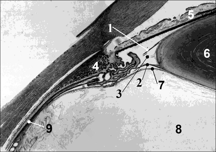
Although accommodation determines high-quality vision and is one of the most important sensory systems in humans, there is still no generally accepted theory of accommodation in ophthalmology. Accommodation standards are not fully developed and not introduced into clinical practice. This is due to the fact that over the past two centuries, many eye researchers, when explaining the results of morphological studies of the executive structures of the accommodation mechanism, did not fully take into account the laws of mechanics. The widespread use of the latest methods for studying the structures of the human eye based on high-resolution electron and ultrasound microscopy did not improve, but, on the contrary, confused the idea of the accommodation mechanism even more, because in the morpho-physiological interpretation of the structural features of the structure of the Zonula Fibers (ZF), the laws of mechanics were not always respected again. Paradoxically, even some leading ophthalmologists so far have not had a clear idea of the exact location and fixing in each portion of the ZF, although an adequate idea of the operation of the main intraocular systems: accommodation and outflow of aqueous humor depends on understanding this issue. An interdisciplinary approach for assessing morphological results using the simultaneous application of research methods in ophthalmology, physiology, biomechanics and control theory allows us to obtain “at the intersection of sciences” an adequate modern understanding of the accommodation mechanism. And this mechanism should be based on an in-depth understanding of the morphological, functional and design features of its key link - ZF (Figure 1).
1- Front Portion of the Fibers (FPF); 2 - Posterior Portion of the Fibers (PPF); 3 - Cilio-Equatorial Portion of Fiber (CEF); 4 - Ciliary muscle; 5 - Iris; 6 - Lens; 7 - Anterior hyaloid Membrane (AGM) of the vitreous chamber; 8 - vitreous chamber.
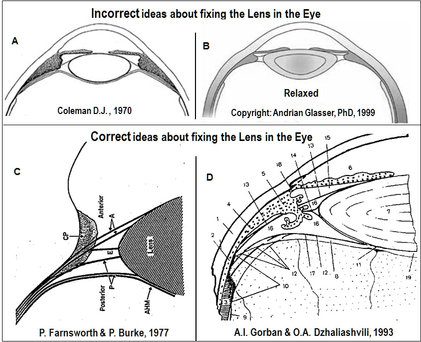
Our ideas (1994-2005) [2-8] about the functional role of the vitreous chamber, which is a necessary executive element in all phases of accommodation, coincided with the opinion of a number of authors [9-13]. All of these authors also considered the vitreous chamber as a possible actuator to assist in the rounding of the lens capsule during near accommodation. As we did before, Lütjen-Drecoll E, et al. [13], recently designated the vitreous chamber as a structure that is capable of damping lens vibrations. It is worth noting separately the views of D.J. Coleman (1970,1986) [12,14], according to which the lens rests in an elastic suspension of zonular fibers, the spatial architectonics of which ensures their functioning like a cable-stayed chain line at suspension bridges (Figure 2).
When looking closer, the vitreous chamber directionally supports the lens from behind and helps it round.
This idea is still used in a significant number of research works in which various mechanical experiments were carried out to stretch the bag of the lens in the equatorial region. On the basis of these experiments, finite element models were considered for calculating the shape of the lens and the functioning of the accommodation mechanism [15-22]. However, unfortunately, all these works, as a rule, have one significant drawback: the boundary conditions for the forceful fixation of the lens in the eye with the help of the fibers of the zonula, choroid, vitreous chamber and ciliary muscle were chosen incorrectly. And this objectively leads to erroneous results of calculations and their interpretation.
According to H. Helmholtz (1855) [23], to enhance eye refraction when working near the Muscle Ciliary (MC) contracts, while weakening the tension of the fibers of ZF and allowing the lens capsule to round. In the absence of accommodation during distance vision, the MC relaxes, the zonula fibers are stretched again, the lens is flattened for distance vision, there is no accommodation. The executive mechanisms of accommodation at Helmholtz are: active MC and ZF together with an elastic lens capsule. Note that choroidea and Vitreous Camerale (VC) are not involved in the accommodation mechanism according to H. Helmholtz [24]. The theory of accommodation was not finally completed by H. Helmholtz and could not explain a number of clinical observations. Therefore, to date, various “new” theories of accommodation are proposed, among which it is necessary to especially highlight the results of the following researchers: Gullstrand (1909) [25], who introduced the theoretical concept of the lens nucleus with greater refractive power to explain the total accommodation volume observed in practice of 10 diopters or more;
M. Tscherning (1898) [26] and A. Pflugk (1935) [27], who considered the force on the lens from the vitreous chamber to be more important in the accommodation mechanism than tension of ZF;
J.W. Rohen (1969-1979) [28,29], who proposed his own interpretation of the functioning of various portions of ZF during Helmholtz accommodation and was widely used by ophthalmologists, but contrary to the laws of mechanics.
R.A. Schachar (1992-2001) [30-33], who presented an interpretation of the accommodation mechanism opposite to H. Helmholtz and an interpretation of the functioning of ZF opposite to J.W. Rohen. Unfortunately, the views of R.A. Schachar today received active support from some eye researchers and ophthalmic surgeons, although the main hypotheses of his “new” theory of accommodation are clearly not adequate to the laws of mechanics, including his understanding of the role ofthe cilio-equatorial portion of fibers (CEF) as leading in the act of accommodation [34,35].
In-depth studies of ZF, which we conducted over 20 years, allowed us to draw a number of important practical conclusions and to develop the H. Helmholtz accommodation theory, which is consistent with the laws of mechanics in all phases of accommodation.
The structure and functioning of the supporting apparatus of the lens
Today, there are reliable studies of the ZF structure at the beginning and end of the 20th century, striking not only with the fundamental approach, but also with the depth of interpretation of the results. The use of high-resolution electron and ultrasound microscopes as a research tool made it possible to obtain unique micrographs of the ZF structure [13,28,29,36,37-42].
However, in a number of cases, due to errors in the formulation of the research problem, as well as in the interpretation of the results, some authoritative researchers were significantly confused with the initially correct ideas about the functional role of various portions of the fibers of the ZF. This led to fundamental disagreements in understanding the accommodation mechanism in different ophthalmological schools, but also “ideologically” prepared the appearance of the “extravagant” theory of accommodation R.A. Schachar [32,33]. It was only possible to cut this truly “tight” knot recently - after conducting ZF structure studies by experts in the field of ophthalmology, morphology, physiology, biomechanics, and control theory [35,38,39].
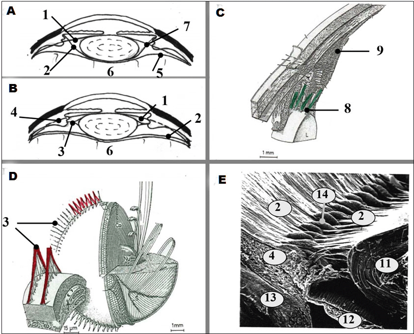
At the beginning of our research [43,44], we went the traditional way and calculated the dynamics of breakage of ZF during lens subluxation after concussion based on the scheme for fixing the lens in the tendon of Zinn presented to us by the leading ophthalmologists of the USSR: this traditional scheme involves fixing the lens (in terms of biomechanics) “in an elastic suspension”, when the Anterior (APF) and Posterior (PPF) portions of ZZ fibers intersect (Figure 3a).
If we accept the scheme of fixing the lens in the eye using only ZF structures and crossing its Anterior Portion of the Fibers (APF) and its Posterior Portion of (PPF), then with such a constructive design of the lens attachment in the eye, it turns out to be virtually unattached due to the "relaxation" of the ZF at the moments of accommodation in the near distance. And this should lead to his constant trembling (phacodonesis) or frequent subluxations, which is not observed in practice. This means that there must be another, more reliable mechanism for fixing the lens in the eye without crossing the ZF (Figure 3b), but to detect this mechanism, we needed a more in-depth understanding of the morphological and physiological features of the ZF structure.
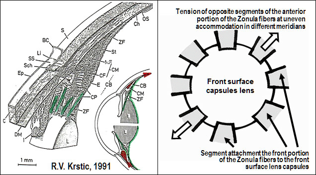
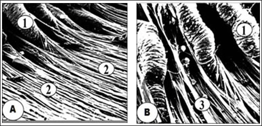
The results of direct studies of the structure and functioning of ZF were unexpected [3-8,12,13,28,29,35-45,46-50]. It should be noted the important role of Russian researchers [24,34,35,39,43-47,51] in the study of this issue. In particular, it can be considered finally established that the powerful FPF, going from the lens bag to the ora cerrata, does not forcefully attach directly to the Ciliary Body (CB), but passes between its processes and is woven by force into the choroid in its dentate region lines. These relatively flat “reins” fibers are located in groups between the processes of the CB and are attached at separate fixed points on the anterior surface of the lens capsule in a circle not uniformly, but in segments (Figures 3c,4) [37, p. 511]. The segmental architectonics of FPF attachment to the anterior surface of the lens sac makes it possible to neutralize induced or natural physiological astigmatism. MC must be able to non-uniform (segmental) contraction of its circular portion of muscle fibers (Muller muscle) and its meridional portion of muscle fibers (Brucke muscle) for this.
Based on this structure of FPF, the conclusion that different parts of the ciliary muscle can work asynchronously, i.e. not only independently of each other, but also with different intensities. Perhaps for the first time the presence in the human eye of uneven accommodation in various meridians was predicted by the Russian military ophthalmologist Professor V.I. Dobrovolsky [52]. In order to create an uneven tension of FPF segments in the required meridians, the ciliary muscle must provide contraction (or relaxation!) of its corresponding spatial portions in these meridians. Although we note that these our ideas contradict one of the persistent myths of ophthalmology: the ciliary muscle is supposedly “single and indivisible” and is unable to contract “in parts”. But it is the biomechanical and structural features of the FPF that convince us of the opposite. And it is worth noting that the presence of uneven accommodation in children for the first time in the world was confirmed in the clinic by professors from Ukraine V.I. Serdiuchenko and I.A. Viasovsky according to their original method [53,54].
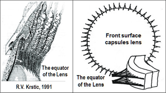
A - with crossing, B - without crossing [13,16,20]; C - attachment of the anterior portion of the fibers (8) of the zonula of Zinn according to Krstic [37, p. 511]; D - attachment of cilio-equatorial fibers (3) of the zonula of Zinn according to Krstic [37, p. 525]; E - attachment of the posterior portion of the fibers (2) of the zonula of Zinn according to R.J. Rochen (uv. 50) [28,29].
1 - anterior portion of the fibers of the zonula of Zinn; 2 - the posterior portion of the fibers of the zonula of Zinn; 3 – cilio-equatorial portion of the fibers of the zonula of Zinn; 4 - ciliary body; 5 - anterior hyaloid membrane of the vitreous chamber; 6 - vitreous chamber; 7 - the apparent intersection of the anterior and posterior portions of the fibers of the zonule of Zinn, 8 - the anterior portion of the fibers is fixed on the anterior surface of the lens capsule; 9 - the anterior portion of the fibers of the ciliary gland is woven into the dentate line of the choroid; 11 - a crystalline lens; 12 - iris; 13– sclera; 14 - processes of the ciliary body.
Cilio-Equatorial Fibers (CEF), on the contrary, are located arched and evenly along the line of their attachment along the equator of the lens capsule (Figure 3d) [37, p. 525]. The posterior portion of fibers (PPF) of the zonula of Zinn, thinner and branched, actually like a “spider web” covers the dome of the vitreous chamber of the vitreous chamber - anterior hyaloid membrane (AGM). On the one hand, PPF is woven into the Wigeri ligamentum (Wiger circular ligament) on the back surface of the lens capsule, and on the other hand, into the choroid in the dentate line, but apparently radially closer to the inside than the FPF is located (Figures 3e, 5) [28].
Under the action of multidirectional elastic forces: on the one hand - the elastic capsule of the crystalline lens, and on the other - the elastic choroid, FPF and PPF in all phases of accommodation are in a constantly stretched state of dynamic equilibrium, and the degree of tension during accommodation is close to minimum (according to G. Helmholtz FPF and PPF are "weakened"), and in the absence of accommodation - maximum.
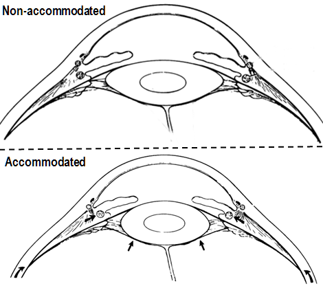
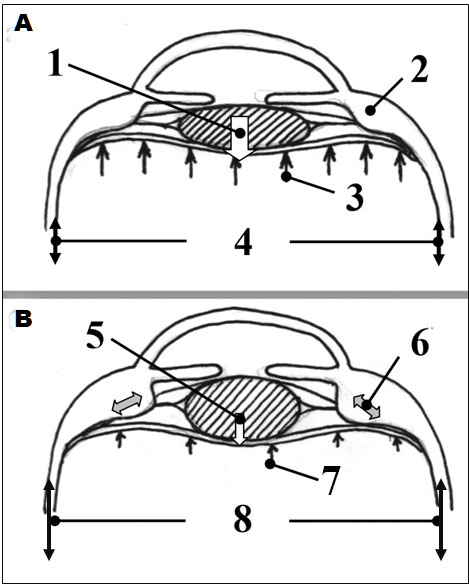
The white arrow shows the arched space between the CEF portions, which are attached to the anterior and posterior surfaces of the lens capsule like a bridle uniformly at the equator. Figuratively speaking, the elastic tissue of the lens capsule and the spatially elastic vascular structure of the choroid are two “teams” involved in the “tug of war”. The role of ropes is played by the practically low-stretch FPF and PPF. It is important to note that the ciliary muscle itself does not directly alter the FPF and PPF tension, but only produces the necessary joint displacement through the movement of the dentate line, increasing or weakening the elastic tension of the elastic structures of the choroid, this kind of “biological spring” (Figure 6) [6,7,24,38,45].
A. No accommodation: 1 - the resulting force exerted on the lens by pulling the anterior portion of the fibers of the zonula of Zinn as much as possible (wide white arrow); 2 - ciliary muscle relaxed; 3 - pressure in the vitreous chamber is increased (long arrow); 4 - choroid is compressed (short double arrows).
B. Accommodation closer: 5 - the resultant force acting on the crystalline lens from the tension of the anterior portion of the fibers of the zonula of Zinn is minimal (narrow white arrow); 6 - ciliary muscle is tense (double gray arrow); 7 - pressure in the vitreous chamber is reduced (short arrows); 8 - choroid extended (long double arrows).
In fact, we must admit that in the absence of accommodation the lens is “squeezed” between the “powerful” stretched FPF of the zonula of Zinn and additionally “tense” from the increasing pressure in the vitreous chamber by the surface of the Anterior hyaloid membrane (AGM). This response increase in the “rigidity” of the AGM due to an increase in pressure in the vitreous chamber occurs when MC relaxes when the lens moves posteriorly and is pressed into the vitreous chamber by the increased force from the FPF. This effect is the basis of our developed method for early diagnosis of OAG using HRT-II, which allows to objectively evaluate the mobility of the Optic disk (OD) by increasing the volume of excavation after MC relaxation with a short-acting mydriatic [55]. At this moment, the tone of the MC is minimal, and the choroid is not stretched (Figure 3) [50].
With the aforementioned ZZ structure, a change in the tension of the APF and PPF of the zonula of Zinn is associated only (!) with the displacement of the dentate line of the choroid depending on the force exerted on it by the MC. Such an organization of work of MC allows to reduce the magnitude of the effort and the amount of work performed by it as an actuating element in the intraocular accommodation mechanism. Speaking in the language of biomechanics, such a design of the accommodation mechanism makes it possible for the MC not only to get tired and save the energy consumed by the eye as an organ, but also to minimize the transition time from passive accommodation when looking completely into active accommodation at medium and short distances.
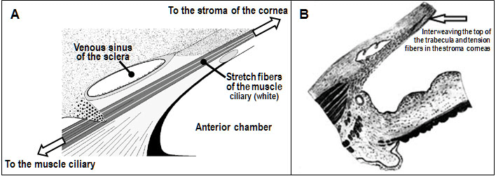
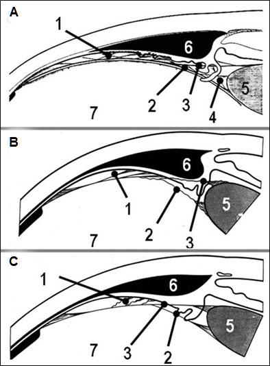
In order to fully understand the whole scheme of the power fixation of the lens in the eye, it is necessary to understand how the muscle ciliary is anchored in the eye. The question of attaching the MC inside the eye is not as simple as it seems. The traditional idea is that the back of the ciliary muscle is woven by its fibers into the choroid a (quote) “In its anterior section, the Brücke longitudinal muscle is attached to the scleral spur and trabecular plates and ... according to histological examination, the anterior ends of the meridional and radial fibers are attached to the scleral spur and trabecular plates” [51]. But this, unfortunately, is not quite the right idea. As objectively pointed out by the studies of A.V. Zolotarev, B.T. Gabelt, et al. Е. Lütjen-Drecoll, et al. E. R. Tamm, et al. [56-59] The ciliary muscle is attached on one side by end-to-end tension fibers to the corneal stroma and on the other to the choroid and through it to the posterior pole of the sclera (Figure 7A). According to the Atlas of Gonio-biomicroscopy by E. Van Boiningham [60], the apex of the trabecular tissue has grown significantly anteriorly above the Descemet's sheath along the through stretching fibers into the corneal-scleral tissue (Figure 7b).
А – the location of tension fibers in the anterior part of the eye, by which the muscle ciliary is attached to the corneal stroma [56]. B - anterior attachment of the trabecular network to the corneal stroma [60].
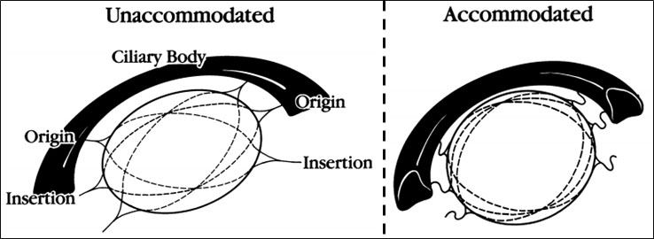
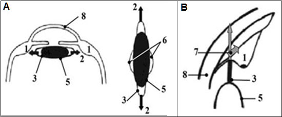
Now we will be able to discuss the question of what is the role of the cilio-equatorial fibers in the lens accommodation mechanism, and whether these concepts are adequate to the laws of mechanics. Several extravagant accommodation theories are based on the concept of the central role of the CEF in the act of accommodation, and these concepts themselves are supported by many ophthalmologists. In particular, RS Wilson [15] (Figure 8), as well as RA Shachar [32,33] (Figure 9) believe that CEF is the main executive link in the accommodation mechanism, and all other structures that hold the lens in the eye in the natural position for optical beams, are auxiliary.
Reducing the diameter of the ciliary ring when looking closer leads to relaxation of the cilio-equatorial fibers, and the lens is able to round. When looking into the distance, the diameter of the ciliary ring increases, the CEF portion stretches and makes the lens flat. FPF and PPF do not participate in the act of accommodation.
The R. A. Shachar's point of view on the role of cilio-equatorial fibers and his theory of accommodation are based on premises that do not fully correspond to the laws of mechanics [7,8,24,34,35,38]. He rejects all accommodation theories that existed before him and proposes a fundamentally different mechanism of accommodation. In his opinion, the tension of the CEF at the moment of relaxation of the ciliary muscle pulls and collapses the equatorial zone of the lens capsule, tightens its peripheral part and forces the lens masses to move to the central region of the lens bag to increase its thickness here. FPF and PPF do not participate in the act of accommodation [24,38].
A and B. Accommodation closer: Ciliary muscle (1) relaxed; the stretching (2, black arrows) of the lens capsule (5) by cilioequatorial fibers (3) leads to an increase in its roundness in the central part (6). B. To ensure accommodation, it is necessary to move outside and behind the ciliary muscle (7, gray arrows); the anterior and posterior portions of fibers according to R.A. Shahar are auxiliary, do not participate in accommodation, therefore, are not shown in the diagram; 8– cornea.
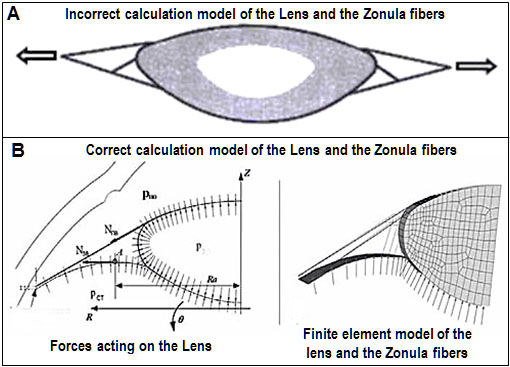
Taking into account all the above, it now becomes clear how incorrect the widely used schemes for fixing the lens in the eye with a cross of fibers going to the front and back surfaces of the lens can be. Based on these incorrect ideas about the course of the ciliated girdle fibers, extensive mechanical calculations of the lens shape in different phases of accommodation are performed (Figure 9a).
According to these representations of RA Schachar, CEF tension compresses the lens capsule evenly along the equator, moving the lens masses into the central part of the lens bag, which causes it to round. And I must say that from the point of view of mechanics, such a view has every right to exist. Indeed, if we take a rubber shell filled with water in the form of an ellipse and begin to stretch it with threads fixed anatomically in the same way as the cilio-equatorial fibers, then water from the equatorial part of the shell will move to its central part. And this will lead to stretching and an increase in the thickness of the shell in the center and its partial rounding. Such a transitional state of the spatial shape of the shell (or capsule of the lens) will be adequate to the laws of mechanics, but under the initial conditions of fixing the lens in the eye using only CEF. Conclusion: CEFs, which are rather weak from a mechanical point of view, should play a major role in the lens mechanism of accommodation. At the same time, powerful FPF and PPF are not basic, but only auxiliary, which do not play any role in the process of accommodation.
According to the extravagant theory of accommodation RA Schachar, a full close look is carried out with the complete relaxation of the ciliary muscle, when the CEFs are maximally stretched and the lens should be rounded. On the contrary, the gaze completely into the distance should then be carried out with the maximum tone of the ciliary muscle, when the CEFs are minimally stretched and the lens should become flat. And then Helmholtz's theory should be considered deeply erroneous. To put it quite simply, according to the theory of accommodation RA Schachar, the spasm of accommodation, as well as the maximum blood consumption of the ciliary muscle, should be observed when looking completely into the distance, which contradicts a huge number of clinical facts.
However, R.A. Schachar, et al. created the concept of "zonular compression" theory and innovative, in their opinion, medical technology to combat presbyopia based on their own computational mathematical models and even carry out clinical practice based on this theory [30]. It is worth emphasizing once again that from the point of view of mechanics, such calculations are fair and reliable if the method of fixing the lens in the eye and the initial conditions for the mathematical calculation are determined in the above manner.
The creation of a computational mathematical platform for confirming the theory of accommodation RA Schachar is exactly the case when ophthalmologists set a task for mechanics to calculate, indicating incorrect boundary conditions for the force fixation of the lens in the eye. There is an obvious error in the initial formulation of the problem for mechanical calculation due to the lack of a clear understanding of the function of all portions of zonular fibers, as well as the role of the elastic structures of the lens capsule, anterior and posterior portions of zonular fibers, choroid and vitreous chamber in the functioning of the accommodation mechanism.
The fallacy of the "zonular compression" theory is as follows. The lens is force-fixed in the eye with the help of powerful FPF and PPF, and in all phases of accommodation it is pressed against the vitreous chamber. Therefore, the boundary conditions when setting the problem for calculating the shape of the lens during accommodation should be fundamentally different and include all external structures that constantly keep the lens in its natural position [15,24,35,38,50]. Normally, with the maximum tone of the ciliary muscle, the tension of the cilio-equatorial fibers should be minimal, or absent altogether, since the structures of the ciliary muscle are displaced at this moment anteriorly and inwardly. This allows the lens bag to become more rounded even at the equator. But if a portion of CEF is relaxed, and other portions of fibers do not participate in the lens rounding process, then the lens is actually not fixed in the eye. However, in practice this is not the case, and we do not observe normal lens phacodenesis in all phases of accommodation.
The preliminary result of our analysis suggests that over the centuries the results of studying the structure of the ZZ, its participation and role in the act of accommodation, gradually approached the state adequate to the laws of mechanics. Despite the fact that many ideas about lens accommodation were based on incorrect assumptions, they all gave a powerful impetus forward to reveal the truth. In our opinion, the European researchers H. Martin, O. Stachs, R. Guthoff, et al. (2018) recently approached the understanding of the role of the ciliary band of the lens in the act of accommodation [61].
In 1993, Russian professors A.I. Gorban and O.A. Dzhaliashvili publish a detailed study taking into account data previously obtained by N.I. Bastrikov and A.A. Leonov [46,47], a diagram of the mutual arrangement of the fibers of the lens support apparatus. This publication has so far most accurately described the spatial structure of ZZ (Figure 10d) [45].
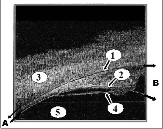
The final clinical confirmation of the correctness of the results of the study of the structure of the ciliary girdle by different authors was obtained by A. Glasser in 1999, when in vivo, using high-resolution ultrasound biomicroscopy of the anterior part of the eye in monkeys (rhesus monkeys) it was possible to obtain photomicrographs of the relative spatial location of FPF and PPF [36]. The main conclusion: these portions of fibers do not really intersect, and APF and PPF of the zonula of Zinn pass between the CB processes to the dentate line of the choroid (Figure 11).
A - the direction of the fibers of the zonula of Zinn to the dentate line of the choroid (two narrow black arrows);
B - the direction of the fibers of the zonula of Zinn towards the lens capsule (two wide black arrows).
To obtain a complete picture of the lens mechanism of accommodation, it is necessary to complete the study of the morphological structure of ZF and practically clarify only the last two questions:
- how are the cilio-equatorial fibers attached to the dentate line and to the recesses of the ciliary body;
- how are all parts of the ZZ fibers anchored structurally in the choroid.
These questions are waiting for their researchers and undoubtedly relate to actual morphological problems of ophthalmology.
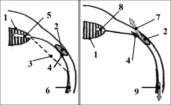
A. No accommodation: ciliary muscle (2) relaxed; the maximum stretching (5, long white arrow) of the elastic lens capsule (1) by the force compressed like a spring of a choroid (6, short gray arrow) provides at this moment the maximum tension of the fibers of the zonula of Zinn and flattening of the lens; 3 - a rectilinear arrangement of the course of the fibers of the zonula of Zinn not observed in practice, but corresponding to the laws of mechanics if they are stretched only by the elastic forces of the lens capsule and choroid; 4 - practically observed curvature of the course of the fibers of the zonula of Zinn under the action of additional forces from the "pulling" fibers;
B. Accommodation closer: the active force (7, gray arrow) of the ciliary muscle (2) additionally pulls the anterior choroid line anteriorly, while reducing the tension of the fibers of the zonula of Zinn and the tensile force (8, short white arrow) of the lens capsule, as a result of which the lens is rounded ; the force (9, the long gray arrow) from the side of the choroid stretched like a spring at this moment is maximum and equal to the sum of two forces: the maximum effort (7) of the ciliary muscle and the minimum tensile force (8, short white arrow) of the elastic lens capsule.
As J.W. Rochen discovered, the cilio-equatorial fibers located along the equator of the lens were also somehow fixed in the recesses of the ciliary body. These ideas led J.W. Rohen to the hypothesis that the lens capsule, as well as the ciliary muscle, which, in his opinion, provides tension or relaxation of the FPF and PPF through the displacement of the dentate line anteriorly with the help of thinner abducting fibers, are involved in the act of accommodation. Simply put, the contraction of the ciliary muscle leads, according to J.W. Rohen, to the contraction of the elastic capsule of the lens only as a result of additional effort from the abducting fibers. Moreover, the ciliary muscle itself is located as part of this mechanism in front of the FPF and PPF and their area of attachment to the choroid, as shown in Figures 13,14. That is, according to J.W. Rohen, the ciliary muscle transmits its force to the choroidea only through tension fibers, which, when looking into the distance, completely relax, and, accordingly, in no way can take part in damping the vibrations of the lens (Figure 13b).
According to J.W. Rohen's rather thin “tension fibers” must transmit force from the elastic structures of the lens capsule through the powerful FPF to the ciliary body. But for this, the diameter of the "tension fibers" and their number must be larger than that of the FPF, respectively. This is not confirmed by electron micrographs: the "tension fibers" are much thinner and quantitatively less than those of FPF. Therefore, they cannot play the main role in the mechanism of accommodation, but, possibly, play an important, but auxiliary role. In particular, they keep the FPF bundles in the grooves between the processes of the ciliary body, dampen shock loads acting on the lens, and also pull the FPF along an arc, like a bowstring, so as not to cut the surface of the anterior hyaloid membrane (Figure 13a and 14) [28,29].
1 - the processes of the ciliary body, 2 - the posterior portion of the fibers, 3 - the anterior portion of the fibers.
A. Attachment of the fibers of the zonula of Zinn: 1 - "pulling" fibers; 2 - the back portion of the fibers of the zonule of Zinn; 3 - the anterior portion of the fibers of the zonula of Zinn; 4 – cilio-equatorial fibers of the zonula of Zinn; 5 - a crystalline lens; 6 - vitreous chamber.
B. Accommodation closer: Ciliary muscle tense (7); tensioning fibers (8) are tensioned; the anterior (9) and posterior (10) portions of the fibers of the zonula of Zinn are relaxed;
C. No accommodation: Ciliary muscle relaxed (11); the lens (5) is compressed between the stretched anterior (13) and posterior (14) portions of the fibers of the zonule of Zinn, 12 - the tension fibers are relaxed.
Thus, this spatial bending of FPFs is necessary so that they cannot “cut” into the AGM of the vitreous chamber in the absence of accommodation. According to the J.W. Rohen scheme, it is at this moment that the “tension fibers” are not tense, but relaxed (Figure 14c). Although it is at this moment that the tension of these abducting fibers should be maximum. Note also that with such an action from the FPF and PPF, the lens must move posteriorly, and in order to hold it in place, a force response from the vitreous chamber is required. And this was fundamentally ignored by J.W. Rohen, since he, like H. Helmholtz, does not include the vitreous chamber among the executive mechanisms of accommodation.
This tragic mistake led J.W. Rohen to misinterpret the functioning of the accommodation mechanism in general, since the powerful forces from the FPF and PPF are transmitted only through the thinner and naturally weaker mechanically weaker "tension fibers". All of the above does not provide sufficient grounds to consider such an interpretation of the functioning of ZF portions adequate for the reliable implementation of the accommodation process, since it does not agree with the laws of mechanics.
Biomechanical interaction of the elastic lens capsule with the fibers of the zonula of Zinn in different phases of accommodation
The physiological nature of the pressure inside the lens and in the vitreous chamber, different from the IOP, has not been understood by most ophthalmologists until now. Therefore, this issue needs to be discussed in detail. It is characteristic that most researchers by default did not include the elastic structures of the lens capsule in the actuating mechanisms of lens accommodation. However, as described in RF Fisher (1971) [63], S. Krag and Tr.T. Andreassen (2003) [64] the elastic properties of the lens bag allow not only to ensure its rounding when looking closer, but also create intralental pressure Inside the Lens Pressure (ILP). The physiological nature of the occurrence of pressure inside the lens and in the vitreous chamber, other than IOP, has not been understood by most ophthalmologists until today. Therefore, this issue should be discussed in detail.
Note that, just like aqueous humor, all structures inside the lens are incompressible and unable to change their volume when the pressure inside the lens capsule changes. From a biomechanical point of view, these structures physically behave normally like water. But here it should be clearly understood that pressure in the lens can arise only when its incompressible structures are compressed from the outside by the elastic capsule of the lens. This is precisely the picture we observe on the enucleated lens: it is rounded as much as possible due to the work of compression of its elastic outer capsule, when there is no constant stretching effect on it from the anterior and posterior portions of zonular fibers. At this point, the pressure inside the lens of the ILP is at its maximum. It should be noted that similarly, IOP is created in the eye only due to external compression of its internal structures by a relatively rigid but elastic fibrous sheath [65].
An excellent illustration of the discussion of this problem is the recent numerous attempts to ensure the filling of the lens sac with an inert transparent biopolymer after extraction of cataract masses in order to restore its ability to accommodate. O. Nishi (1998, 2007) [65,66] has made the furthest advance here. It is characteristic that when filling the sac of the lens with a biopolymer, the volume of which was almost equal to the removed volume of cataract masses, the ability of the lens to accommodate was not restored.
The lens also could not accommodate when filling his pouch at 45% or less of its presbyopic volume. But on the other hand, when the lens bag was filled with a biopolymer, the volume of which was 50-55% of the volume of the cataract lens, its ability to accommodate was restored! And the reasons for this were difficult for many researchers to understand.
However, from the point of view of mechanics, everything can be explained quite simply. The fact is that the presbyopic lens has its volume about 2 times larger than normal [67]. And this is due to the accumulated and not removed slags inside his bag. These still transparent slags begin to expand from the inside and stretch the lens capsule. In fact, the presbyopic lens is an overstretched and already inelastic shell filled with incompressible cataract masses. Therefore, filling the lens bag with biopolymer by 75-98% of its volume before the operation, we actually overstretch its capsule again in comparison with its normal volume, i.e. we artificially transform the lens into a presbyopic one.
Therefore, filling the lens bag with biopolymer by 75-98% of its volume before the operation, we actually overstretch its capsule again in comparison with its normal volume, i.e. we artificially transform the lens into a presbyopic one. At the same time, in the presbyopic lens, the fibrous structures of his bag are stretched as much as possible, the rigidity of the bag itself increases because of this, which leads to a response jump in ILP. The increased ILP makes it difficult for fresh aqueous humor to pass into the lens through the outer epithelium. In fact, the lens loses its ability to "breathe", i.e. loses the functional ability to micro volume fluctuations. And this happens, in particular, because the pressure inside the presbyopic lens is higher than normal.
We have already found out that FPF and PPF, as well as the CEF portion, stretch the lens capsule as much as possible when there is no accommodation (looking completely into the distance). And the capsule of the lens is minimally stretched with the tension of accommodation (look completely close). Moreover, the process of active accommodation itself takes much less time than the process of diffusion of aqueous humor through the epithelium of the anterior capsule into the lens. Therefore, the normal volume of the lens during accommodation remains practically constant [68].
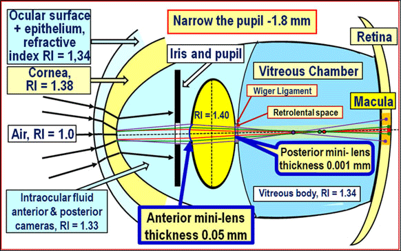
But precisely when looking completely close up, the elastic capsule of the lens is less stretched by the fibers of the zonule. And this allows the lens bag to round and, due to the reduction of its elastic structures, to squeeze the incompressible lens masses from the outside as much as possible. Moreover, the ILP will be maximum. This leads to the stability of the anterior and posterior mini-lenses of the crystalline lens, increasing its refractive power for rays passing near the optical axis (Figure 15). This important mechanism of additional accommodation is described in [69].
The increase in pressure in the rounded lens when looking up close causes the surface of the anterior and posterior mini-lens to stand.
On the contrary, when looking completely into the distance, the lens capsule will be maximally stretched by the zonule fibers, which will reduce its ability to compress the lens masses due to its elasticity. And this will lead not only to a decrease in ILP, but also to the appearance of waviness (shrinking) of the tissue of the lens capsule in its equatorial part, when the tissue of the capsule in this area is not fully stretched. Because of this, it becomes necessary to exclude the overflow of lens masses into the unstretched (wrinkled) equatorial part of the lens sac in order to keep these masses near the optical axis. The function of keeping the lens masses along the optical axis of the lens when looking completely into the distance is performed by the cilio-equatorial fibers.
As it has already become clear, the main question that must be answered when creating any theory of accommodation is the following: when the pressure inside the lens is maximum - when looking close or when looking into the distance? Unfortunately, the traditional answer of most ophthalmologists is: “the maximum pressure inside the lens will be when looking into the distance, when the lens is flattened” [see, for example, [70]. And this answer does not quite agree with the laws of mechanics. Apparently, this is an implicit error connected with such a seemingly correct idea: in moments of lack of accommodation, the lens becomes flatter, since it is “compressed” by the ZZ fibers.
And since the substance of the lens is incompressible, the pressure inside the lens should increase. With the external correctness of such an explanation, the main circumstance is not taken into account that during the initial transition from accommodative stress to relaxation of the ciliary muscle, microstretching of the lens sac and micro increase (!) of its internal volume occur. Let us explain this.
The inner volume of the lens bag when looking closer is minimal: the elastic capsule of the lens squeezes the intralenticular substance as much as possible, so the pressure inside the lens is maximum. In the transition from fully close gaze to full distance gaze, FPF and PPF begin to stretch the lens capsule, thereby increasing its internal volume. an increase in the internal volume of an elastic shell when an external tensile load is applied to it follows from the theory of shells and is described in a number of modern works [71-73]. Since the interfiber intracranial substance does not behave like a gas, but like a liquid (that is, it cannot expand when pressure drops), then with a micro increase in the internal volume of the lens bag, the pressure in it when looking into the distance will be less than when looking close (Figure 16) [38].
With a micro increase in the internal volume of the lens bag when looking into the distance, a shift towards the equator of the intra-lens masses can occur, which is equivalent to decentration of the lens as a whole. In our opinion, CEFs serve to prevent this potential bias. This is the essential role of the CEF in ensuring the accommodation process is normal. In case of lens subluxation, this mechanism does not normally work, since partially torn FPF or PPF do not provide stretching of the lens capsule in this meridian, which is confirmed by the clinic: displacement of intracranial masses and an increase in the thickness of the entire lens in the area of breakage of ZZ fibers was observed by O.V. Svetlova [2] and other researchers in the clinic with his subluxation [3].
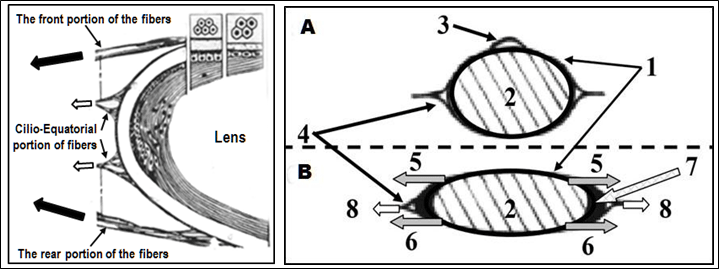
A. Accommodation closer: The lens capsule (1) squeezed the intracranial masses as much as possible (2), the pressure in the lens increased as much as possible and a mini-lens stand formed in the center (3); the tension of the cilio-equatorial (4) fibers of the zonula of Zinn is weakened.
B. There is no accommodation: The anterior (5) and posterior (6) portions of the fibers of the zonula of Zinn stretched the elastic capsule of the lens (1) and its internal volume increased by the amount of additional volume (7, painted black and shown with a gray long arrow); the volume of intracranial masses (2) is constant and incompressible; the tension (8) of the cilio-equatorial fibers ensured a “flattening” of an additional volume (7) along the equator of the lens bag (1).
Taking into account all the above, it is now clear how correct the widely used schemes for fixing the lens in the eye with the cross-location of FPF and PPF should be (Figure 17a) [76,77]. Therefore, many researchers today understand how important it is to observe adequate boundary conditions when performing mechanical calculations of the lens shape in various phases of accommodation [20,22,76,77].
It is worth noting that the correct scheme for fixing the lens in the eye presented in Figure 17b is completely adequate to the laws of mechanics.
It should be noted that a generalized model of the accommodation mechanism that is fully adequate to the laws of mechanics was created at the interdisciplinary level in Russia in 1999 [5]. Its main difference from the models of accommodation mechanisms considered here by other authors is that it does not contradict all known clinical facts, evolutionarily develops the classical theory of accommodation by H. Helmholtz and allows to clearly define the boundary conditions for carrying out specialized calculations of the interaction of intraocular structures in the process of accommodation. This latest knowledge made it possible to create a generalized classification of all accommodation mechanisms in the human eye, as well as to develop and test the metabolic theory of adaptive myopia with ten-year clinical observations [78-81].
In general, the refined morphological structure of the ciliary girdle and the physiological characteristics of its main elements, consistent with the laws of mechanics, made it possible to clearly understand not only the functional purpose of each portion of the fibers, but also to determine its key role in the executive mechanisms of accommodation and outflow. In addition, in our opinion, it has become practically possible to make a significant step forward in the refinement and development of H. Helmholtz's theory of accommodation, as well as to advance in the description of previously unknown mechanisms of regulation of the outflow of intraocular fluid.
Conclusion
An in-depth anatomical and physiological analysis of the structure and structure of the elements of the zonula of Zinn allows us to note the following:
The executive mechanisms of lens accommodation include: muscle ciliary, fibers of the zonula of Zinn, Lens. vitreous chamber, choroidea.
The role of the anterior and posterior portions of the fibers of the zonula of Zinn is functionally main and corresponds to the Helmholtz accommodation mechanism.
The role of the cilio-equatorial fibers of the zonula of Zinn is functionally auxiliary, and their morphological structure cannot serve as a sufficient basis for ideas about the dominant role of these fibers in the executive mechanism of accommodation.
- Salzman M (1913) Anatomy and histology of the human eye in its normal state, its development and withering M. Publishing House of Dankinа and Khomutovа 231 .
- Svetlova OV (1994) Laser treatment of subluxation of the lens. Abstract of the dissertation of the candidate of medical sciences, PhD. St. Petersburg, Medical Academy of Postgraduate Education 26.
- Volkov VV, Kotlyar KE, Koshits IN, Svetlova OV, Smolnikov BA (1997) Biomechanical features of interaction of accommodation and drainage regulatory systems of the eye in normal and in contusion subluxation of the lens. Vestn ophthalmol 113: 5-7.
- Svetlova OV, Kotliar KE, Smolnikov BA (1998) Contemporary biomechanical conceptions about zonula and choroidea functioning. Experimental eye research 67: 26.
- Kotliar KE, Svetlova OV, Skoblikov AS, Smolnikov BA (1999) Biomechanical modeling of the accommodative system based on some contemporary conceptions of lens supporting apparatus functioning. Vision Science and Its Applications 156-164. Link: https://bit.ly/39jPdKm
- Svetlova OV, Makarov FN, Kotliar KE, Zaseeva MV, Koshits IN (2003) Morpho-functional characteristics of lens ciliary body as a key mechanism of accommodation in human eye. Morfologiia 123: 7-16. Link: https://bit.ly/2BpXoZa
- Koshits IN, Svetlova OV (2003) Development of the Helmholtz theory of accommodation based on the results of studies of Executive mechanisms of accommodation. Bulletin of the Russian Academy of Medical Sciences 2: 3-12.
- Koshits IN, Makarov FN, Svetlova OV, Kotlyar KE (2005) Biomechanical and morphological features of fastening and functioning of the ciliary lens fibers – a key link in the Executive mechanism of accommodation systems and outflow of watery moisture. In sat Biomechanics of the eye 2005, Helmholtz Moscow research Institute of eye diseases 3-20.
- Tscherning M (1898) Optique physiologique, dioptriiqe oculaire, functions de la retine, les moivements oculaires et la sivion binoculaire, lecons professes a la Sorbonne par le Dr Tscherning. Paris J Sarre et C Naud 335.
- Pflugk A (1935) Neue Wege zur Erforschung der Lehre von der Akkomodation. Arch f Ophth 339-351.
- Pau H (1951) Akkomodative Verschiedung von Cholesterinkrisstallen im Auge. Klin Monatsbl f Augenh 119: 424-427.
- Coleman DJ (1970) Unified model for accommodative mechanism. Am J Ophthalmol 69: 1063-1079. Link: https://bit.ly/3eRuy1t
- Lütjen-Drecoll E, Kaufman PL, Wasielewski R, Ting-Li L, Croft MA (2010) Morphology and accommodative function of the vitreous zonule in human and monkey eyes. Invest Ophthalmol Vis Sci 51: 1554-1564. Link: https://bit.ly/32FFRHy
- Coleman DJ (1986) On the hydraulic suspension theory of accommodation. Tr Am Ophthalmol Soc 84: 846-868. Link: https://bit.ly/2ZMIbdZ
- Wilson RS (1993) A new theory of human accommodation: cilio-zonular compression of the lens equator. Trans Am Ophthalmol Soc 91: 401-416. Link: https://bit.ly/2E8gqnP
- Ziebarth NM, Borja D, Arrieta E, Aly M, Manns F, et al. (2008) Role of the Lens Capsule on the Mechanical Accommodative Response in a Lens Stretcher. Investigative Ophthalmology & Visual Science October 49: 4490-4496. Link: https://bit.ly/2CTOmDZ
- Croft MA, Mc Donald JP, Nork MT, Kaufman PT (2013) Extralenticular and Lenticular Aspects of Accommodation and Presbyopia in Human Versus Monkey Eyes. Investigative Ophthalmology Visual Science 54: 5035-5048. Link: https://bit.ly/2E8fzU9
- Pour HM, Kanapathipillai S, Zarrabi K, Manns F, Ho A (2015) Stretch-dependent changes in surface profiles of the human crystalline lens during accommodation: A finite element study. Clin Exp Optom 98: 126-137. Link: https://bit.ly/2BjnyMY
- Schachar RA, Huang T, Huang X (1993) Mathematical proof of Schachar's hypothesis of accommodation. Ann Ophthalmol 25: 5-9. Link: https://bit.ly/2ZNECEb
- Goldberg DB (2015) Computer-animated model of accommodation and presbyopia. Cataract Refract Surg 41: 437-445. Link: https://bit.ly/3eWAPJm
- Wang K, Venetsanos DT, Wang J, Augousti AT, Pierscionek BK (2017) The importance of parameter choice in modelling dynamics of the eye lens. Sci Rep 7: 16688. Link: https://go.nature.com/2ZODG2r
- David G, Pedrigi RM, Humphrey JD (2017) Accommodation of the human lens capsule using a finite element model based on nonlinear regionally anisotropic biomembranes. Comput Methods Biomech Biomed Engin 20: 302-307. Link: https://bit.ly/3hs5znc
- Helmholtz H (1855) Űber die Akkomodation des Auges. Albrecht von Graefe`s Arch Ophthalmol 1: 1-74. Link: https://bit.ly/32HLULJ
- Svetlova OV, Koshits IN (2001) Modern biomechanical concepts of the theory of accommodation Helmholtz. In the book: “Biomechanics of the Eye 2001”. Moscow Research Institute of Eye Diseases named after Helmholtz, 142-160.
- Gullstrand A (1912) Wieich den intrakapsularen Akkommodations-mechaismus fand. Arch f Augenh 72: 169-190.
- Tscherning M (1898) Optique physiologique, dioptriiqe oculaire, functions de la retine, les moivements oculaires et la sivion binoculaire, lecons professes a la Sorbonne par le Dr Tscherning, Paris J. Sarre et C Naud 335.
- Pflugk A (1935) Neue Wege zur Erforschung der Lehre von der Akkomodation. Arch. Ophthalmol 133: 339-351.
- Rohen JW (1979) Scanning electron microscopic studies of the zonular apparatus in human and monkey eye. Invest Ophthalmol Vis Sci 18: 133-141. Link: https://bit.ly/3jshinp
- Rohen JW, Rentch FJ (1969) Architecture and function of the zonula apparatus in the human eye. Graefes Arch Clin and Exp Ophthalmol 176: 1-19. Link: https://bit.ly/2WJl6qt
- Schachar RA (1992) Cause and treatment of presbyopia with a method for increasing the amplitude of accommodation. Ann Ophthalmol 24: 445-452. Link: https://bit.ly/39fKRUD
- Schachar RA (1994) Pathophysiology of Accommodation and Presbyopia, Understanding the Clinical Implications. J J Fla Med Assoc 81: 268-271. Link: https://bit.ly/3fTlPgC
- Schachar RA (1999) Original article "Is Helmholtz's Theory of accommodation Correct?". Annals of Ophthalmology 31: 10-17.
- Schachar RA, Siganos DS, Knorz M (2000) The Mechanism of accommodation. Manual of Refractive Surgery. San-Diego, ECRS 39-41.
- Bauer SM (2001) On the need to build an adequate mathematical model of the theory of accommodation. In: Biomechanics of the Eye – 2001. M., Moscow Research Institute of Eye Diseases named after Helmholtz 137-139.
- Svetlova OV, Koshits IN (2002) Modern ideas about the theory of accommodation Helmholtz. Tutorial. Saint-Petersburg, Medical Academy of Postgraduate Education 46.
- Glasser A, Croft MA, Kaufman PL (1999) A morphometric ultrasound biomicroscopic study of the aging rhesus monkey ciliary region. Vision Science and Its Applications. Santa-Fe, Optical Society of America 126-129. Link: https://bit.ly/3fPtRH3
- Krstic RV (1991) Human Microscopic Anatomy. Berlin, Springer-Verlag. 197.
- Koshits IN, Svetlova OV, Gorban AI (2016) The functioning of the executive mechanisms of accommodation and the development of the theory of accommodation of Helmholtz. Normal physiology of the eye: a monograph. - 2nd ed., St. Petersburg 264.
- Koshits NI (2018) The Physiological Causes for the Eye’s Optical Axis Growth and Executive Mechanisms in the Metabolic Theory of Adaptive Myopia and the Theory of Retinal Defocus. Ophthalmology and Vision Science 2: 251-268.
- Streeten BW (2000) Anatomy of the Zonular Apparatus. Link: https://bit.ly/3eMiIWp
- Bornfeld N, Spitznas M, Breipohl W, Bijvank GJ (1974) Scanning electron microscopy of the zonule of Zinn. I. Human eyes. Albrecht Von Graefes Arch Klin Exp Ophthalmol 192: 117-129. Link: https://bit.ly/39gD5JW
- McCulloch C (1954) The zonule of Zinn: its origin, course, and insertion, and its relation to neighboring structures. Trans Am Ophthalmol Soc 52: 525-585. Link: https://bit.ly/3hiJx62
- Svetlova OV, Ignatiev SN, Smolnikov BA (1997) Assessment of traumatic displacements of the lens with partial breakage of the elements of its attachment in the human eyeball. In the book: "Mechanics and control processes." Saint-Petersburg, Ed. State Polytechnic University 172-177.
- Svetlova OV, Ignatiev SN, Smolnikov BA, Kugleev AA (1996) The displacement of the lens during contusion breakage of the fibers of the zonule of Zinn. In the book: Actual problems of ophthalmology. Kirov, Russia, 19-20.
- Gorban AI, Dzhaliashvili OA (1993) Eye Microsurgery. Errors and complications. Petersburg, Hippocrates. 256 .
- Bastrikov NI (1974) Clinical and morphological characteristics of the supporting apparatus of the lens. Abstract of the dissertation of the candidate of medical sciences 32 .
- Leonov AA (1986) Clinical significance of fakovitreal connections in cataract microsurgery. Abstract of the dissertation of the candidate of medical sciences PhD, M 29 .
- Glasser A, Kaufman PL (1999) The mechanism of accommodation in primates. Ophthalmology 106: 863-872. Link: https://bit.ly/3hnXL5F
- Koshits IN, Svetlova OV, Makarov FN, Guseva MG (2013) On the physiological and biomechanical postulates of new theories of accommodation. Eye M 3: 15-21.
- Svetlova O, Koshits I (2018) Theory- Actuating mechanisms of accommodation and development of the theory of accommodation by Helmholtz. Proceedings of 3rd Global Pediatric Ophthalmology Congress, London, UK. J Clin Exp Ophthalmol 9: 66-70. Link: https://bit.ly/3hq3ZSB
- Buzykin MA (2004) Ultrasonic anatomical and physiological picture of the accommodation apparatus of the eye in young people in vivo. Abstract of the dissertation of the candidate of medical sciences PhD, St. Petersburg, Military Academy, 28 .
- Dobrovolsky VI (1868) On various changes in astigmatism under the influence of accommodation. Military Medical Journal 103: 34-54.
- Serdiuchenko VI, Viasovsky IA (1987) The new device for determination of absolute accommodation volume of the eye. Ophthalmol Zhurnal, Ukraine 1: 12-15. Link: https://bit.ly/2WIMODX
- Serdiuchenko VI, Viasovsky IA (2004) The study of accommodation in various meridians of the eye and a modified method of treatment of its disorders in hypermetropic amblyopia. Conference proceedings: "Biomechanics of the eye Moscow, Russia 33-37.
- Svetlova O, Koshits I, Guseva M, Makarov F (2017) Two key managers in the pathogenesis of OAG and innovative methods of early diagnosis. 7-th World Glaucoma Congress: Abstract book, 532.
- Zolotaryov AV (2000) Non-penetrating surgery of primary open-angle glaucoma: a histotopographic approach: Full medical dissertation Sciences 192.
- Gabelt BT, Gottanka J, Lutjen-Drecoll E, Kaufman PL (2003) Aqueous humor dynamics and trabecular meshwork and anterior ciliary muscle morphologic changes with age in rhesus monkeys. Invest Ophthalmol Vis Sci 44: 2118-2125. Link: https://bit.ly/2CE1HR9
- Lutjen-Drecoll E, Tamm E, Kaufman PL (1988) Age-related loss of morphologic responses to pilocarpine in rhesus monkey ciliary muscle. Arch Ophthalmol . 106: 1591-1598. Link: https://bit.ly/32GGIYp
- Tamm ER, Lutjen-Drecoll E (1996) Ciliary body. Microsc Res Tech 33: 390-439. Link: https://bit.ly/30GBFVw
- Textbook and atlas (1934) "Eye diseases". Krukmann: “Vascular tract disease” Axenfeld 935. Ed. 8th; / Cit. by: E. Van Boyningham. Atlas of goniobiomicroscopy M. Medicine 29.
- Martin H, Stachs O, Guthoff R, Garbow N, Juneman A (2018) Biomechanische Untersuchungen zur Akkommodation des Auges. Der Ophthalmologe 115: 649-654. Link: https://bit.ly/3juUuUb
- Farnsworth PN, Burke P (1977) Three-dimensional Architecture of the Suspensory Apparatus of the Lens of the Rhesus Monkey. Exp Eye Res 25: 563-576. Link: https://bit.ly/3hiIoeK
- Fisher RF (1971) The Elastic Constants of The Human Lens. J Physiol 212: 147-180. Link: https://bit.ly/2E2uHSN
- Krag S, Andreassen TT (2003) Mechanical Properties of the Human Posterior Lens Capsule. Investigative Ophthalmology & Visual Science 44: 691-696. Link: https://bit.ly/3fT1FTN
- Koshits IN, Svetlova OV (2019) The essence of the physiological intraocular pressure. Part 1. Criteria: IOP, rigidity and fluctuation. National Journal Glaucoma 18: 59-72. Link: https://bit.ly/32OPbsu
- Okihiro N, Naoki Y, Kayo N, Yutaro N (2007) Contact inhibition of migrating lens epithelial cells at the capsular bend created by a sharp-edged intraocular lens after cataract surgery. J Cataract Refract Surg 33: 1065-1070. Link: https://bit.ly/3fOuNvm
- Scammon R, Hesdorffer M (1937) Growth in mass and volume of the human lens in postnatal life. Arch Ophthalmol 17: 104-112. Link: https://bit.ly/3fQYCLY
- Stepanova LV, Sychev GM, Kratasyuk VA, Svetlova OV (2017) Physiological features of water exchange processes in animal lenses taking into account the phase of accommodation. Ophthalmol Zhurnal (Ukraine) 6: 36-49.
- Koshits IN, Svetlova OV, Guseva MG, Pevko DV, Egemberdiev MB (2017) Optical features of passing light through the refractive structures of the eye. Ophthalmol Zhurnal (Ukraine) 4: 60-74. Link: https://bit.ly/3fWjzVW
- Maltsev EV (1988) The lens. M., Medicine 194.
- Chernyshev GN (1963) On the action of concentrated forces and moments on an elastic shell of arbitrary shape. Applied mathematics and mechanics27: 126-134. Link: https://bit.ly/2WEaOrR
- Bauer SM, Tovstik PE (1998) Buckling of Spherical Shells under Concentrated Load and Internal Pressure. Technische mechanik 18: 135-139.
- Bushnell D (1967) Bifurcation phenomena in spherical shells under concentrated and ring loads. American Institut of Aerocraft and Aerospace Journal 5: 2034-2040. Link: https://bit.ly/3hpHwF8
- Weber HA (1999) A finite elements model of accommodation; first results. Vision Science and Its Applications 113-116. Link: https://bit.ly/2ONAPke
- Weber HA, Martin H (2001) Finite Elements Simulation of Accommodation. In.: Guthoff R., Ludwig K. Current Aspects of Human Accommodation / Kaden Verlag 135.
- Wang K, Venetsanos DT, Wang J, Augousti AT, Pierscionek BK (2017) The importance of parameter choice in modelling dynamics of the eye lens. Sci Rep 7: 16688. Link: https://go.nature.com/2ZODG2r
- Martin H, Guthoff R, Stachs O, Grabow N (2016) Finite-Elemente-Modellierung des Auges zur Untersuchung der Akkommodation. Klin Monbl Augenheilkd 233: 1357-1361. Link: https://bit.ly/3jrqZT2
- Koshits IN, Svetlova OV, Egemberdiev MB, Guseva MG (2018) Traditional and new mechanisms of accommodation and their classification. Russian children's ophthalmology 3: 20-36.
- Koshits IN, Svetlova OV (2011) A mechanism of formation of the eye adequate length in the norm and metabolic theory of pathogenesis of acquired myopia. Ophthalmol Zhurnal (Ukraine) 5: 4-23. Link: https://bit.ly/32D9tW2
- Kaczurowsky MI (1964) Zonular fibers of the human eye. Am J Ophthalmol 58: 1030-1047. Link: https://bit.ly/3htmQwd
- OkihiroN, KayoN (1998) Accommodation Amplitude After Lens Refilling With Injectable Silicone by Sealing the Capsule With a Plug in Primates. Arch Ophthalmol116: 1358-1361. Link: https://bit.ly/3hsBJ1R

Article Alerts
Subscribe to our articles alerts and stay tuned.
 This work is licensed under a Creative Commons Attribution 4.0 International License.
 Save to Mendeley
