Journal of Clinical Research and Ophthalmology
Prevalence of age Related Macular Degeneration in A Tertiary Care centre
Jayashree MP1, Harika JVL2*, Arathi C3, Brijesh A Patil4 and Niveditha RK5
2Junior Resident, Department of Ophthalmology, S.Nijalingappa Medical College, Bagalkot, RGUHS, Banglore, Karnataka, India
3Assistant Professor, Department of Ophthalmology, S.Nijalingappa Medical College, Bagalkot, RGUHS, Banglore, Karnataka, India
4Proffesor and HOD, Department of Ophthalmology, S.Nijalingappa Medical College, Bagalkot, RGUHS, Banglore, Karnataka, India
5Junior Resident, Department of Ophthalmology, S.Nijalingappa Medical College, Bagalkot, RGUHS, Banglore, Karnataka, India
Cite this as
Jayashree MP, Harika JVL, Arathi C, Patil BA, Niveditha RK (2019) Prevalence of age Related Macular Degeneration in A Tertiary Care centre. J Clin Res Ophthalmol 6(1): 007-010. DOI: 10.17352/2455-1414.000056Background: Age-related macular degeneration (AMD) is one of the leading cause of irreversible blindness in elderly population affecting the quality of life and there by general health. AIM: The purpose of the present study was to estimate the prevalence, risk factors and subtypes of AMD in a hospital population attending to ophthalmology outpatient department.
Materials and Methods: This observational study was carried out at tertiary eye institute between November 2017 and April 2018. Newly registered patients of both the sex attending the hospital with complaints of diminution of vision for various reasons were selected for detailed ophthalmic examination and evaluation.
Results: Among the patients attending to our outpatient department, 120 patients were diagnosed with Age related macular degeneration. As the age of subjects increased prevalence also increased and most of them were in the age group of 61-70. Male to female ratio was 1:2.33. Early AMD was seen in 71.7% (n = 86) and Late AMD (n= 34) was seen in 28.3%. Dry AMD was seen in 75.84% (n=91), Wet AMD was seen in 24.16% (n=29).
Conclusion: Study reports prevalence of AMD increases with age. Higher prevalence was noted in females. Early AMD was seen in more cases than Late AMD and Dry AMD was noted more than Wet AMD. So complete ophthalmic evaluation is necessary in order to diagnose and stabilize the progression of AMD especially in the elderly age group of patients who complains of recent onset central visual loss.
Introduction
Age-related macular degeneration (AMD) is the leading cause of irreversible blindness apart from cataract and glaucoma especially in elderly population. The disease adversely affects quality of life and activities of daily living, causing many affected individuals to lose their independence in their retirement years. It accounts for 8.7% of all blindness worldwide and is the most common cause of blindness in developed countries [1-5], particularly in people older than 60 years.
Age- related macular disorder is a degenerative disorder affecting macula, characterized by presence of drusens and RPE changes. Conventionally it is classified in to two types mainly Dry (non-exudative, non-neovascular) form which is more common and presents as Geographical atrophy (GA) in its advanced stage. Wet (exudative, neovascular) form which is relatively less common but is associated with rapid progression to vision loss. Choroidal neovascualarisation (CNV) and Pigment epithelial detachment (PED) are its main manifestations. According to a Recent clinical classification it is divided in to Early AMD (drusen size more than 63μm less than 125μm), Intermediate AMD ( drusen size more than 125μm), Late AMD (Neovascualr AMD and geographical atrophy) [6].
Although, there is considerable information on visual impairment in the Western world, to date there are only few studies on the prevalence and risk factors of AMD in Indian subcontinent [7]. It was also included in the action plan of the World Health Organization, to address avoidable blindness in VISION 2020 program [8]. Differences in the prevalence of AMD based on gender, race, or geographic area have been observed. With increasing longevity & shift in demographic profile, prevalence of the disease is expected to rise dramatically in India also.
Recent studies from India account the prevalence of AMD among 70 years and above as 2% and 3.7% which was comparable to Western countries [7,9]. Therefore, the investigation of risk factors of AMD is important to suggest preventive measures that can retard or control AMD progression. Risk factors such as age, hypertension, smoking, diabetes mellitus, cardiovascular disease, obesity, female sex, sunlight exposure, dietary factors and positive family history [9-14] and their contributions to AMD had already been documented in literature. The purpose of the present study was to estimate the prevalence and identify risk factors and subtypes of AMD in a hospital population. The Early AMD included the presence of drusen and/or retinal pigment epithelium (RPE) abnormalities; and Late AMD, included geographic atrophy of RPE or choroidal neovascular complex.
Materials and Methods
This current observational study was carried out at tertiary care centre, SNMC & HSK hospital, Bagalkot, Karnataka. Study was conducted between November 2017 and April 2018. The institutional review board approved this study and an informed written consent was obtained from the subjects as per the Declaration of Helsinki. Before examination, the study was explained in detail to all the participants. The study population included the patients attending the outpatient department. Data regarding personal, medical history and lifestyle factors was collected and documented. Self-reported hypertension or diabetes and its duration from diagnosis were recorded. Lifestyle factors like smoking and alcohol consumption were explored by means of interview and were categorized as never smokers and smokers. Cases were considered as non-smokers if duration of smoking was less than one year.
All the ophthalmic examination was performed by investigating ophthalmologists. The examination was conducted according to a standardized protocol that included visual acuity for distance by snellens chart, jaeger chart for near vision, autorefractometer (POTEC PRK - 7000), computerized noncontact tonometer (NIDEK NT-510) for intraocular pressure measurement and anterior segment evaluation using slit lamp biomicroscopy, followed by dilation of pupil with tropicamide (0.8%) and phenylephrine (5%). For grading lens opacity, each eye was compared with the Lens Opacities Classification System (LOCS III) photographs [15]. Patients with small pupils, dense corneal and lens opacities precluding the view of retina are excluded from the study. Fundus findings were noted by using Direct, Indirect ophthalmoscopy using 20 D lens and in slitlamp biomicroscopy using 90 D lens. Findings were classified according to an International classification and grading system of Age related macular degeneration [16]. The features were examined for hard and soft drusens, changes in the retinal pigment epithelium (RPE), geographic atrophy, choroidal neovascular membrane, and disciform scar. AMD was classified as late (neovascular or geographic atrophy) or early (soft drusen or retinal RPE abnormalities). The cases of AMD, thus, detected were also confirmed by the principal ophthalmologist. When the both eyes of participants had lesions of different severity, the grade assigned was that of the more severely involved eye. Retinal lesions considered due to high myopia and inflammation were excluded.
Statistical analysis
The data were analysed using the Microsoft excel version MS 2007 software packages. Descriptive statistics was used to determine mean and percentages. Significance was derived by using pearson chi square test. P value <0.05 was considered as significant.
Results
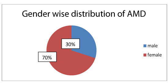
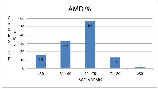
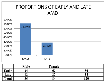
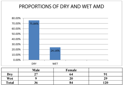
Among the patients attending the out patient department in 6 months duration, 120 subjects were diagnosed with Age related macular degeneration. Prevalence was 0.46%. 36 were males and 84 were females. Male: female ratio was 1: 2.33 (Figure 1). As the age of subjects increased prevalence also increased and most of them were in the age group of 61-70 (47.5%) (Figure 2). Prevalence rate was 0.46 %. Early AMD was diagnosed in 86 cases (71.7%) and Late AMD was seen in 34 cases (28.3%) (Figure 3). Dry AMD was seen in 91cases (75.84%), Wet AMD was seen in 29 cases (24.16%) (Figure4). Geographical atrophy was seen in 5 cases.
Out of 120 patients Early AMD was seen in 86 out of which 24 were males and 62 were females. Late AMD was seen in 34 cases, out of which 12 were males and 22 were females. 91 cases has shown Dry AMD changes out of which 27 were males and 64 were females. 29 cases shown Wet AMD out of which 9 were males and 20 were females (Table 1).
Out of 120 subjects 62 were hypertensive (p = 0.560), 33 were diabetics (p = 0.418), 23 were smokers (p=0.493) 11 were alcoholics (p = 0.23). P values for all were not significant.
Discussion
Age related macular degeneration is the leading cause of visual disturbance and blindness especially among the elderly population affecting their quality of life. It accounts for 8.7 % of the total blindness globally. As the aging population is rapidly increasing, age related blinding causes will be more important in coming years.
Various previous studies reported the prevalence and riskfactors of AMD. Overall prevalence of AMD was 1.82 % in Andhra Pradesh Eye Disease Study [9], 1.51 % in Beaver Dam Eye Study [7], 1.81 % in Blue Mountain Eye Study [2], 1.38 % in a hospital based study [17] and 2.31% in a study done in Central India [18]. In our study prevalence rate was 0.46%. Differences in this prevalence rates may be due to the differences in the type of study undertaken like community based or hospital based, genetic factors and environment.
As the age of subjects increased prevalence also increased and most of them were in the age group of 61-70. This result is consistent with other studies [2,4,7,9,19]. Increasing age is strongly associated with AMD.
Pooled data from the Beaver Dam Eye Study, Blue Mountains Eye Study, and the Rotterdam Study revealed no sex differences in AMD risk. However, recent analyses from the Blue Mountains Eye Study suggest that the 5-year incidence of Neovascular AMD among women is double that of men (1.2% vs. 0.6%). Females were having higher risk of AMD than males in our study. However, the difference is not statistically significant (p= 0.486). This was also reported by several other studies. However, there were studies showing prevalence of AMD more in males that is related to smoking habits, sunlight exposure and better longevity among females.
The Aravind Comprehensive Eye Study reported the prevalence of Early AMD and Late AMD were 2.7% and 0.6% respectively [7]. It was 2 % and 1.6% in the INDEYE population [20]. Kulkarni et al had reported the proportion of early and late AMD as 1.34% and 0.37 % respectively [21]. According to Gupta et al it was 47% and 1.4% respectively [22]. The proportion of early and late AMD in south Korean population was 5.1% and 0.34% respectively [23]. In the current study early AMD % was more than late AMD (71.7% and 28.3%) which is similar to forementioned studies.
Hypertension and cigarette smoking were identified as risk factors for AMD constantly apart from age, prior cataract surgery, CVD, obesity and dietary factors. Smoking is an important, independent, modifiable risk factor for AMD. Mechanisms by which smoking may increase the risk of developing AMD include its adverse effect on blood lipids by decreasing levels of high-density lipoprotein (HDL) and increasing platelet aggregability and fibrinogen, increasing oxidative stress and lipid peroxidation, and reducing plasma levels of antioxidants. In the present study 62 cases were noted as hypertensives (p= 0.560), 23 were smokers (p=0.493) but the p value was not statistically significant. And this may be due to the large number of female cases in the study. 33 were diabetics (p= 0.418), 11 were alcoholics. To date the evidence suggests that alcohol doesnot affect the AMD. Many studies have investigated the relationship between diabetes and/or hyperglycemia and AMD, and most have found no significant relationships and this may be due to the coexisting diabetic retinopathy. Limitations of the study includes small sample size and we could not associate the risk factors like smoking, alcohol, diabetes and hypertension as the study was observational and controls were not included in the study and odds ratio was not calculated.
Conclusion
Age related macular degeneration is a common and potential cause of blindness in elderly population that often goes unnoticed without a clear ophthalmological examination. So early detection of cases is important to reduce the progression. Study reports prevalence of AMD increases with age. Higher prevalence was noted in females. Early AMD was common than Late AMD.
- Klein R, Klein BE, Cruickshanks KJ (1999) The prevalence of age-related maculopathy by geographic region and ethnicity. Prog Retin Eye Res 18: 371-389. Link: https://tinyurl.com/yybzqr9d
- Mitchell P, Smith W, Attebo K, Wang JJ (1995) Prevalence of age-related maculopathy in Australia. The Blue Mountains Eye Study. Ophthalmology 102: 1450-1460. Link: https://tinyurl.com/yyuxq9mh
- Klaver CC, Assink JJ, van Leeuwen R (2001) Incidence and progression rates of age-related maculopathy: the Rotterdam Study. Invest Ophthalmol Vis Sci 42: 2237-2241. Link: https://tinyurl.com/y6zp8led
- Kawasaki R, Yasuda M, Song SJ (2010) The prevalence of age-related macular degeneration in Asians: a systematic review and meta-analysis. Ophthalmology 117: 921-927. Link: https://tinyurl.com/yxnbadrl
- Wong TY, Chakravarthy U, Klein R, Mitchell P, Zlateva G, et al. (2008) The natural history and prognosis of neovascular age-related macular degeneration: a systematic review of the literature and meta-analysis. Ophthalmology 115: 116-126. Link: https://tinyurl.com/y6r4zrbc
- Kanski J, Bowling B (2016) Kanski clinical ophthalmology. 8th edition, China, Elsevier 928. Link: https://tinyurl.com/y5m544xy
- Nirmalan PK, Katz J, Robin AL, Tielsch JM, Namperumalsamy P, et al. (2004) Prevalence of vitreoretinal disorders in a rural population of Southern India: The Aravind Comprehensive Eye Study. Arch Ophthalmol 122: 581-586. Link: https://tinyurl.com/y2472wh9
- World Health Organization (2020) Age Related Macular Degeneration: Disease control and prevention of visual impairment in global initiative for the elimination of Avoidable Blindness Action Plan 2006–2011. Printed in France. 32–33. Link: https://tinyurl.com/y488bo2g
- Krishnaiah S, Das T, Nirmalan PK, Nutheti R, Shamanna BR, et al. (2005) Prevalence of vitreoretinal disorders in a rural population of southern India: The Aravind Comprehensive Eye Study. Invest Ophthalmol Vis Sci 46: 4442-4449.
- Age-Related Eye Disease Study Research Group (2000) Risk factors associated with age-related macular degeneration-A case-control study in the age-related eye disease study: Age-Related Eye Disease Study report number 3. Ophthalmology 107: 2224-2232. Link: https://tinyurl.com/yybtrl6a
- Cackett P, Wong TY, Aung T, Saw SM, Tay WT, et al. (2008) Smoking, cardiovascular risk factors and age-related macular degeneration in Asians: The Singapore Malay Eye Study. Am J Ophthalmol 146: 960-967. Link: https://tinyurl.com/y3rw3co4
- Klein R, Klein BE, Wong TY, Tomany SC, Cruickshanks KJ (2002) Association of cataract and cataract surgery with the long-term incidence of age-related maculopathy: The Beaver Dam Eye Study. Arch Ophthalmol 120: 1551-1558. Link: https://tinyurl.com/yy6vn8t7
- Wang JJ, Klein R, Smith W, Klein BE, Tomany S, et al. (2003) Cataract surgery and the 5-year incidence of late stage age-related maculopathy: Pooled finding from the Beaver Dam and Blue Mountains eye studies. Ophthalmology 110: 1960-1967. Link: https://tinyurl.com/y4lcmpd8
- Klein R, Peto T, Bird A, Vannewkirk MR (2004) The epidemiology of age-related macular degeneration. Am J Ophthalmol 137: 486-495. Link: https://tinyurl.com/yxu5abkt
- Chylack LT, Wolfe JK, Singer DM, Leske MC, Bullimore MA, et al. (1993) The Lens Opacities Classification System III. The longitudinal study of cataract study group. Arch Ophthalmol 111: 831-836. Link: https://tinyurl.com/y2t2jdqe
- Bird AC, Bressler NM, Bressler SB, Chisholm IH, Coscas G, et al. (1995) An international classification and grading system for age-related maculopathy and age-related macular degeneration. The International ARM Epidemiological Study Group. Surv Ophthalmol 39: 367-374. Link: https://tinyurl.com/y2kvme88
- Klein R, Klein BE, Linton KL (1992) Prevalence of age-related maculopathy. The Beaver Dam Eye Study. Ophthalmology 99: 933-943. Link: https://tinyurl.com/y37x3rxy
- Kulkarni SR, Aghashe SR, Khandekar RB, Deshpande MD (2013) Prevalence and determinants of age-related macular degeneration in the 50 years and older population: A hospital based study in Maharashtra, India. Indian J Ophthalmol 61: 196-201. Link: https://tinyurl.com/yxk6va6x
- Singare RP, Deshmukh S, Ughade SN (2015) Age-related macular degeneration: Prevalence and Risk factors in elderly population (aged >60 years) in Central India. International Journal of Scientific and Research Publications 5. Link: https://tinyurl.com/y5mb25bh
- Krishnan T, Ravindran RD, Murthy GV, Vashist P, Fitzpatrick KE, et al. (2010) Prevalence of early and late age-related macular degeneration in India: the INDEYE study. Invest Ophthalmol Vis Sci 51: 701-707. Link: https://tinyurl.com/y3svsqf6
- Pokharel S, Malla OK, Pradhananga CL, Joshi SN (2009) A pattern of age-related macular degeneration. JNMA J Nepal Med Assoc 48: 217-220. Link: https://tinyurl.com/yy9czdvl
- Gupta SK, Murthy GV, Morrison N, Price GM, Dherani M, et al. (2007) Prevalence of early and late age-related macular degeneration in a rural population in northern India: The INDEYE feasibility study. Invest Ophthalmol Vis Sci 48: 1007-1011. Link: https://tinyurl.com/y3y6vsem
- Song SJ, Youm DJ, Chang Y, Yu HG (2009) Age-Related Macular Degeneration in a Screened South Korean Population: Prevalence, Risk Factors, and Subtypes. Journal Ophthalmic Epidemiology 16: 304-310. Link: https://tinyurl.com/y33q28kb

Article Alerts
Subscribe to our articles alerts and stay tuned.
 This work is licensed under a Creative Commons Attribution 4.0 International License.
 Save to Mendeley
