Journal of Clinical Research and Ophthalmology
Ocular Fundus changes in pregnancy induced hypertension – A case series study
Jayashree MP1, Niveditha RK2*, NG Kuntoji3, Vishalakshi Bhat4, Shravan GM5, Brijesh A Patil6 and Harika JVL7
2Junior Resident, Department of Ophthalmology, S. Nijalingappa Medical College, Bagalkot, RGUHS, Banglore, India
3Professor, Department of Ophthalmology, S. Nijalingappa Medical College, Bagalkot, RGUHS, Banglore, India
4Assistant professor, Department of Ophthalmology, S. Nijalingappa Medical College, Bagalkot, RGUHS, Banglore, India
5Assistant Professor, Department of Ophthalmology, S. Nijalingappa Medical College, Bagalkot, RGUHS, Banglore, India
6Professor and HOD, Department of Ophthalmology, S. Nijalingappa Medical College, Bagalkot, RGUHS, Banglore, India
7Junior resident, Department of Ophthalmology, S. Nijalingappa Medical College, Bagalkot, RGUHS, Banglore, India
Cite this as
Jayashree MP, Niveditha RK, NG Kuntoji, Bhat V, Shravan GM, et al. (2018) Ocular Fundus changes in pregnancy induced hypertension – A case series study. J Clin Res Ophthalmol 5(2): 037-041. DOI: 10.17352/2455-1414.000054Back ground: Pregnancy Induced Hypertension is a challenging stigma in the field of obstetrics and one of major contributors to maternal and perinatal mortality. PIH is a hypertensive disorder in pregnancy that occurs after 20 weeks of pregnancy in the absence of other causes of elevated blood pressure.
Aims and objectives:
· To determine the proportion of different groups of PIH patients i.e., gestational hypertension, preeclampsia and eclampsia having retinal changes.
· To study the correlation of retinal changes with severity.
Methods: It is a is a Prospective (hospital based) study done on 150 patients of PIH Age, gravida, gestation period, B.P. and proteinuria were noted. Ocular examination done using direct and indirect ophthalmoscope. The findings were noted and analysed using chi square test.
Results: A total of 150 patients were examined. The mean age of patients was 24.34±4.01 years. The gestation period ranged from 28 to 40 weeks. 72(48%) were primigravida, 74(49.3%) were multigravida and 4 (2.7%) were grand multi gravida. Out of all patients, most of the patients had severe preeclampsia i.e., 71(47.3%), 34(22.6%) had mild preeclampsia, 32(21.3%) had eclampsia and 13 (8.7%) had gestational hypertension.
In our study, 32 patients (21.3%) had arteriolar attenuation which included generalised arteriolar attenuation in 12(37.5%) and focal arteriolar attenuation in 20 (62.5%) which is the most common retinal finding. Retinal detachment was seen in 1 patient. There was statistically significant positive association of retinal changes with blood pressure (p) and severity of PIH (p).
Conclusion: Fundus examination in PIH is important in monitoring and managing cases as it correlates with severity as it indirectly implies severity of changes in placental micro-circulation that can help to predict the foetal outcome and ocular morbidity.
Introduction
Pregnancy Induced Hypertension is a challenging stigma in the field of obstetrics and one of major contributors to maternal and perinatal mortality. Pregnancy is associated with a group of physiological and pathological changes. Pregnancy Induced Hypertension (PIH) is a hypertensive disorder in pregnancy that occurs after 20 weeks of pregnancy in the absence of other causes of elevated blood pressure (B.P.) i.e., >140/90 mm Hg measured 2 times with at least of 6-hour interval. PIH is the hypertension that develops as a direct result of gravid state. Hypertensive disorders complicate 5-10% of all pregnancies [1]. Most important pathologies accompanying pregnancy is pregnancy induced hypertension which includes gestational hypertension, preeclampsia and eclampsia. Gestational hypertension is diagnosed when blood pressure reach140/90mm Hg or greater for the first time after mid pregnancy [1]. Gestational hypertension associated with significant proteinuria is termed as preeclampsia (>300mg/ 24hr).
Preeclampsia complicated by generalised tonic clonic convulsion is termed as Eclampsia. Hypertension is the most significant primary sign. Oedema occurs initially in the lower legs but may progress too massive oedema or anasarca. PIH is a multisystem disorder which include cardiovascular, haematological, hepatic, renal, neurological abnormalities and ocular manifestations [2,3]. Severe toxaemia is the main cause for visual system involvement. The retinal vascular changes majority of times but not always correlate with systemic hypertension. The constriction of vessels may take days to develop and may last for weeks to months.
Retinal changes are liable to occur when the systolic pressure raises above 160 and the diastolic above 100mm hg and are marked when these limits reach 200/130 mm of Hg [4,5]. Choroid is also frequently affected in the disease; choroidal ischemia and infarction may occur. The ischemia of occipital lobe and optic nerve may occur and recovery usually occurs unless there is significant infarction. Sometimes visual disturbances may be the presenting symptom; less common symptoms include amaurosis, photopsia, scotomata, diplopia, achromatopsia and hemianopia. The abnormalities of the retina and the retinal vasculature are most frequent though the conjunctiva, choroid, optic nerve and the visual cortex may be affected. Visual loss as a result of vascular involvement is usual. Vision threatening conditions involve central retinal artery occlusion, secondary optic atrophy, macular tear, central serous retinopathy, retinal detachment, central retinal vein occlusion, choroidal ischemia and haemorrhage. Spontaneous vitreous haemorrhage may occur in cases of HELLP syndrome.
Progression of retinal changes correlates with progression of PIH, maternal outcome and alsowith the foetal mortality due to similar vascular ischemic changes in the placenta.
Methods and Materials
It is a hospital based Case Series study. All Pregnant women attending OPD and admitted in the obstetric ward, S. Nijalingappa Medical College and Sri Hanagal Kumareshwara Hospital, Bagalkot b/w the study period of November 2016 to June 2018 in the age group of 18 to 35 years with more than 20 weeks of gestation having systolic blood pressure of more than 140 mm of Hg and Diastolic Blood pressure of more than 90 mm of Hg were included in the study. The study was conducted on 150 diagnosed PIH ases. This study was approved by the institutional ethical committee.
Methodology
After taking informed consent, baseline data for all patients was recorded. All the patients were initially evaluated by an obstetrician. Detailed history, general physical examination and systemic examination were done and noted down in the case sheet. History of eye symptoms and visual acuity was evaluated. As most of the patients were admitted and examination was done on the bed side, counting finger at 3 metres is considered as normal. Bedside ocular examination was done using torch light to see ocular alignment and motility, pupillary examination and to exclude gross anterior segment pathology. Patients were dilated with Tropicacyl plus eye drops (Tropicamide 0.8%W/V Phenylephrine 5%W/V). Fundus examination was done using direct ophthalmoscope/indirect ophthalmoscope and retinopathy in both eyes was noted. Fundus photos were taken using Volk in view fundus camera. The age, B.P. values, gravida, para, severity and level of disease was noted down. The patients were divided into 2 groups based on their mean B.P. values at the time of presentation whether greater than or less than 160/110 mm Hg for analysis. The findings were noted down in the case sheet. The PIH was graded as gestational hypertension, mid preeclampsia, severe preeclampsia and eclampsia. Proteinuria was tested using dipstick method was graded as + = 30mg/dl, ++ = 100mg/dl, +++ = 300mg/dl, ++++ >= 2000mg//dl.
Inclusion Criteria:
▹ patients diagnosed with pregnancy induced hypertension in the age group of 18 – 35 years
▹ with > 20 weeks of gestation
Exclusion criteria:
▹ Patients with pre-existing hypertension
▹ Pre-existing Diabetes
▹ Renal pathology
▹ Ocular pathologies like glaucoma, cataract, corneal opacities and history of ocular trauma, surgery or previous laser treatment and having hazy media which hinders the fundus examination were excluded from the study.
▹ Patients with cardio-vascular diseases.
▹ Patients with anaemia, connective tissue disorders, vasculitis or any other systemic disease.
▹ Patients with malignancy, leukaemia.
The clinical course of the disease in the more acute cases may be divided into 3
stages*:
This classification is used in this study.
· Spastic stage arterial irritation during which the toxin excites angiospastic phenomena.
· Stage of sclerosis when organic changes appear in the vessels.
· Stage of retinopathy when oedema, haemorrhages and damage to tissue occur.
Statistical analysis:
Ocular fundus changes was evaluated using unpaired t-test.
Categorical data’s analysed using Chi-square test.
P value <0.05 is considered significant.
Data was collected and tabulated in excel sheet.
Results
A total of 150 patients were studied. The age ranged from 18 to 35 with a Mean ± SD: 24.34±4.01 years.72 (48%) were primigravida, 74(49.3%) were multigravida and 4 (2.7%) were grand multigravida. Headache was the most common complaint reported in 42(28%) cases and blurring of vision is seen in 38(25.3%) cases.
Most of the patients had severe preeclampsia 71(47.3%), 34(22.6%) had mild preeclampsia, 32(21.3%) had eclampsia and 13 (8.7) had gestational hypertension. Out of 150 patients 32(21.3%) patients had 1+ protein, 75(50%) had 2+ protein, 30(20%) had more than 3+ proteinuria. Patients with proteinuria more than 3+ had greater chances of developing retinopathy.
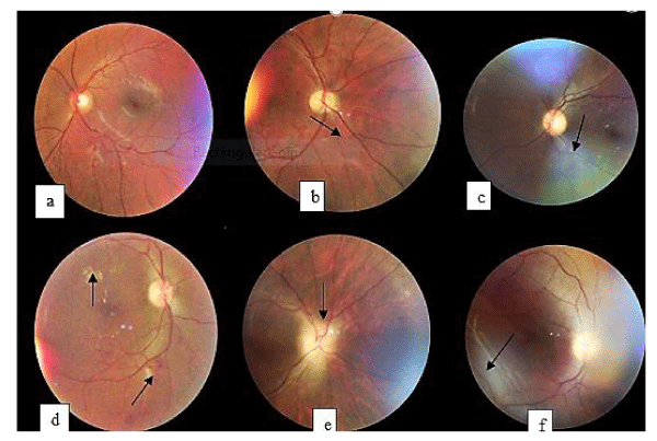
4 patients had blurred margins. Focal arteriolar attenuation was present in 20 patients, generalised arteriolar attenuation was present in 12 patients and silver wiring, AV crossing changes was present in 5 cases. Mean Pre SBPin patients with normal fundus is 159.77±14.13 and mean Pre DBP in patients with normal fundus is103.48±10.63.In patients with BP <160/110 mm Hg majority had normal fundus 74(85.1%) with stage I being an important change 10(11.5%). In patients with BP >160/110 mm Hg stage I change is seen in 20(31.7%), stage II in 1(1.6%) and stage III in 4(6.3%).Out of 150 patients, no changes were seen in 112 patients and 38 patients had retinopathy changes. primigravida showed the most fundus changes 18(47.36%). Multigravidas with changes were 16(42.1%) and grand multigravidas with changes were 4 (10.5).Stage 3 changes are seen in 2 eclampsia patients and 5 Severe preeclampsia patients. Stage 1 was the most common finding which was present in 23 patients (76.7%) in severe PE patients. Gestational hypertension and mild PE patients did not show any retinopathy changes (Figure 1 and Tables (1-7).
Disscussion
This study was undertaken to evaluate the fundus changes in patients of pre-eclampsia and eclampsia. PIH is one of the most common causes of morbidity and mortality in obstetrics.
In our study of 150 patients, hypertensive retinopathy changes were seen in 38 patients. Majority of them were in the age group of 20 – 30 years (72.7%). Age in our study was not associated with retinopathy changes which is similar to the results quoted by Reddy et al, [4].
Mean age of the cases in our study was 24.34±4.01 years. In study conducted by Shukla et al.[6], they examined 20 cases of preeclampsia and eclampsia and noted incidence of retinal changes in 70% of the cases in different age group. In their study, 60% cases aged less than 25 years. Mean age group of patients in the present study matches with the studies by Karki et al. [7] and Shukla et al, [6].
In our study 72 (48%) were primigravida, 74 (49.3%) were multigravida and 4(2.7%) were grand multigravida. In S.C. Reddy et al. [4], study, 34 (43.6%) were primigravidae, 27 (34.6%) were multigravida and 17 (21.8%) were grand multigravidas.
In our study 13(8.7%%) had gestational hypertension, 34 (22.6%) had mild preeclampsia, 71(47.3%) had severe preeclampsia and 32 (21.3%) had eclampsia. In S.C. Reddy et al study, 30(38.5%) had mild preeclampsia, 46 (59%) had severe preeclampsia and 2 (2.5%) had eclampsia [8]. In Tadin.I.et al study, all cases 40 (100%) had severe preeclampsia [9]. In Z. Kurdoglu et al study all cases 148 (100%) were preeclamptic [10]. In Jaffe and Schatz study 17 cases had mild preeclampsia and 14 cases had severe preeclampsia [11].
According to Duke Elder the most common retinal changes is attenuation of retinal arterioles, occurring in approximately 60% of patients with pre-eclampsia [12]. In our study, 32 patients (21.3%) showed arteriolar attenuation which included generalised arteriolar attenuation in 12(37.5%) and focal arteriolar attenuation in 20 (62.5%). From above, it is seen that arteriolar attenuation is the major retinal change seen in PIH. AV crossing changes and silver wiring was present in 5 cases; out of which 4 had associated hard exudates and therefore staged as stage 3.
Overall 7(4.6%) patients had stage 3 retinopathy; 5 with hard exudates; 1 case had superficial haemorrhage and 1 patient had serous retinal detachment. Fry W [13] have stated in their study that the serous retinal detachment occurs in approximately 1% of patients with pre-eclampsia and 10% of those with eclampsia. Reddy et al., found 6 cases (3%) with retinal haemorrhages and 6 cases (3%) with cotton wool spots belonging to severe pre-eclampsia [8].
Francis et al., found 5% of cases with cotton wool spots and retinal haemorrhage [14]. These studies showed observations close to what we found in our study.
Karki et al., mentioned optic nerve head changes in 8 cases [7]. Shukla et al., found 10% case of papilledema [6]. In our study 4 cases (2.6%) had papilledema. Decline in the percentage regarding papilledema in our study could be due to early and prompt obstetrical and medical management of PIH.
Headache was the most common symptom seen in our patients i.e., 42/ 150 (28%) whereas in Z. Kurdoglu et al. [10], study showed headache, floating specks or blurred vision was most common while blurring of vision was seen in 3 cases in S.C. Reddy et al study [4]. In our study blurred vision was seen in 38(25.3%) cases.
In our study fundus findings were seen in 38/150 (25.33%) cases with Pregnancy Induced Hypertension. Tadin I. et al., from Croatia had reported 45% of retinal changes in their study of 40 patients with PIH (9). Reddy et al., in a study of 275 cases of preeclampsia and 125 cases of eclampsia found an incidence of 53.4% in preeclampsia and 71.6% in eclampsia of hypertensive retinopathy [8]. S.C. Reddy et al., studied 78 cases with PIH and found a prevalence rate of 59% [4].
Zehra Kurdoglu et al., studied 148 cases of PIH retrospectively in Turkey [10]. The prevalence rate of hypertensive retinopathy in their study was 48%. Karki et al from Nepal have reported 13.7% of fundus changes in their study of 153 subjects with PIH [7].
In our study, 38/150 cases Stage I changes were seen in 30 cases (79%), Stage II changes were seen in 1 case (2.6%) and Stage III changes were seen in 7cases (18.4%). Papilledema was seen in 4 cases.
In our study there was a statistically significant correlation between the retinopathy changes and the degree of blood pressure and the severity of the disease. S.C. Reddy et al., found a statistical correlation between proteinuria, blood pressure and hypertensive retinopathy [8]. The degree of retinopathy was directly proportional to severity of preeclampsia with which our study was in accordance. Tadin I. et al found that the degree of retinopathy was directly related to the blood pressure and to the severity of disease [9]. They stated that hypertensive retinopathy is a valid and reliable prognostic factor in determining the severity of preeclampsia.
Z. Kurdoglu et al., found no significant correlation with severity of disease which was against our observation [10]. Karki et al., assessed the foetal outcome in these patients and concluded that retinal and optic nerve head changes were associated with low birth weight; choroidal and optic nerve head changes were associated with low Apgar score; and fundus evaluation in patients with PIH is an important procedure to predict adverse foetal outcomes [7].
Vision is important criteria to be seen in these patients including on follow-up after delivery. Most cases of late onset severe eclampsia present with exudative retinal detachment which usually resolves with termination of pregnancy.
There is a significant positive correlation with the levels of hypertension and with the severity of the disease. This observation was similar to that of Tadin I et al. [9], study and S.C. Reddy et al. [9], study.
Mean systolic BP in the group without changes, that is, with normal fundi, was 159.7±14.13 mm of Hg and mean diastolic BP was 103.48±10.63 mm of Hg. In the group with finding, the mean systolic B.P. was 170±13.39 mm of Hg and the mean diastolic B.P. was 110±8.28mm of Hg. The mean B.P. level in .Kurdoglu et al., study was 174/107 mm of Hg [10].
Vision and the retinal findings documented need to be followed up after the termination of pregnancy and any residual optic nerve affection should be correlated.
Conclusion
The retinal vascular changes have been said to correlate with the severity of hypertension. Many studies have considered the progression of retinal vascular changes as a sign of increasing severity of PIH and have correlated them with foetal mortality as well as maternal outcome. These changes help as a guideline for termination pregnancy as they may reflect similar ischemic vascular changes in the placenta.
Ophthalmoscopy is a simple tool that can help the obstetrician in assessing the severity of disease in cases of PIH. In general, it is believed that the presence of changes in the retinal arterioles and retinal haemorrhages may indicate similar changes in the placenta. Since the wellbeing of the foetus depends on the placental circulation, ophthalmoscopic examination of mother’s fundus may give a clue to similar microcirculation changes in the placenta and indirectly to the foetal wellbeing and maternal outcome. Fundus examination in patients with PIH is an important clinical evaluation to predict adverse foetal outcome (31, 32).
- Gary FC, Leveno KJ, Bloom SL (2014) Williams Obstetrics. 24th ed. Hypertensive disorders Vol.1. United States of America: MC Graw-Hill Education 728-730.
- ACOG Practice bulletin committee (2002) Diagnosis and management of preeclampsia and eclampsia. Obstetgynecol 99:159-167. Link: https://goo.gl/oPpH1p
- Drife JO, Magowan BA. Clinical obstetrics and gynaecology 367-370.
- Reddy SC (2012) Fundus changes in pregnancy induced hypertension. International journal of ophthalmology 5: 694-697. Link: https://goo.gl/HzLTQd
- Duke-elder S, Debree JM (1967) System of ophthalmology. Volume X. Disease of the retina. United States of America: CV Mosby Company 350.
- Kishore N, Tandon S (1965) Significance of biochemical and ophthalmoscopic changes in toxaemia of pregnancy. Journal of Obstetrics and Gynaecology 15: 551-559.
- Karki P, Malla KP, Das H, Uprety DK (2010) Association between pregnancies induced hypertensive fundus changes and foetal outcome. Nepal Journal of Ophthalmology 2: 26–30. Link: https://goo.gl/DUFrss
- Reddy SC (1989) Ocular fundus changes in toxaemia of pregnancy. The Antiseptic 86: 367-372.
- Tadin I, Bojić L, Mimica M, Karelović D, Dogas Z (2001) Hypertensive retinopathy and preeclampsia. Coll Antropol 25: 77–81. Link: https://goo.gl/wJ7f2e
- Kurdoglu Z, Kurdoglu M, Ay EG, Yasar T (2011) Retinal Findings in Preeclampsia. Perinatal Journal 19: 60-63.
- Jaffe G, Schatz H (1987) Ocular manifestations of preeclampsia. American Journal of Ophthalmology 103: 309–315. Link: https://goo.gl/gS8DdN
- Duke E (1971) System of Ophthalmology. In: Sir Stewart, editor. Diseases of Retina. 2nd ed. Vol. X. St Louis: CV Mosby 136.
- Fry W (1929) Extensive bilateral retinal detachment in eclampsia with complete reattachment: Report of two cases. Arch Ophthalmology 1: 609-614. Link: https://goo.gl/x64Nsy
- Francis O (1959) An analysis of 1150 cases of abortions from the Government R. S. R. M. lying in Hospital, Madras. J of Obstetrics and Gynaecology 10: 62-70. Link: https://goo.gl/NQ2Qiw

Article Alerts
Subscribe to our articles alerts and stay tuned.
 This work is licensed under a Creative Commons Attribution 4.0 International License.

 Save to Mendeley
