Journal of Clinical Research and Ophthalmology
Cutaneous Horn of the Eyelid: Anatomoclinical Implications
1Department of Ophthalmology, “Grigore T. Popa” University of Medicine and Pharmacy, Iaşi, Romania
2IInd Ophthalmology Clinic, “Prof. Dr. Nicolae Oblu” Emergency Hospital, Iaşi, Romania
3Pathology Laboratory, “Prof. Dr. Nicolae Oblu” Emergency Hospital, Iaşi, Romania
4Department of Anatomy, “Grigore T.Popa” University of Medicine and Pharmacy, Iaşi, Romania
5Neurosurgery Unit II, “Prof. Dr. Nicolae Oblu” Emergency Hospital of Iaşi, Romania
6IIIrd year student, “Grigore T. Popa” University of Medicine and Pharmacy of Iaşi, Romania
7Department of Neurosurgery, “Grigore T. Popa” University of Medicine and Pharmacy of Iaşi, Romania
Author and article information
Cite this as
Costea CF, Dimitriu G, Sava A, Chihaia M, Dancă C, et al. (2017) Cutaneous Horn of the Eyelid: Anatomoclinical Implications. J Clin Res Ophthalmol. 2017; 4(1): 1-5. Available from: 10.17352/2455-1414.000035
Copyright License
© 2017 Costea CF, et al. This is an open-access article distributed under the terms of the Creative Commons Attribution License, which permits unrestricted use, distribution, and reproduction in any medium, provided the original author and source are credited.Introduction
The cutaneous horn (cornu cutaneum) is a circumscribed,conical and keratotic lesion, which can hide benign or malignant lesions [1-3]. The clinical diagnosis is established based on its appearance, the lesion being classified as solitary or multiple, straight, curved or twisted, white or yellow [4,5], most often located at the level of the skin on the patient’s face [4,6,7]. While the cause leading to the formation of cutaneous horns [8] is unknown, UV radiations are believed to be the trigger of this condition [9]. Usually, the cutaneous horn occurs in people over the age of 50, in both genders [10-14].
The cutaneous horn can occur in any part of the body: the malar or frontal areas, dorsum of nose, neck, lips [3], upper eyelids [2,9,12], lower eyelid [15], external ear [3,16], scalp [3,4], upper limbs [3,11,17,18], chest [9], lower limbs [1] and penis [19].
The cutaneous horn (cornu cutaneum) is a circumscribed, conical and keratotic lesion, which can hide benign or malignant lesions [1-3]. The clinical diagnosis is established based on its appearance, the lesion being classified as solitary or multiple, straight, curved or twisted, white or yellow [4,5], most often located at the level of the skin on the patient’s face [4,6,7]. While the cause leading to the formation of cutaneous horns [8] is unknown, UV radiations are believed to be the trigger of this condition [9]. Usually, the cutaneous horn occurs in people over the age of 50, in both genders [10-14].
The cutaneous horn can occur in any part of the body: the malar or frontal areas, dorsum of nose, neck, lips [3], upper eyelids [2,9,12], lower eyelid [15], external ear [3,16], scalp [3,4], upper limbs [3,11,17,18], chest [9], lower limbs [1] and penis [19].
The treatment of choice is the surgical excision of the lesion to the healthy tissue [17,20,21], followed by a histopathological examination in order to confirm the diagnosis [6, 13, 18, 22, 23], the real point of interest being not the cutaneous horn, but the underlying lesion [1].
The cutaneous horn can develop on benign (seborrheic keratosis, viral warts, histiocytoma, inverted follicular keratosis, verrucous epidermal nevus, moluscum contagiosum, etc), premalignant (solar keratosis, arsenical keratosis, Bowen’s disease) or malignant lesions (squamocellular carcinoma, rarely, basal cell carcinoma, renal metastatic carcinoma, granular cell tumor, sebaceous carcinoma or Kaposi’s sarcoma) [2,3,14].
We are presenting two clinical cases of cutaneous horns of the eyelid diagnosed in a 19 and a 78 year old patients, respectively, which developed on an association of preexisting lesions: chalazion and inclusion cyst, along with moderate dysplasia of the epidermis in both cases.
Case 1
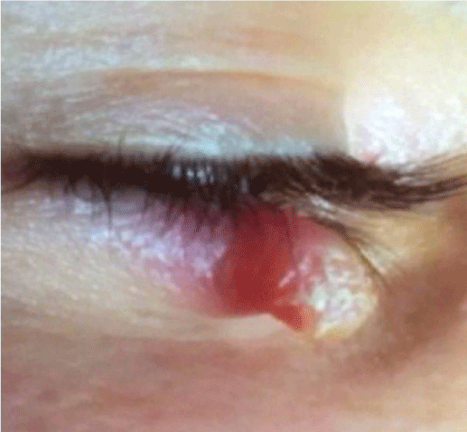
A 19-year-old male patient, resident in a the rural area, was admitted to the Ophthalmology Clinic for a solitary firm horn on the lower eyelid, which had gradually progressed over the course of two months. One year before, the patient noticed a focal swelling of the inferior eyelid treated empirically, on which the cone shape growth developed progressively. The patient’s medical and ocular history was not significant.
The clinical examination revealed a solitary cone shape hyperkeratotic growth mesuring 1.0/0.6 cm in size, with an inflamed nodular base, located in the middle 1/3rd of the inferior right eyelid (Figure 1). There was no regional lymphadenopathy. The clinical diagnosis was that of solitary inferior right eyelid cutaneous horn. The lesion was excised completely with local anesthesia, and the defect was closed by sliding the skin of the inferior eyelid and sutured with Vicryl (gauge 6.0).
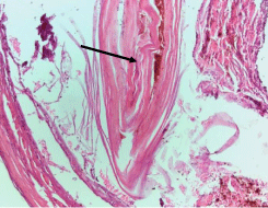
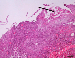
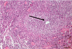
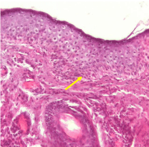
The resection specimen was evaluated histologically, revealing an association of three vertically overlapped lesions: compact acellular keratin, with a ”dome” shape (Figure 2), overlying an hyperplastic epithelium showing an infection with human papilloma virus (Figure 3A) and a moderate dysplasia of the adjacent epidermis (Figure 4). Underneath the hyperkeratosis and the hyperplastic epidermis, a chalazion could be identified into the deep dermal structure (Figure 3B). The postoperative evolution was favorable: no scar formation and no clinical relapse for six months.
Case 2
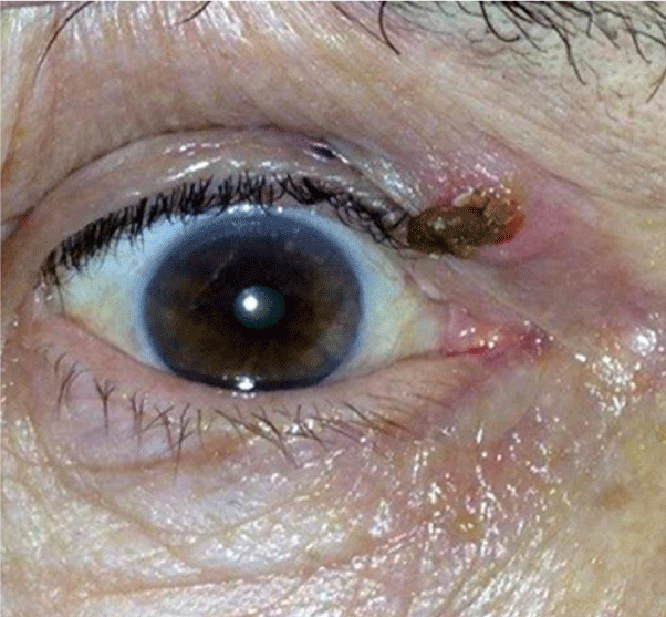
A 78-year-old male patient, resident in a rural area, was hospitalized for a cone shape growth on the upper right eyelid which had gradually progressed for six months. The patient’s medical ocular and general history was not significant. An ophthalmological clinical examinations demonstrated a cone shape hyperkeratotic growth developed on a well-defined inflamed nodular lesion, with ulcerated horn implantation edges, mesuring 0.6/0.3 cm in size, in the internal 1/3rd of the upper right eyelid (Figure 5). Moreover, multiple pigmented lesions were noticed on the patient’s face. The biomicroscopic examination diagnosed a incipient cataract in both eyes. There was no regional lymphadenopathy. The clinical diagnosis was that of a cutaneous horn located on a cystic sebaceous lesion on the right upper eyelid. The tumor growth was excised completely through ellipsoidal incision and primary closure of the defect with Vicryl suture (gauge 6.0) was done.
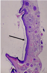
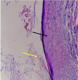
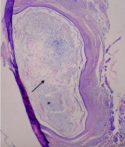
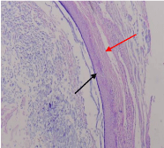
The resection specimen was evaluated histopatologically, revealing an association of three lesions: cutaneous horn (which “detached” from the basis of the underlying lesion, just before the surgical intervention), moderate dysplasia of the underlying epidermis and epidermal inclusion cyst located in the deep dermal structure (Figure 6A-D). The postoperative evolution was favorable: no scar formation and no clinical relapse for a year.
Discussion
The cutaneous horn is a clinical diagnosis which refers to a tumor located on the surface of the skin [14,24] with a hyperkeratotic cone shape [3], white-yellowish in colour, ranging from a few millimeters to a few centimeters in size, which can hide other benign or malignant underlying lesions [1].
This tumor is common in Caucasians, less frequent in Asian and Arabic populations, and rare in the African population [25].
The age of onset of the cutaneous horn is between 60-70 years old, and the underlying malignant lesions identified are common in people over the age of 70 [14].
The first case of cutaneous horn was reported in London, in 1588, in an elderly Welsh woman [8,9].
The cutaneous horn is made of compact keratin. The basis can be flat, nodular or „crater-like”. Clinical aspects can not give any clue to differentiate a benign or a malignant lesion, but the indurated and bleeding base of a large tumor pleads in favour of malignancy [14]. The cutaneous horn develops most often on sun exposed skin, on a preexisting lesion, such as benign warts or seborrheic keratosis. However, the real pathobiology of the developing of a cutaneous horn on the surface of these lesions remains unknown [14].
From a histological perspective, the cutaneous horn is made of compact hyperkeratosis, which can be either orthokeratotic (an anuclear keratin layer, as in the normal epidermis [26]) or parakeratotic (retention of nuclei in the cells of the stratum corneum of the epidermis [27]).
Moreover, it is often associated with acanthosis; the base of the cutaneous horn will highlight a pathological process which is responsible for its formation [1,11,14].
The most common types of lesions that can be identified histopathologically at the base of the cutaneous horn include: actinic keratosis, keratoacanthoma, seborrheic keratosis, pyogenic granuloma, discoid lupus erythematosus, verruca vulgaris, epidermal nevus, trichilemmal cyst, trichilemmoma, prurigo nodules, intradermal nevi, Bowen’s disease, basal cell carcinoma and squamous cell carcinoma [14,18,28-30].
The clinico-pathological study of 48 cases with cutaneous horns of the eyelid conducted by Mencía-Gutiérrez et al. (2004) shows that 77.1 % of them were associated with benign lesions, 14.6% with premalignant lesions, and 8.3 % with malignant lesions. They also searched for the association between cutaneous horn with a certain lesion regarding the malignancy. So, they found the following correlation: seborrheic keratosis as a benign lesion, actinic keratosis as the premalignant lesion, and basal cell carcinoma and squamous cell carcinoma as malignant lesions were associated more commonly with the cutaneous horn [20].
The 19-year-old patient stated that the first lesion he noticed was the chalazion, which was treated empirically and on the surface of which, the cutaneous horn developed later, over a period of two months, in Case 1. The lesion on which the cutaneous horn developed was a benign one, but the epidermis adjacent to it, showed a moderate dysplasia, so it required a larger excision of the base of the cutaneous horn to the healthy tissue.
In the second clinical case, the patient also came from the rural area, being exposed to solar radiations, and the lesion on which the cutaneous horn developed was a benign one (epidermal inclusion cyst), but again the epidermis adjacent to it showed a moderate dysplasia, and required a larger excision of the base of the cutaneous horn to the healthy tissue.
In these two clinical cases of cutaneous horns that we have presented here, associations of bening lessions were noticed, as well as the presence of a premalignant lesion, which highlight the fact that the histopathological examination is essential for identifying the nature of the lesion at the base of the cutaneous horn. The clinical appearance of the lesion can mask the premalignant or malignant potential of the underlying lesions and, for this purpose, the excision to the healthy tissue is essential, in order to prevent relapse.
In the case of the patients with benign lesions, subsequent monitoring is unnecessary.
However, if an association of the cutaneous horn with a malignant lesion is found, the patient must be assessed in order to diagnose potential relapse. In the case of patients with squamous cell or basal cell carcinomas at the base of the cutaneous horn, a screening must be performed to prevent relapses every 3-12 months for first 2 years, every 6-12 months for 3 years, and then at least annually for life [31].
Conclusions
The cutaneous horn is a lesion which, from a clinical perspective, cannot give the physician any indication as to its nature, i.e. benign or malignant; it can mask several benign, premalignant, as well as malignant lesions, and can only be diagnosed through a histopathological examination, which is essential in later therapeutic conduct.
- Mantese SA, Diogo PM, Rocha A, Berbert AL, Ferreira AK, et al. (2010) Cutaneous horn: a retrospective histopathological study of 222 cases. An Bras Dermatol 85: 157-163. Link: https://goo.gl/k8WvwK
- Thappa DM, Laxmisha C (2004) Cutaneous horn of eyelid. Indian Pediatr 41: 195. Link: https://goo.gl/WqAHhs
- Copcu E, Sivrioglu N, Culhaci N (2004) Cutaneous horns: are these lesions as innocent as they seem to be? World J Surg Oncol 2: 18. Link: https://goo.gl/5L9IB8
- Michal M, Bisceglia M, Di Mattia A, Requena L, Fanburg-Smith JC, et al.(2002) Gigantic cutaneous horns of the scalp: Lesions with agross similarity to the horns of animals: A report of four cases. Am J Surg Pathol 26: 789-794. Link: https://goo.gl/Rcs7zJ
- Nthumba PM (2007) Giant cutaneous horn in an African woman: a case report. J Med Case Reports 1: 170. Link: https://goo.gl/Os8ddk
- Duncan KO, Leffell DJ (2003) Epithelial precancerous lesions. In: Freedberg IM, Eisen AZ, Wolff K, Austen KF, Goldsmith LA, Katz SI. Fitzpatrick's Dermatology in General Medicine. 6th edition. New York: McGraw Hill 719-721.
- Souza LN, Martins CR, de Paula AM (2003) Cutaneous horn occurring on the lip of a child. Int J Paediatr Dent 13: 365-367. Link: https://goo.gl/djVynn
- Bondeson J (2001) Everard Home, John Hunter, and cutaneous horns: a historical review. Am J Dermatopathol 23: 362-369. Link: https://goo.gl/D49Cjr c
- Padmini HR, Pandey N (2015) Eyelid Cutaneous Sebaceous Horn: A Rare and Interesting Case Report. International Journal of Scientific Study 3: 134-136. Link: https://goo.gl/1I5hwz
- Bart RS, Andrade R, Kopf AW (1968) Cutaneous horns. A clinical and histopathologic study. Acta Derm Venereol 48: 507-515. Link: https://goo.gl/c1y1zK
- Yu RCH, Pryce DW, Macfarlane AW, Stewart TW (1991) A histopathological study of 643 cutaneous horns. Br J Dermatol 124: 449-452. Link: https://goo.gl/9HYRMc
- Festa Neto C, Falda S, Rivitti EA (1995) Corno cutâneo: estudo retrospectivo de 514 casos. An Bras Dermatol 70: 21-25. Link: https://goo.gl/a799ay
- Castillo D, Zerpa O, Loyo N, López C, Oliver M (2002) Histopatologia del cuerno cutáneo: estudio retrospectivo de 77 casos. Derm Venez 40: 65-69. Link: https://goo.gl/fTRNig
- Rush PS, Cockerell CJ, Butler DF, Chan EF, James WD, et al. (2015) Cutaneous Horn: Background, Pathophysiology, Epidemiology. [online] Emedicine.medscape.com. Link: https://goo.gl/aBtRxU
- Sadeghi E, Johari HG, Deylami HN (2012) Eyelid cutaneous horn. J Cutan Aesthet Surg 5: 153-154. Link: https://goo.gl/p1savP
- Markie RM (2004) Epidermal Skin Tumours. In: Champion RH, Burton JL, Ebling FJG. Rook/Wilkinson/Ebling Textbook of Dermatology. 5th edition Oxford: Blackwell Scientific Publications.
- Aquino LL, Wu JJ, Murase JE, Dyson SW, Jeffes EW (2008) Cutaneous horn on the finger. Clin Exp Dermatol 33: 529-530. Link: https://goo.gl/R7GdNr
- Vano-Galvan S, Sanchez-Olaso A (2008) Images in clinical medicine. Squamous-cell carcinoma manifesting as a cutaneous horn. N Engl J Med 359: e10. Link: https://goo.gl/9t9LRm
- Cruz Guerra NA, Sáenz Medina J, Ursúa Sarmiento I, Zamora Martínez T, Madrigal Montero R, et al.(2005) Malignant recurrence of a penile cutaneous horn. Arch Esp Urol 58 :61-63. Link: https://goo.gl/7IMQKJ
- Mencía-Gutiérrez E, Gutiérrez-Díaz E, Redondo-Marcos I, Ricoy JR, García-Torre JP (2004) Cutaneous horns of the eyelid: A clinicopathological study of 48 cases. J Cutan Pathol 31: 539–543. Link: https://goo.gl/1bPece
- Cristóbal MC, Urbina F, Espinoza A (2007) Cutaneous horn malignant melanoma. Dermatol Surg 33: 997-999. Link: https://goo.gl/umKJbW
- Azulay RD, Azulay DR (2006) Dermatologia. 4 edition Rio de Janeiro: Guanabara Koogan 430-431.
- Sampaio SAP, Rivitti EA (2007) Dermatologia. 3 edition São Paulo: Artes Médicas 1157- 1162.
- Durkee S (1866) Human Horn (Cornu Cutaneum of Rokitansky). Boston Med Surg J 74 :9-11. Link: https://goo.gl/yOeRm6
- Oludiran OO, Ekanem VJ (2011) Cutaneous horns in an African population. J Cutan Aesthet Surg 4: 197-200. Link: https://goo.gl/CCGRnr
- The Free Dictionary.com. (2012) orthokeratosis. Link: https://goo.gl/AvNYqU
- The Free Dictionary. com. (2012) parakeratosis. [online]. Link: https://goo.gl/dstE3F
- Findlay RF, Lapins NA (1983) Pyogenic granuloma simulating a cutaneous horn. Cutis 31: 610–612. Link: https://goo.gl/HLI2e8
- Dabski K, Stoll HL (1985) Cutaneous horn arising in chronic discoid lupus erythematosus. Arch Dermatol 121:837–8. Link: https://goo.gl/E3vfC1
- Gould JW, Brodell RT (1999) Giant cutaneous horn associated with verruca vulgaris. Cutis 64: 111–112. Link: https://goo.gl/uuRxB1
- National Comprehensive Cancer Network (2016) Squamous Cell Skin Cancer (Version 1.2017). [online]. Link: https://goo.gl/HS9uzn



 Save to Mendeley
Article Alerts
Subscribe to our articles alerts and stay tuned.
 This work is licensed under a Creative Commons Attribution 4.0 International License.