Journal of Clinical Research and Ophthalmology
Atypical Presentation of Uveal and Conjunctival Melanoma in the Anterior Segment
1Department of Ophthalmology, Northwell Health, Great Neck, New York; Hofstra Northwell School of Medicine, Hempstead, New York. North Shore Long Island Jewish Health System, USA
2Mount Sinai School of Medicine, New York, USA
3Mount Sinai Hospital, New York Eye and Ear Infirmary of Mount Sinai, New York, USA
Author and article information
Cite this as
Djougarian A, Rodgers IR, Friedman AH (2016) Atypical Presentation of Uveal and Conjunctival Melanoma in the Anterior Segment. J Clin Res Ophthalmol. 2016; 3(2): 41-43. Available from: 10.17352/2455-1414.000034
Copyright License
© 2016 Djougarian A, et al. This is an open-access article distributed under the terms of the Creative Commons Attribution License, which permits unrestricted use, distribution, and reproduction in any medium, provided the original author and source are credited.Ocular melanoma is a rare malignancy arising from melanocytes of the uvea, the conjunctiva, and the orbit. Uveal melanoma is the most common intraocular malignancy in adults, accounting for 85-95% of ocular melanomas [1-3]. The conjunctival form accounts for 5% of ocular melanomas. Uveal and conjunctival melanoma are very distinct from each other biologically. We describe a case of conjunctival melanoma and a uveal melanoma both presenting as large masses of the anterior segment.
Case Report
Patient 1
75 year old female from Haiti presented with spontaneous bleeding from her left eye for 1 day. She had a black mass present on the surface of her left eye which was noted by her family 4 months prior when she arrived from Haiti. The patient did not seek any medical care until bleeding began. She did not complain of pain. She denied past ocular trauma or surgery or a history of anticoagulation therapy. On examination, vision was 20/40 in the right eye, light perception in the left eye. The right pupil was reactive. The mass prevented the visualization of the left pupil. In the left eye, there was 3+ diffuse conjunctival injection, and a large black and somewhat bloody mass measuring 12x12x4mm that precluded visualization of the entire cornea and anterior segment (Figure 1A). Computed Tomography (CT) scan demonstrated a formed and normal sized globe with a solid mass overlying an intact cornea.
Ocular melanoma is a rare malignancy arising from melanocytes of the uvea, the conjunctiva, and the orbit. Uveal melanoma is the most common intraocular malignancy in adults, accounting for 85-95% of ocular melanomas [1-3]. The conjunctival form accounts for 5% of ocular melanomas. Uveal and conjunctival melanoma are very distinct from each other biologically. We describe a case of conjunctival melanoma and a uveal melanoma both presenting as large masses of the anterior segment.
Case Report
Patient 1
75 year old female from Haiti presented with spontaneous bleeding from her left eye for 1 day. She had a black mass present on the surface of her left eye which was noted by her family 4 months prior when she arrived from Haiti. The patient did not seek any medical care until bleeding began. She did not complain of pain. She denied past ocular trauma or surgery or a history of anticoagulation therapy. On examination, vision was 20/40 in the right eye, light perception in the left eye. The right pupil was reactive. The mass prevented the visualization of the left pupil. In the left eye, there was 3+ diffuse conjunctival injection, and a large black and somewhat bloody mass measuring 12x12x4mm that precluded visualization of the entire cornea and anterior segment (Figure 1A). Computed Tomography (CT) scan demonstrated a formed and normal sized globe with a solid mass overlying an intact cornea. There was no evidence of intraocular extension radiologically (Figure 1B). Blood tests including liver function tests were in normal limits. An examination under anesthesia and exploration was performed.
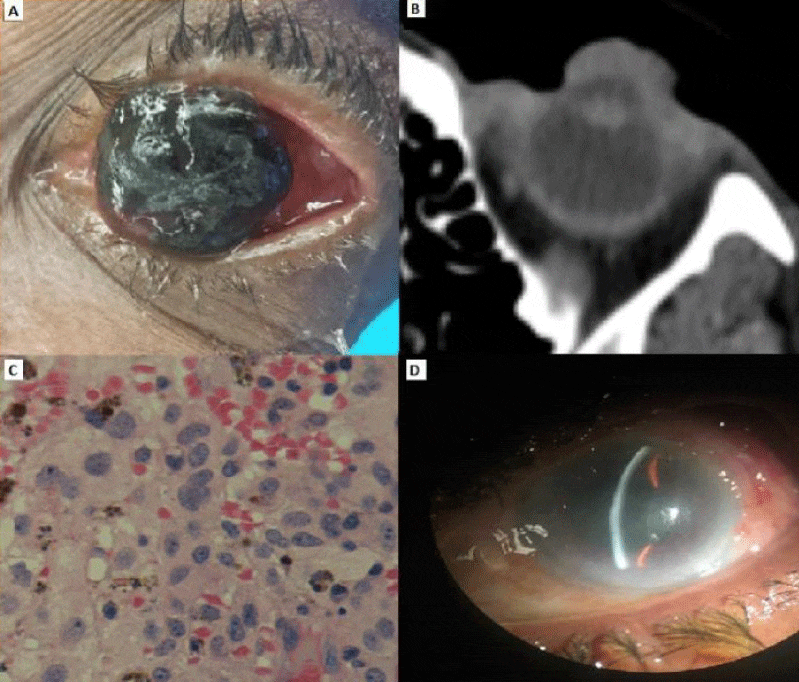
Intraoperatively, the mass was easily removed from the corneal epithelium. It was adherent to the limbal conjunctiva temporally. A small, bleeding limbal blood vessel was cauterized. The corneal epithelium was intact. No other lesions were noted on the anterior aspect of the globe and dilated exam with scleral depression revealed normal anatomy. Histopathology disclosed mixed spindle B and pigmented epithelioid cells (Figure 1C). MHB-45, MART-1, Mel A, and S-100 markers were positive. The final diagnosis was malignant melanoma, clinically arising from the limbal conjunctiva. Postoperatively, the patient’s vision in the left eye was 20/100, attributable to a nuclear cataract. There was a 2 mm area of conjunctival melanosis at 12 o’clock and the eye was otherwise grossly anatomically normal (Figure 1D). There was no evidence of tumor in the angle on gonioscopy. The patient underwent a metastatic workup which was negative. She underwent further cryotherapy of the conjunctival tissue and treatment with interferon topical drops.
Patient 2
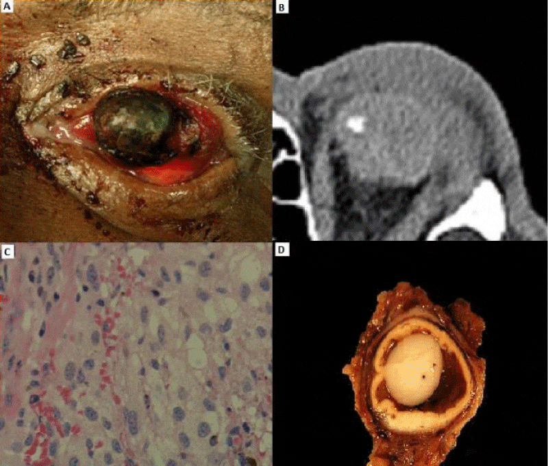
72 year old African-American man presented with spontaneous bleeding from his left eye for 3 days. He had a black mass present on the surface of the left eye for unknown duration of time. He did not complain of pain but reported a foreign body sensation. His past ocular history included enucleation of the right eye in1970 following an automobile accident. His left eye was severely injured in a fight in 1979 which had left him with no light perception vision. He denied any recent trauma. He denied any medical history and was not taking any medications. On examination, the right orbit was anophthalmic. His left eye was blind and phthisical. There was a black and bloody mass protruding from and occupying the corneal surface; measuring 15x15x5mm (Figure 2A). CT scan demonstrated a phthisical eye with bone and soft tissue within the globe (Figure 2B). The patient underwent evisceration as the surgeon clinically believed the pigmented lesion did not originate from the uvea. There was no gross intraocular extension seen intraoperatively. An Acrylic implant was inserted into the scleral shell. Histopathology revealed the presence of both intraocular and extraocular melanoma. The tumor was composed of large and small cells with epithelioid features (Figure 2C). Many of the cells stained for melanin. Fragments of bone and degenerated uveal tissue were present as well. HMB-45, MEP IP and XIAP stains were positive. The final diagnosis was malignant melanoma epithelioid cell type arising from the uveal tract. The patient underwent metastatic work up and no metastases were found. He then underwent a definitive exenteration of the left orbit with histopathology showing no evidence of primary or residual tumor (Figure 2D).
Discussion
Primary ocular melanoma can occur in the uvea, conjunctiva, or orbit. These melanomas are distinct entities although they share a common origin from melanocytes. The annual incidence of uveal melanoma is 5.1 per million [2]. The most common location is of the choroid posterior to the equator in 85% of cases. 15% arise anterior to the equator and involve the iris, ciliary body or the choroid anterior to the equator [4]. Conjunctival melanomas account for only 2-5% of ocular malignancies. Tumor spread occurs through lymphatics and blood stream resulting in metastasis [5,6]. Poor prognostic factors include tumor thickness greater than 2mm, high mitotic rate, and epithelioid cell type. With uveal melanoma, poor prognostic factors have been associated with involvement of the ciliary body, extrascleral extension, pathologic features of epithelioid cytology, and monosomy 3 [7-11]. In both uveal and conjunctival melanomas, involvement of the entire corneal surface is extremely rare. The two cases presented share several striking characteristics. Both individuals had moderate to dark skin pigmentation. Both cases presented as large masses involving the cornea, precluding anterior segment visualization. They appeared to be free of significant intraocular or conjunctival pigmentation elsewhere, at least on clinical evaluation at presentation and on radiologic imaging. The first case was striking as the mass easily peeled off the corneal epithelium with moist cotton tip applicator sticks; there was bleeding at the lateral limbus where the lesion was clinically minimally adherent to the limbal conjunctiva. The differential diagnosis in each case included coagulated blood on the surface of the cornea, ocular surface squamous neoplasia, and a ruptured globe with exposed uvea. The corneal epithelium can be involved when conjunctival melanoma involves the limbus.
Bowman’s layer may provide a barrier to deeper invasion [12].
Conclusion
Uveal and conjunctival melanoma presenting as a pigmented large mass overlying the cornea is rare. The mass may resemble exposed uveal tissue from a ruptured globe, large hematoma or squamous neoplasia. The management can be complex. Wide margins should be taken from the visible lesion since positive margins can be associated with higher reoccurrence rates. Tumor cells should not be irrigated. Care must be taken to not penetrate Bowman’s layer during excision to prevent further tumor spread. Post-operatively, patients must be monitored closely since new pigmentation may be a sign of reoccurrence. In summary, melanoma must be considered for any pigmented lesion obscuring the cornea and prompt management should be undertaken, including a work-up to exclude metastatic disease.
- McLaughlin CC, Wu XC, Jemal A, Martin HJ, Roche LM, et al. (2005) Incidence of noncutaneous melanomas in the U.S. Cancer 103: 1000-1007. Link: https://goo.gl/3o9bOk
- Singh AD, Turell ME, Topham AK (2011) Uveal melanoma: trends in incidence, treatment, and survival. Ophthalmology 118: 1881-1885. Link: https://goo.gl/NeQQxr
- Blum ES, Yang J, Komatsubara KM, Carvajal RD (2016) Clinical management of uveal and conjunctival melanoma. Oncology 30: 29-32, 34-43, 48. Link: https://goo.gl/UU0LbO
- Shields CL, Kaliki S, Furuta M, Mashayekhi A, Shields JA (2012) Clinical spectrum and prognosis of uveal melanoma based on age at presentation in 8,033 cases. Retina 32: 1363-1372. Link: https://goo.gl/rbHJkZ
- Yu GP, Hu DN, McCormick S, Finger PT ((2003) Conjunctival melanoma: is it increasing in the United States? Am J Ophthalmol 135: 800-806. Link: https://goo.gl/0w5Aiv
- Shields CL, Markowitz JS, Belinsky I, Schwartzstein H, George NS, et al. (2011) Conjunctival melanoma: outcomes based on tumor origin in 382 consecutive cases. Ophthalmology 118: 389-395. Link: https://goo.gl/xWgJYc
- Wang Z1, Nabhan M, Schild SE, Stafford SL, Petersen IA, et al. (2013) Charged particle radiation therapy for uveal melanoma: a systematic review and metaanalysis. Int J Radiat Oncol Biol Phys 86: 18-26. Link: https://goo.gl/5dn1H9
- Collaborative Ocular Melanoma Study G (2006) The COMS randomized trial of iodine 125 brachytherapy for choroidal melanoma: V. Twelve year mortality rates and prognostic factors: COMS report No. 28. Arch Ophthalmol 124: 1684-1693. Link: https://goo.gl/vaPq6L
- Harbour JW, Onken MD, Roberson ED, Duan S, Cao L, et al. (2010) Frequent mutation of BAP1 in metastasizing uveal melanomas. Science 330: 1410-1413. Link: https://goo.gl/z5HdQa
- Refaian N, Schlereth SL, Koch KR, Notara M, Hos D, et al. (2015) Comparing the hem- and lymphangiogenic profile of conjunctival and uveal melanoma cell lines. Invest Ophthalmol Vis Sci 56: 5691-5697. Link: https://goo.gl/5PFgw7
- Wong JR, Nanji AA, Galor A, Karp CL (2014) Management of conjunctival malignant melanoma: a review and update. Expert Rev Ophthalmol 9: 185-204. Link: https://goo.gl/S3MUi7
- Tuomaala S, Aine E, Saari KM, Kivelä T (2002) Corneally displaced malignant conjunctival melanomas. Ophthalmology 109: 914-919. Link: https://goo.gl/Psq3QG



 Save to Mendeley
Article Alerts
Subscribe to our articles alerts and stay tuned.
 This work is licensed under a Creative Commons Attribution 4.0 International License.