Journal of Clinical Research and Ophthalmology
Comparison of the Contact Lens-Shell Vs the Soft-Shell Technique for Corneal Endothelial Protection during Phacoemulsification in High-Risk Cataracts
Ophthalmology Department, Aastha Hospital, Anand, Gujarat, 388001, India
Author and article information
Cite this as
Maru DN. Comparison of the Contact Lens-Shell Vs the Soft-Shell Technique for Corneal Endothelial Protection during Phacoemulsification in High-Risk Cataracts. J Clin Res Ophthalmol. 2026; 13(2): 42-47. Available from: 10.17352/2455-1414.000115
Copyright License
© 2026 Maru DN. This is an open-access article distributed under the terms of the Creative Commons Attribution License, which permits unrestricted use, distribution, and reproduction in any medium, provided the original author and source are credited.Purpose: To compare the efficacy of the contact lens-shell technique (CST) and the conventional soft-shell technique (SST) in reducing corneal endothelial cell loss during phacoemulsification in eyes with high-risk cataracts.
Setting: Single private eye care centre, Anand, Gujarat, India.
Design: Prospective, randomized comparative study.
Methods: Forty eyes with high-risk cataracts, including mature cataracts, hard cataracts, small pupils, or shallow anterior chambers, were randomized to undergo phacoemulsification using either the CST (20 eyes) or SST (20 eyes). The CST employed an Etafilcon A hydrogel bandage contact lens as a temporary mechanical endothelial protector, whereas the SST used dispersive and cohesive ophthalmic viscosurgical devices. Uncorrected and corrected distance visual acuity, intraocular pressure (IOP), central corneal thickness (CCT), endothelial cell density (ECD), and endothelial cell loss (ECL) were evaluated during postoperative follow-up.
Results: Postoperative visual acuity and central corneal thickness were comparable between the two groups at all follow-up visits (P>.05). On postoperative day 1, mean IOP was significantly lower in the CST group than in the SST group (13.2 ± 2.92 vs 16.06 ± 6.32 mmHg; P=.02). At 1 month, the CST group demonstrated significantly lower endothelial cell loss than the SST group (3.0% ± 5.3% vs 8.8% ± 12.8%; P=.03), despite a longer effective phacoemulsification time.
Conclusion: The contact lens-shell technique was associated with significantly lower endothelial cell loss than the soft-shell technique during phacoemulsification in high-risk cataracts while providing comparable visual and anatomical outcomes. Larger studies with longer follow-up are warranted to confirm these findings.
Phacoemulsification is the preferred surgical technique for cataract extraction because it provides rapid visual rehabilitation and excellent refractive outcomes [1]. However, ultrasound energy, thermal effects, turbulent fluidics, lens fragments, and free radical generation during surgery can damage the corneal endothelium, particularly in eyes with limited endothelial reserve or surgically challenging cataracts [2]. Since corneal endothelial cells have minimal regenerative capacity, excessive endothelial cell loss (ECL) may result in persistent corneal oedema and, in severe cases, irreversible corneal decompensation [3].
The risk of endothelial injury is substantially greater in high-risk cataracts, including dense nuclear cataracts, mature cataracts, small pupils, shallow anterior chambers, and eyes with pre-existing endothelial compromise. Although refinements in phacoemulsification technology and surgical techniques have reduced endothelial trauma, protecting the corneal endothelium during complex cataract surgery remains a significant clinical challenge.
Several strategies have been developed to minimize endothelial damage, including optimization of phacoemulsification parameters, femtosecond laser-assisted cataract surgery, improved nucleus-fragmentation techniques, ophthalmic Visco surgical devices (OVDs), and mechanical barrier techniques such as the soft-shell technique.4,5,6,7,8,9 Experimental approaches, including lens anterior capsule disc and polymeric endothelial shields, have also demonstrated encouraging results [3-11]. Nevertheless, many of these techniques provide incomplete protection during prolonged phacoemulsification because dispersive OVDs may gradually wash out of the anterior chamber, while several mechanical protective devices have been evaluated only in experimental animal models [10].
Mechanical endothelial protection using a soft hydrogel contact lens has recently emerged as an alternative approach. Experimental studies have demonstrated that hydrogel contact lenses can function as a temporary physical barrier between the phacoemulsification tip and the corneal endothelium while attenuating the effects of ultrasound energy, fluid turbulence, cavitation bubbles, and lens fragment impact [10]. Etafilcon A is a hydrophilic hydrogel material with excellent optical transparency, flexibility, biocompatibility, and adherence to ophthalmic viscosurgical devices, making it a suitable candidate for intracameral mechanical endothelial protection [12].
To our knowledge, no prospective clinical study has evaluated the use of an Etafilcon A contact lens-shell technique in human eyes undergoing phacoemulsification for high-risk cataracts or directly compared it with the conventional soft-shell technique. Therefore, the present prospective randomized comparative study was designed to compare the efficacy of the Contact Lens-Shell Technique (CLST) and the Soft-Shell Technique (SST) in reducing postoperative endothelial cell loss while evaluating visual outcomes, intraocular pressure, central corneal thickness, and endothelial cell density in eyes with high-risk cataracts.
Methods
This was an exploratory, prospective, randomized comparative study conducted at Aastha Hospital, Anand, Gujarat, India, between January 2025 and June 2025. The study was approved and prospectively registered by the local ethics committee (165/ethics/2024) and was conducted in accordance with the tenets of the Declaration of Helsinki. Written informed consent was obtained from all patients before surgery. Forty eyes from 40 patients were included, with 20 eyes assigned to CLST and 20 to SST. No formal a priori sample-size calculation was performed.
High-risk cataracts included mature cataracts, hard cataracts with nuclear opalescence grade greater than 3 according to the Lens Opacities Classification System III, cataracts associated with a small pupil after pharmacological dilatation, and cataracts with a shallow anterior chamber. A pupil was considered small when its diameter was less than 5 mm after dilatation with tropicamide 0.8% and phenylephrine 5%; the diameter was measured automatically using an auto-keratorefractometer (Grand Seiko, Japan) and confirmed intraoperatively with a manual caliper. A shallow anterior chamber was defined as an anterior chamber depth of less than 2.8 mm, measured using the IOLMaster 700. Eyes with intraoperative or postoperative complications, uveitis, glaucoma, optic nerve disorders, previous ocular trauma, macular degeneration or other retinal disease, corneal scarring or other corneal pathology, amblyopia, or inability to complete the required examinations were excluded.
All eyes underwent a detailed ophthalmic evaluation by trained technicians and hospital physicians, with study data masked during assessment. Preoperative and postoperative assessments included uncorrected visual acuity, best-corrected visual acuity, intraocular pressure, and central corneal thickness, which were recorded preoperatively and on postoperative days 1, 7, and 30. Endothelial cell density was measured preoperatively and on postoperative days 7 and 30. Intraocular pressure was measured using a noncontact tonometer (Shin Nippon NCT 200, Rexxam Co., Ltd., Osaka, Japan). Central corneal thickness and endothelial cell density were measured using a noncontact specular microscope (EM-4000, Tomey Corporation, Japan). Endothelial cell loss was calculated as the percentage decrease in central corneal endothelial cell density from the preoperative value.
Patients were randomly assigned in a 1:1 ratio to either the CLST or SST group using a computer-generated randomization sequence. Outcome assessors were masked to the treatment allocation.
All surgeries were performed by a single surgeon under either topical or peribulbar anesthesia.
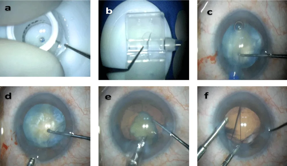
For the contact lens-shell technique, before surgery, an Etafilcon A hydrophilic hydrogel soft bandage contact lens (FreshKon 58; Oculus Private Limited, Singapore) was placed in a bottle containing solution prepared using 3 cc (mL) of moxifloxacin 0.5% combined with 7 cc (mL) of sterile balanced salt solution (BSS) for five minutes, then it was transferred onto a sterile Teflon block. A disposable 9.5-mm corneal trephine was used to create a circular disc. The disc was folded and loaded into a 2.8-mm butterfly cartridge attached to an injector (Biotech, India). The disc should be handled atraumatically and kept hydrated.
After creation of two side ports and a 2.8-mm temporal clear corneal incision, the anterior capsule was stained with trypan blue when required, particularly in mature cataracts, and a continuous curvilinear capsulorhexis was performed. Hydrodissection was then performed, followed by filling of the anterior chamber with ophthalmic viscosurgical device.
In the CLST group, the preloaded contact lens disc was introduced into the anterior chamber using an IOL injector and positioned beneath the corneal endothelium, with a layer of ophthalmic viscosurgical device maintained between the contact lens and the endothelium.
In the SST group, a dispersive ophthalmic viscosurgical device (hydroxypropyl methylcellulose 2.4%; Biotech, India) was applied to coat the corneal endothelium, followed by a cohesive ophthalmic viscosurgical device (sodium hyaluronate 1.4%; Biotech, India) to maintain anterior chamber stability during phacoemulsification.
In both groups, the nucleus was managed by phacoemulsification using the stop-and-chop technique with the Compact Intuitiv system (Johnson & Johnson, USA). After phacoemulsification, the contact lens disc in the CLST group was removed with curved forceps and a dialer under continuous irrigation through an anterior chamber maintainer or with an ophthalmic viscosurgical device. Cortical material was then aspirated, and the intraocular lens was implanted in the capsular bag using hydroimplantation. The side ports were hydrated, and an intracameral antibiotic was administered after surgery.
The primary outcome was endothelial cell loss at postoperative month 1, calculated as the percentage decrease in central corneal endothelial cell density from baseline. Secondary outcomes included endothelial cell density, best-corrected visual acuity, intraocular pressure, central corneal thickness, and effective phacoemulsification time (EPT).
Statistical analysis
Data were analysed using Microsoft Excel statistical software. Continuous variables are presented as mean ± standard deviation. Categorical variables are summarized as counts. Distributional assumptions were assessed using the Shapiro–Wilk test. The longitudinal outcomes were analyzed using linear mixed-effects models with treatment group, time, and Group×Time interaction as fixed effects and patient as a random intercept. Baseline-adjusted postoperative models included the baseline value of the outcome as a covariate and postoperative time as a repeated factor. The primary endpoint, month-1 ECL, was analyzed using a baseline-ECD-adjusted mixed-effects model with treatment group as the fixed effect and patient as a random intercept. Where the Group×Time interaction was significant, model-based between-group contrasts were examined with Bonferroni adjustment for the number of postoperative time points. A two-sided P value <0.05 was considered statistically significant.
Results
Study population and baseline characteristics
A total of 40 eyes from 40 patients with high-risk cataracts were enrolled and completed the study. Twenty eyes were randomized to the Contact Lens-Shell Technique (CLST) group and 20 eyes to the Soft-Shell Technique (SST) group.
TThe outcomes included various types of cataracts: hard cataracts (CLST-2, SST-5), hard cataracts with small pupils (CLST-3, SST-6), hard cataracts with shallow anterior chamber (A/C) (CLST-1, SST-1), mature cataracts (CLST-6, SST-1), mature cataracts with shallow A/C (CLST-3, SST-2), cataracts with small pupils (CLST -0, SST-2), and cataracts with both small pupils and shallow A/C (CLST-5, SST-3). The SST group showed a higher prevalence of hard cataracts, while the CLST group had more mature cataracts, small-pupil cataracts, and shallow A/C.
The mean age was 62.3 ± 9.8 years in the CLST group and 63.0 ± 11.8 years in the SST group (P=0.32). Sex distribution was comparable (7 men/13 women vs 8 men/12 women). Preoperative anterior chamber depth, lens thickness, central corneal thickness, and endothelial cell density did not differ significantly between groups (P>0.05). However, the SST group had a significantly greater axial length (23.37 ± 1.18 mm vs 22.53 ± 0.97 mm; P=0.01), better preoperative best-corrected visual acuity (0.67 ± 0.68 vs 1.34 ± 0.79 log MAR; P<0.001), and higher preoperative intraocular pressure (20.30 ± 9.63 mmHg vs 15.80 ± 4.27 mmHg; P=0.03). Baseline demographic and ocular characteristics are summarized in Table 1.
Baseline axial length, best-corrected visual acuity, and intraocular pressure differed statistically between groups. Because these variables were not balanced at baseline, postoperative comparisons should be interpreted with caution, and the final inferential analysis should use baseline-adjusted longitudinal models rather than relying solely on separate between-group comparisons at each time point.
EPT from the phaco-time field: CLST 12.46 ± 6.34 seconds; SST 8.38 ± 4.67 seconds; Welch t-test, P = 0.026.
Operative outcomes
Mean effective phacoemulsification time (EPT) was significantly longer in the CLST group than in the SST group (P<.03). No intraoperative complications related to either endothelial protection technique were observed. The contact lens disc was successfully inserted and removed in all eyes without adverse events.
Intraocular pressure
Mean intraocular pressure was significantly lower in the CLST group on the first postoperative day (13.20 ± 2.92 mmHg) compared with the SST group (16.06 ± 6.32 mmHg), unadjusted (P=0.02). At postoperative week 1 and month 1, no statistically significant differences were observed between the two groups (P=0.47 and P=0.14, respectively). Intraocular pressure measurements are summarized in Table 2. The longitudinal mixed model showed a significant Group×Time interaction (P=.003). In the baseline-adjusted postoperative model, the interaction remained significant (P=.003); however, no individual postoperative contrast remained significant after Bonferroni correction. Thus, the day-1 lower IOP observed with CLST should not be presented as a statistically significant isolated time-point difference.
Central corneal thickness
Both groups demonstrated an increase in central corneal thickness on postoperative day 1, followed by a gradual reduction during follow-up. Mean central corneal thickness did not differ significantly between the CLST and SST groups at baseline, postoperative day 1, week 1, or month 1 (all P>0.05). These findings are presented in Table 3. The baseline-adjusted Group×Time interaction was significant (P<.001), driven by a lower day-1 CCT in SST (model-adjusted difference SST minus CLST −29.26 µm; Bonferroni-adjusted P=.001). The groups were similar at week 1 and month 1.
Best-corrected visual acuity
Best-corrected visual acuity improved substantially in both groups following surgery. The SST group demonstrated significantly better BCVA at postoperative day 1 (0.20 ± 0.20 vs 0.31 ± 0.20 log MAR; P=0.05) and postoperative week 1 (0.10 ± 0.10 vs 0.16 ± 0.11 log MAR; P=0.04). By postoperative month 1, BCVA was comparable between the CLST and SST groups (0.10 ± 0.10 vs 0.12 ± 0.10 log MAR; P=0.28). Detailed visual outcomes are shown in Table 4. The baseline-adjusted mixed model showed a significant Group×Time interaction (P=.013), driven by a day-1 difference favoring SST (adjusted difference −0.21 logMAR; Bonferroni-adjusted P=.001). The week-1 and month-1 contrasts were not significant after adjustment.
Endothelial cell loss and endothelial cell density
At postoperative week 1, mean endothelial cell loss was 5.95 ± 8.17% in the CLST group and 9.13 ± 11.63% in the SST group; the difference was not statistically significant (P=0.16). At postoperative month 1, endothelial cell loss was significantly lower in the CLST group (3.02 ± 5.31%) than in the SST group (8.81 ± 12.78%) (P=0.03). The prespecified baseline-ECD-adjusted mixed-effects primary analysis showed 6.46 percentage points greater ECL in SST than CLST (95% CI, 1.32–11.59; P=0.01370384387627). This adjusted result supports lower month-1 ECL with CLST.
Preoperative endothelial cell density was comparable between groups (2573.25 ± 363.53 cells/mm2 vs 2695.55 ± 280.84 cells/mm2; P=0.12). Endothelial cell density remained comparable at postoperative week 1 (2428.40 ± 434.19 vs 2458.90 ± 439.75 cells/mm2; P=0.41) and month 1 (2502.65 ± 410.48 vs 2466.05 ± 446.88 cells/mm2; P=0.39). These data are summarised in Table 5. The full longitudinal ECD model, including baseline, day 1, week 1, and month 1, did not show a significant Group×Time interaction (P=.321).
Results
The present prospective randomized comparative study evaluated the efficacy of the Contact Lens-Shell Technique (CLST) compared with the conventional Soft-Shell Technique (SST) for corneal endothelial protection during phacoemulsification in eyes with high-risk cataracts. The principal finding was that the CLST was associated with significantly lower endothelial cell loss at one month despite a longer effective phacoemulsification time. Postoperative visual acuity and central corneal thickness were comparable between the two groups, while the CLST group demonstrated significantly lower intraocular pressure on the first postoperative day.
The apparent reduction in calculated endothelial cell loss between week 1 and month 1 in the CLST group should not be interpreted as endothelial cell recovery. Because endothelial cell density was measured by noncontact specular microscopy, repeatability and sampling variability may influence serial measurements, particularly when small changes are observed. The measurement protocol used the same noncontact specular microscope for follow-up; however, the study did not include repeated same-session measurements or a formal repeatability analysis. Accordingly, the month-1 ECL finding should be interpreted as an observed between-group difference rather than evidence of endothelial cell regeneration.
Phacoemulsification is a globally accepted surgical technique for cataract removal, and it is known for its efficiency and minimal invasiveness [13]. The overall success rate of phacoemulsification is high, particularly in uncomplicated cases [14]. Corneal endothelial injury remains one of the major concerns during phacoemulsification in eyes with dense nuclei, mature cataracts, small pupils, shallow anterior chambers, or compromised endothelial reserve. Endothelial cell loss results from a combination of ultrasound energy, acoustic cavitation, turbulent fluidics, mechanical trauma from lens fragments, thermal injury, and oxidative stress [15-19]. Although improvements in phacoemulsification technology and ophthalmic Visco surgical devices (OVDs) have reduced endothelial trauma, effective protection during prolonged or technically challenging surgery remains difficult [7-9,19].
The significantly lower endothelial cell loss observed with CLST suggests that a temporary hydrogel contact lens positioned beneath the corneal endothelium may provide additional mechanical protection beyond that achieved with OVDs alone. Unlike dispersive OVDs, which may gradually wash out during prolonged phacoemulsification, the contact lens remains in position throughout nucleus emulsification and may reduce direct endothelial trauma from lens fragments, ultrasound-generated turbulence, and cavitation. Although the present study was not designed to investigate the exact mechanism of endothelial protection, the observed reduction in endothelial cell loss supports further evaluation of this concept.
Other endothelial protection techniques, such as the LACD [3] (lens anterior capsule disc) technique and PGD (Poly glycerol dodecanedioate) based shape memory polymeric shield technique [11], demonstrated reduced damage to the endothelium and corneal oedema by ultrasonic energy during phacoemulsification.
Experimental work by Kim et al. demonstrated that a Senofilcon A hydrogel contact lens could protect the corneal endothelium during phacoemulsification in rabbit eyes by reducing endothelial damage and postoperative inflammation [10]. Our study extends these experimental observations by evaluating a similar mechanical protection strategy using an Etafilcon A hydrogel contact lens in human eyes undergoing cataract surgery. However, direct comparison between the two hydrogel materials should be interpreted cautiously because they differ in composition, physical properties, and study design [10,12].
The CLST group also demonstrated significantly lower intraocular pressure on the first postoperative day. This finding may be explained by the smaller quantity of OVD required during CLST, thereby reducing retained viscoelastic material within the anterior chamber and minimizing transient obstruction of aqueous outflow. Similar observations have been reported with the soft-shell technique, where retained OVD has been implicated in early postoperative intraocular pressure elevation [9].
The lower postoperative day-1 intraocular pressure in the CLST group may be related to differences in ophthalmic Visco surgical device use or retention; however, OVD volume or residual OVD was not directly measured in this study. This explanation therefore remains a hypothesis rather than an established mechanism.
Although effective phacoemulsification time was significantly longer in the CLST group, endothelial cell loss remained significantly lower. This finding suggests that the additional mechanical protection provided by the contact lens may compensate for the potential adverse effects of prolonged ultrasound exposure. Nevertheless, this hypothesis requires confirmation through larger mechanistic studies.
Both groups demonstrated significant improvement in postoperative visual acuity. Although early postoperative BCVA was slightly better in the SST group, this difference was no longer present at the 1-month follow-up. The early difference is likely attributable to baseline differences in cataract severity, as the CLST group included a greater proportion of mature cataracts, shallow anterior chambers, and small pupils rather than reflecting inferior performance of the technique.
The present study has several strengths. To our knowledge, it is among the first prospective randomized clinical studies to evaluate an intracameral hydrogel contact lens as a mechanical endothelial protector in human eyes undergoing phacoemulsification for high-risk cataracts. Furthermore, all procedures were performed by a single experienced surgeon using a standardized surgical technique, thereby minimizing inter-surgeon variability.
Several limitations should be acknowledged. The study included a relatively small sample size with short-term follow-up, limiting assessment of long-term endothelial preservation. The distribution of high-risk cataract subtypes differed slightly between the study groups, which may have influenced some postoperative outcomes. The study was performed at a single centre by a single surgeon, potentially limiting the generalizability of the findings. Furthermore, no formal a priori sample size calculation was performed. Future multicentre randomized studies with larger sample sizes, longer follow-up, endothelial morphology analysis, and biomechanical evaluation of intracameral hydrogel protectors are required to validate these findings.
To conclude, the Contact Lens-Shell Technique was associated with lower endothelial cell loss at 1 month than the conventional Soft-Shell Technique in this small prospective comparative study of high-risk cataracts. Because of baseline imbalances and the small sample size, these findings should be considered preliminary. Larger adequately powered studies with longer follow-up are required to establish the clinical benefit and safety of CLST.
Value statement
What was known: The soft-shell technique is widely used to protect the corneal endothelium during phacoemulsification in high-risk cataract surgery, although endothelial cell loss remains a significant concern.
What this paper adds: This study demonstrates that the contact lens-shell technique provides superior endothelial protection, resulting in significantly lower endothelial cell loss and reduced early postoperative intraocular pressure compared with the soft-shell technique.
These findings suggest that the contact lens-shell technique is a simple, cost-effective, and effective alternative for endothelial preservation in challenging cataract cases.
AI use seclaration
During the preparation of this manuscript, the authors used OpenAI’s ChatGPT to assist with language editing, organization, and clarity of the manuscript. The authors critically reviewed and edited all AI-assisted content and take full responsibility for the accuracy, integrity, and final content of the manuscript.
Acknowledgement: I am deeply grateful to my dedicated hospital staff for their invaluable assistance in conducting this study and to my patients for their generous cooperation.
- Moshirfar M, Milner D, Patel BC. Cataract surgery. In: StatPearls. StatPearls Publishing; 2024.
- Hayashi K, Hayashi H, Nakao F, Hayashi F. Risk factors for corneal endothelial injury during phacoemulsification. J Cataract Refract Surg. 1996; 22:1079-1084.
- Wu B, Ding X, Li S, Huo D, Zhang F, Liang W, et al. Mechanical protective effect of lens anterior capsule disc on corneal endothelial cells during femtosecond laser-assisted cataract surgery in a rabbit model. BMC Ophthalmol. 2023; 23:166.
- Vargas LG, Holzer MP, Solomon KD, Sandoval HP, Auffarth GU, Apple DJ. Endothelial cell integrity after phacoemulsification with 2 different handpieces. J Cataract Refract Surg. 2004; 30:478-482. Available from: https://doi.org/10.1016/s0886-3350(03)00620-5
- Berger A, Contin IN, Nicoletti G, Baltar Pazos PF, Baltar Pazos HS, Gomes JA. Middle prechop: Fracturing the middle portion of the nucleus. J Cataract Refract Surg. 2012; 38:564-567. Available from: https://doi.org/10.1016/j.jcrs.2012.02.001
- Conrad-Hengerer I, Al Juburi M, Schultz T, Hengerer FH, Dick HB. Corneal endothelial cell loss and corneal thickness in conventional compared with femtosecond laser-assisted cataract surgery: Three-month follow-up. J Cataract Refract Surg. 2013; 39:1307-1313. Available from: https://doi.org/10.1016/j.jcrs.2013.05.033
- Glasser DB, Katz HR, Boyd JE, Langdon JD, Shobe SL, Peiffer RL. Protective effects of viscous solutions in phacoemulsification and traumatic lens implantation. Arch Ophthalmol. 1989; 107:1047-1051. Available from: https://doi.org/10.1001/archopht.1989.01070020109041
- Arshinoff SA. Dispersive-cohesive viscoelastic soft shell technique. J Cataract Refract Surg. 1999; 25:167-173. Available from: https://doi.org/10.1016/s0886-3350(99)80121-7
- Miyata K, Nagamoto T, Maruoka S, Tanabe T, Nakahara M, Amano S. Efficacy and safety of the soft-shell technique in cases with a hard lens nucleus. J Cataract Refract Surg. 2002; 28:1546-1550. Available from: https://doi.org/10.1016/s0886-3350(02)01323-8
- Kim S, Cha D, Song YB, Choi JY, Han YK. Effects of senofilcon A mechanical protector on corneal endothelial cells during phacoemulsification in rabbit eyes: Pilot study. J Cataract Refract Surg. 2017; 43:394-399. Available from: https://doi.org/10.1016/j.jcrs.2017.01.006
- Liu Y, Li Y, Ji J, Fan Y, Hong J, Wang L. A shape memory polymeric shield for protecting corneal endothelium during phacoemulsification. Transl Vis Sci Technol. 2024; 13:11. Available from: https://doi.org/10.1167/tvst.13.4.11
- Efron N, Brennan NA, Chalmers RL, Jones L, Lau C, Morgan PB, et al. Thirty years of "quiet eye" with etafilcon A contact lenses. Cont Lens Anterior Eye. 2020; 43:285-297. Available from: https://doi.org/10.1016/j.clae.2020.03.015
- Dr Agarwal's Eye Hospital. Phacoemulsification: Modern cataract surgery techniques for clear vision restoration. Published 2023. Accessed July 25, 2024.
- Rosen H, Vernon SA. Unexpected poor vision within 24 h of uneventful phacoemulsification surgery: A review. J Clin Med. 2022; 12:48. Available from: https://doi.org/10.3390/jcm12010048
- Park S, Leonard BC, Raghunathan VK, et al. Animal models of corneal endothelial dysfunction to facilitate development of novel therapies. Ann Transl Med. 2021; 9:1271. Available from: https://doi.org/10.21037/atm-20-4389
- Chen K, Xu WY, Sun SS, Zhou HW. Corneal endothelial cells and acoustic cavitation in phacoemulsification. World J Clin Cases. 2023; 11:1712-1718. Available from: https://doi.org/10.12998/wjcc.v11.i8.1712
- Ramirez-Garcia MA, Khalifa YM, Buckley MR. Vulnerability of corneal endothelial cells to mechanical trauma from indentation forces assessed using contact mechanics and fluorescence microscopy. Exp Eye Res. 2018; 175:73-82. Available from: https://doi.org/10.1016/j.exer.2018.06.005
- Nita M, Grzybowski A. The role of reactive oxygen species and oxidative stress in the pathomechanism of age-related ocular diseases and other pathologies of the anterior and posterior eye segments in adults. Oxid Med Cell Longev. 2016; 2016:3164734. Available from: https://doi.org/10.1155/2016/3164734
- Ben-Eliahu S, Tal K, Milstein A, Levin-Harrus T, Ezov N, Kleinmann G. Protective effect of different ophthalmic viscosurgical devices on the corneal endothelium during a severe phacoemulsification model in rabbits. Ophthalmic Surg Lasers Imaging. 2011; 42:152-156. Available from: https://doi.org/10.3928/15428877-20110125-01

 Save to Mendeley
Article Alerts
Subscribe to our articles alerts and stay tuned.
 This work is licensed under a Creative Commons Attribution 4.0 International License.