Journal of Clinical Research and Ophthalmology
Idiopathic Epiretinal Membrane Treated with QIAPI 1®: Case Report
Director and Founder, Human Photosynthesis(TM) Research Centre, Mexico
Author and article information
Cite this as
Herrera AS, et al. Idiopathic Epiretinal Membrane Treated with QIAPI 1®: Case Report. J Clin Res Ophthalmol. 2026; 13(2): 24-41. Available from: 10.17352/2455-1414.000114
Copyright License
© 2026 Herrera AS, et al. This is an open-access article distributed under the terms of the Creative Commons Attribution License, which permits unrestricted use, distribution, and reproduction in any medium, provided the original author and source are credited.Idiopathic epiretinal membrane (iERM) is a common cause of visual impairment that can lead to distortion and disorganisation of the retinal structure. Vitrectomy has been widely used for the treatment of this disorder. During surgery, the entire vitreous is removed, and the ERM is completely peeled. The internal limiting membrane (ILM) is also peeled if there is any residual ILM at the macular surface extending to the arcades. Surgical complications such as postoperative vitreous haemorrhage, retinal detachment, and endophthalmitis are described. Postoperative cataract remains inevitable.
In terms of postoperative complications, the potential recurrence of ERM remains a primary concern. It was disclosed that the neighbouring remnant membrane and hyperreflective dots on the retinal surface and postoperative inner retinal wrinkling persisting for over 1 month were predisposing OCT findings for ERM recurrence. In some reported series, the recurrence rate with varying degrees of severity reached 84.4%.
Most patients with epiretinal membranes (ERMs) have no symptoms in their initial stages; their ERMs are found incidentally on dilated retinal exam or on retinal imaging such as with ocular coherence tomography (OCT). In such cases, patients typically have normal or near-normal vision. However, ERMs can slowly progress, leading to a vague visual distortion that can be perceived better by closing the non- or less-affected eye.
Patients may notice metamorphopsia, a symptom that causes visual distortion in which shapes that are normally straight, like window blinds or a door frame, look “wavy” or “crooked,” especially when compared to the other eye. In advanced cases, this can lead to severely decreased vision. Less commonly, ERMs may also be associated with double vision, light sensitivity or images looking larger or smaller than they are.
The cause of ERMs is possibly due to a defect in the surface layer of the retina where a type of cell, called glial cells, can migrate by unknown causes through it and start to grow in a membranous sheet on the retinal surface. This membrane can appear like cellophane and over time may contract and cause traction (or pulling) and puckering of the retina, leading to decreased vision and metamorphopsia.
ERMs can be associated with several ocular conditions such as prior retinal tears or detachment, retinal vascular diseases such as diabetic retinopathy or venous occlusive disease; they can also be post-traumatic, occurring following ocular surgery, or be associated with intraocular (inside the eye) inflammation. ERM is called idiopathic (of unknown origin).
Since ERMs are apparently stable after an initial period of growth, they can be monitored if they are not affecting vision significantly. In some circumstances, the membrane will spontaneously be released from the retina, relieving the traction and clearing up the vision. However, if an exam shows progression and/or functional worsening in vision, surgical intervention may be recommended. Supposedly, there are no eye drops, medications or nutritional supplements to treat ERMs [1].
So far, a surgical procedure called vitrectomy is the only option for eyes that require treatment. With vitrectomy, small incisions are placed in the white part of the eye, and the vitreous gel filling the inside of the eye is replaced with saline. This allows access to the surface of the retina where the ERM can be removed with delicate forceps, thereby allowing the macula to relax and become less wrinkled. Visual recovery is slow, and most eyes experience improvement within 3 months, but it may take a year to attain maximal visual acuity improvement.
Oxygen deprivation in retinal diseases
The lack of oxygen in the retina can cause different eye diseases like glaucoma, diabetic retinopathy, and retinal artery occlusions. The retina is one of the most metabolically active organs in the human body [2]. Supposedly, the metabolism of the retina is finely controlled to allow for enough vascular supply to power the neural transmission of light.
Hypoxia leads to the loss of the retinal ganglion cells (RGCs) via either apoptosis or necrosis, preventing the transmission of light to the visual cortex [3]. Retinal hypoxia encompasses a rather broad family of conditions [4].
Anatomically, the retina is supplied by branches of the ophthalmic artery, which comes from the internal carotid artery. The central retinal artery and short posterior ciliary arteries perfuse the inner retina [5]. The retina is supplied by two vascular systems: the choriocapillaris and the central retinal artery. The choriocapillaris supplies the layers of the outer retina, including the retinal pigment epithelium and photoreceptors, which hypothetically are oxygenated by the choriocapillaris through diffusion. The central retinal artery supposedly supplies oxygen to the inner retina, the retinal ganglion cells, and the retinal nerve fibre layers [6]. Thereby, Retinal oxygenation is believed to be regulated by the circulation of the vascular inner and avascular outer layers. As could be expected, the control of oxygen in the retinal arteries and the choroid differs [7].
Retinal pericytes are one key regulator of vascular flow in both the choroid and retina, presumably acting as protective factors against retinal hypoxia. Pericytes sit around the retinal vessels and regulate neurovascular coupling and blood flow within the choroid and the retina through intrapericyte tunnelling nanotubes [8].
Pericytes cover almost 85% of the retinal microvasculature [9]. Even more so, pericytes are essential in the postnatal period to help develop the blood-retinal barrier. Loss of pericytes leads to the breakdown of this barrier, leading to the infiltration of immune cells and the formation of microhemorrhages [10]. Blood-retinal barrier breakdown has been implicated in glaucoma due to damage caused by high pressure and in diabetic retinopathy due to high-glucose-related reactive oxygen species [11].
So far, it is wrongly believed that the retina maintains its oxygen and energy requirements through aerobic respiration and β-oxidation [12]. It is thought that under metabolic stress and hypoxia, the retina relies more heavily on alternate metabolic pathways such as the pentose phosphate pathway and anaerobic metabolism [13]. Strikingly, hypoxia was also found to lead to an increase in β-oxidation, indicating that this pathway is utilized in both normal and pathological conditions [14], which indicates that our current concepts of hypoxia must be modified, given the presence of molecules capable of transforming the power of light into free chemical energy, through the dissociation of the water molecules also present inside our cells [15].
It seems that the primary driver of hypoxia-induced changes in the retina is the activation of hypoxia-inducible factor 1-α (HIF-1α), leading to downstream activation of vascular endothelial growth factor (VEGF) and nitric oxide synthase (NOS) [16]. Additional methods of vascular regulation in the retina are through a local renin-angiotensin system, endothelin-1, and adenosine [17]. Pericytes also play an additional role here, as VEGF not only increases pericyte proliferation but also increases angiogenesis [18]. The top seven genes that HIF-1α (or hypoxia itself) activate promote the conversion of metabolic processes from oxidative phosphorylation to glycolysis that supposedly are essential to helping the cell survive hypoxia [19]. Of course, with each retinal hypoxia pathology, the system is affected slightly differently. Overproduction of both VEGF and NOS is associated with vasodilation and permeability of retinal vessels, allowing for infiltration of immune cells for the removal of lost tissue [20] or proliferation of local cells and collagen. The resulting breakdown of the blood-retinal barrier allows for the pathological accumulation of fluid in extracellular and intracellular spaces, leading to vasogenic or cytotoxic oedema, respectively [21]. Additionally, this increased vascular permeability is associated with endothelial dysfunction, microaneurysms, and leukocyte vascular plugs that can further damage the tissue, especially in diabetic retinopathy [22].
Lung gas exchange
Leaving aside the prevailing concept that the oxygenation of the retina depends on the choroidal blood supply, we will briefly refer to the pulmonary gas exchange as described to date, since the theoretical source of this oxygen would be the atmosphere and therefore would penetrate the blood circulation of the human body through the lung.
Gas exchange in the lung depends on tidal breathing, which brings new oxygen to and removes carbon dioxide from alveolar gas. This maintains alveolar partial pressures that promote passive diffusion to add oxygen and remove carbon dioxide from blood in alveolar capillaries [23].
The mechanism mentioned above, which is described as apparently simple through the passive diffusion of oxygen (atmosphere-lung-bloodstream), which has not been able to be demonstrated even though it has been intensively searched for since the beginning of the past century, however, data on the oxygen concentration and transport in tissues, cells, and subcellular structures are required to understand oxygen-related physiological and pathophysiological phenomena, and to date it has not been possible to determine them with sufficient accuracy, so the explanation of it has been based on completely theoretical mathematical models. On the contrary, the removal of CO2 contained in the blood through the alveolar capillaries, and with a relevant role of the enzyme carbonic anhydrase in this process, has been well established since 1950 [24].
Passive diffusion of oxygen across the cell membrane
It is established that simple fluid-phase lipid bilayers are not barriers to oxygen transport. However, further investigations indicate that many physical and chemical (compositional) factors can significantly decrease this permeation. In biological cell plasma membranes, the lipid bilayer forms the matrix in which integral membrane proteins are immersed, changing the organisation and properties of the lipid matrix. To evaluate oxygen permeability coefficients across these complex membranes, oxygen permeation across all membrane domains and components must be considered [25].
The methods available to measure oxygen permeation across membranes are limited and often complex, normalised to an oxygen concentration corresponding to the sample equilibrated with air at normal 760 mmHg atmospheric pressure.
It is practically impossible to directly measure the oxygen permeability coefficient across the membrane by creating fast-decaying oxygen concentration gradients across the membrane [26]. Worst, the physical state of the PL bilayer membrane (double bond) can significantly (drastically) affect its permeability property. In biological membranes, the double bond is located mainly at the C9–C10 position. It was shown that the presence of a cis or trans double bond at this position decreases the membrane permeability coefficient (PM) as compared with that of saturated membranes (the comparison should be performed at the same temperature and for both membranes in the fluid phase).
Cholesterol (Chol) is a significant lipid component of human plasma membranes. It plays a major role in the determination of membrane properties such as fluidity and permeability, as well as induction of the formation of coexisting membrane phases and membrane domains [27], however, 50 mol% Chol decreases oxygen permeation by a factor of ~5 in saturated membranes and by factor of ~2.5 in unsaturated membranes (the plasma membranes of different types of cells contain different amounts of Chol). This is contradictory given that we would expect an increase in oxygen permeability, but on the contrary, everything seems to oppose atmospheric oxygen being able to penetrate the interior of the thin alveolar wall and, worse, pass through the walls of the alveolar capillaries.
The enormous technical difficulties and the very sophisticated equipment necessary to carry out measurements such as those described have delayed the progress in the study of the passive diffusion of atmospheric oxygen through cell membranes. And to make matters worse, the results are contradictory to what was expected, and despite this, the very distant hope of confirming once and for all, demonstrating the mechanisms that make up Marie and August Krogh´s theory [28] and discarding Christian Bohr's statement, whom rejected the theory that O2 was exchanged purely by diffusion and postulated that active secretion must in part be invoked as a mechanism for O2 transfer from air to blood, and he found support for this view from J. S. Haldane [29].
And almost 125 years later, our observation about the presence of molecules inside human retinal cells, which have the intrinsic property of transforming the power of light into energy that can be used by human eukaryotic cells, allows us to support Christian Bohr and discard Krogh's theory.
Thereby, both the pathophysiology and the current treatments available will have to be rethought considering the unsuspected ability of human eukaryotic cells to transform the power of sunlight into chemical energy through the dissociation of the water molecules present inside the same cells. Given that the current concept is that our body obtains the oxygen it requires, absorbing it through the lungs, from the air that surrounds it, dating from the mid-eighteenth century, has not been verified to date, and on the other hand, the physical and chemical properties of both oxygen and water, and of the cell membranes that line the lung spaces (alveoli), make it impossible. That is: oxygen cannot enter from the outside to the inside of the body, and neither can the oxygen that is produced strictly inside the cells; it can go outside the cells that generate it. All the oxygen that is constantly produced inside every one of the cells that make up the body is used by the same cell that produces it, and in several ways, it is enough to remember that 99.9% of organic molecules contain it (oxygen).
It seems that the way in which all our cells, tissues, organs and systems are interconnected with each other is the correct balance between the intracellular generation of oxygen and the metabolic needs.
The presence of adequate intracellular levels of O2, from the dissociation of water molecules also of intracellular location, in balance with the metabolic requirements of the same cell, leads to a correct functioning of the tissues that make us up, in this case of the tissues involved (vitreous, retina, choroid) in the disease called idiopathic premacular gliosis.
Therefore, rather than delving into the different hypotheses that try to explain the etiopathogenesis of the alterations of the macular region and the adjacent vitreous, as well as the different types of treatment, which include vitrectomy, immunogenic therapy, and even hyperbaric therapy, all with questionable results, we present a case of idiopathic epiretinal membrane, treated with QIAPI 1®, A drug that restores the balance between the generation and consumption of molecular oxygen generated at the intracellular level through the dissociation of water molecules, with surprising results, and it is in the patent phase in several countries.
Case report
Patient name: #####
Date of birth: 27/05/58 TODAY: 07/08/25 GENDER: Female.
Phototype V: Has scattered attention. All her teeth were removed in 2019, intestinal polyposis, moderate chronic obstructive pulmonary disease from smoking since 1980, and congenital spina bifida.
She recorded this consultation in audio. The patient forgot the previous studies. She reports that since 2020, began with blurred vision and a foreign body sensation. Ophthalmologists found elevated IOP, 24-26 mm Hg, and thereby recommended topical eicosanoids in ophthalmic presentation (Latanoprost), but she continued to have discomfort.
To this day, the symptoms persist. In the last six months, three ophthalmological consultations, Dx. Myopia, astigmatism, Glaucoma, and proposed cataract surgery. Currently, the patient is using many lubricants and many eye drops; the prescribed ophthalmic lenses help little.
The IOP was still elevated, and they performed a procedure with laser beams, with which the IOP dropped to 12 mm Hg. But the symptoms continue; they did a study that they did not give her. Then she went to an ophthalmologist in Guadalajara, Jalisco, México (GDL); and they thought of toxoplasmosis, and she had it in 1980, during her second pregnancy, and has chorioretinitis scars.
The ophthalmologist specialist, who is also an immunologist, allowed him to do so.
With dry syndrome, they took a CT scan but did not tell him anything.
They indicated a spray with foam and cream. But it remains the same, with tearing, discharge, and unstable vision. Then he went to another doctor at GDL, who gave him the diagnosis of a cyst in the macula zone.
The right eye does not see well, and there are 3 other health problems. He ruled out dry eye, suggesting that he has poor-quality tears. And cataracts, although they are small and not operable yet.
Surgery is proposed for the macular cyst. Floaters, occasional phosphenes; the lenses do little to help. Persistent conjunctivitis, especially with air, with smoke...
Eye pain, especially the right one, sometimes pungent.
He worries that he has no improvement.
She does not want to have surgery. Her doctor is not assured that surgery will resolve the cyst and improve vision. Cataract surgery was proposed first, and then, a second time, resection of the cyst through vitrectomy.
Examination results (07/08/25)
% SpO2: 91 %
Heartbeat per minute: 77 x´
Objective retinoscopy: ++/++
Figure 1) In photographs of the anterior segment of the right eye, nuclear sclerosis, zonular opacities, and thickening of the central area of the posterior capsule of the lens can be seen. The anterior part of the vitreous body is condensed.
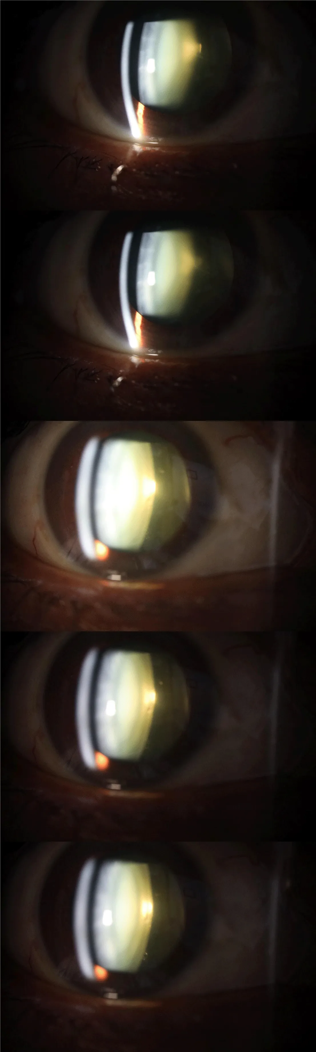
Figure 2) In relation to the left eye, the photographs also show nuclear sclerosis, zonular opacities, and the thickening of the central region of the posterior capsule of the lens. The vitreous body, in its anterior part, is observed with increased density.
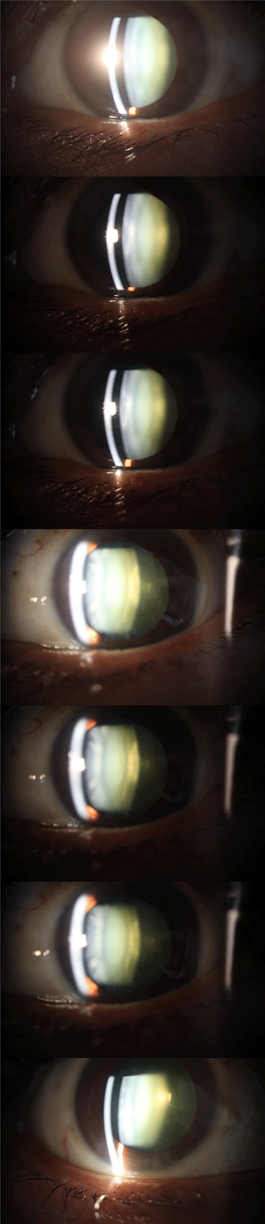
Figure 3) Photographs of the left fundus show arteriovenous tortuosity, moderate venous congestion, and an excavation of the optic nerve of less than 30%. The macular region is seen.
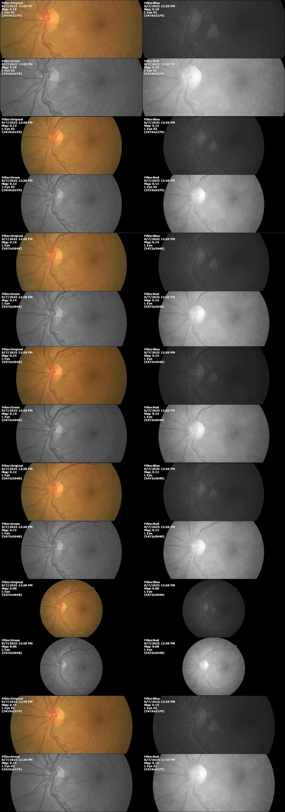
Figure 4) The photographs focusing on the vitreous body show a condensation in the lower right of the image.
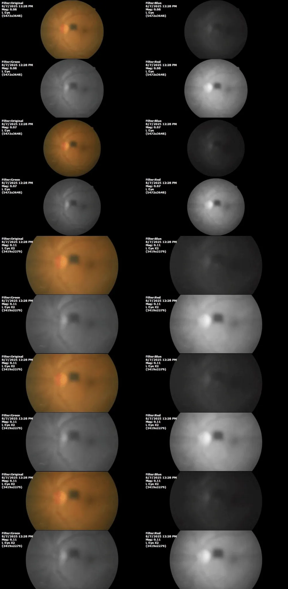
Figure 5) The anterior segment of the left eye does not show significant alterations. Even specular reflection indicates that alterations in the transparency of the lens are not significantly affecting the patient's vision.
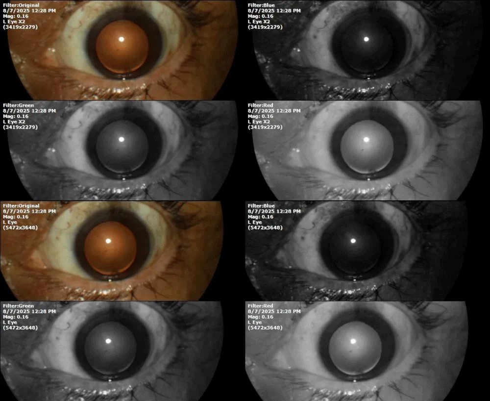
Figure 6) The anterior segment of the right eye does not show important pathological data, and specular reflection indicates that the transparency of the lens is acceptable, despite the changes seen in Figure 1.
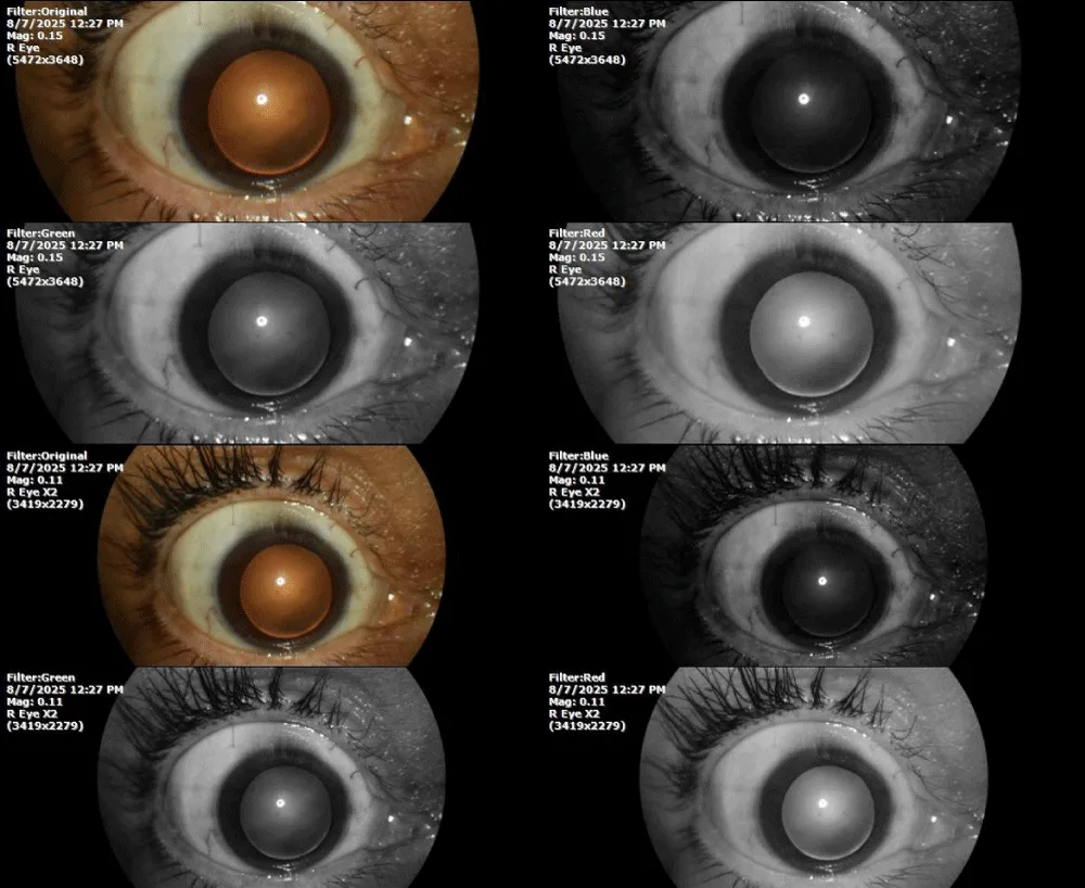
Figure 7) The images correspond to the vitreous body of the right eye, but do not show relevant condensations
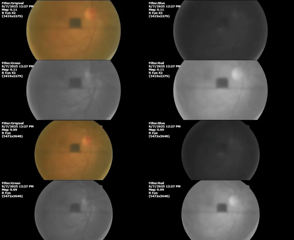
Figure 8) In the retina of the right eye, moderate elevation of the macular umbo is observed, as well as moderate venous congestion.
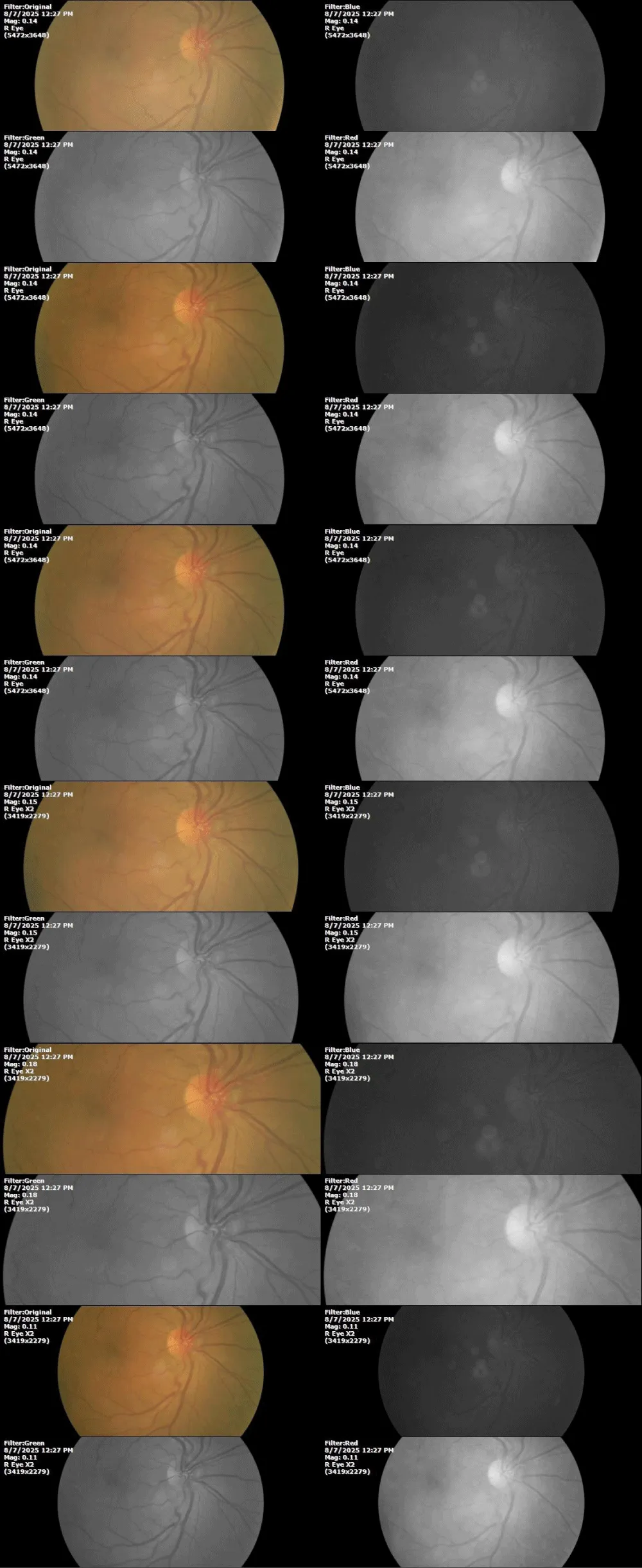
Figure 9) In these images of the retina of the right eye, premacular gliosis is seen as a whitish area that extends into the temporal region, affecting the temporal zone of the macular region.
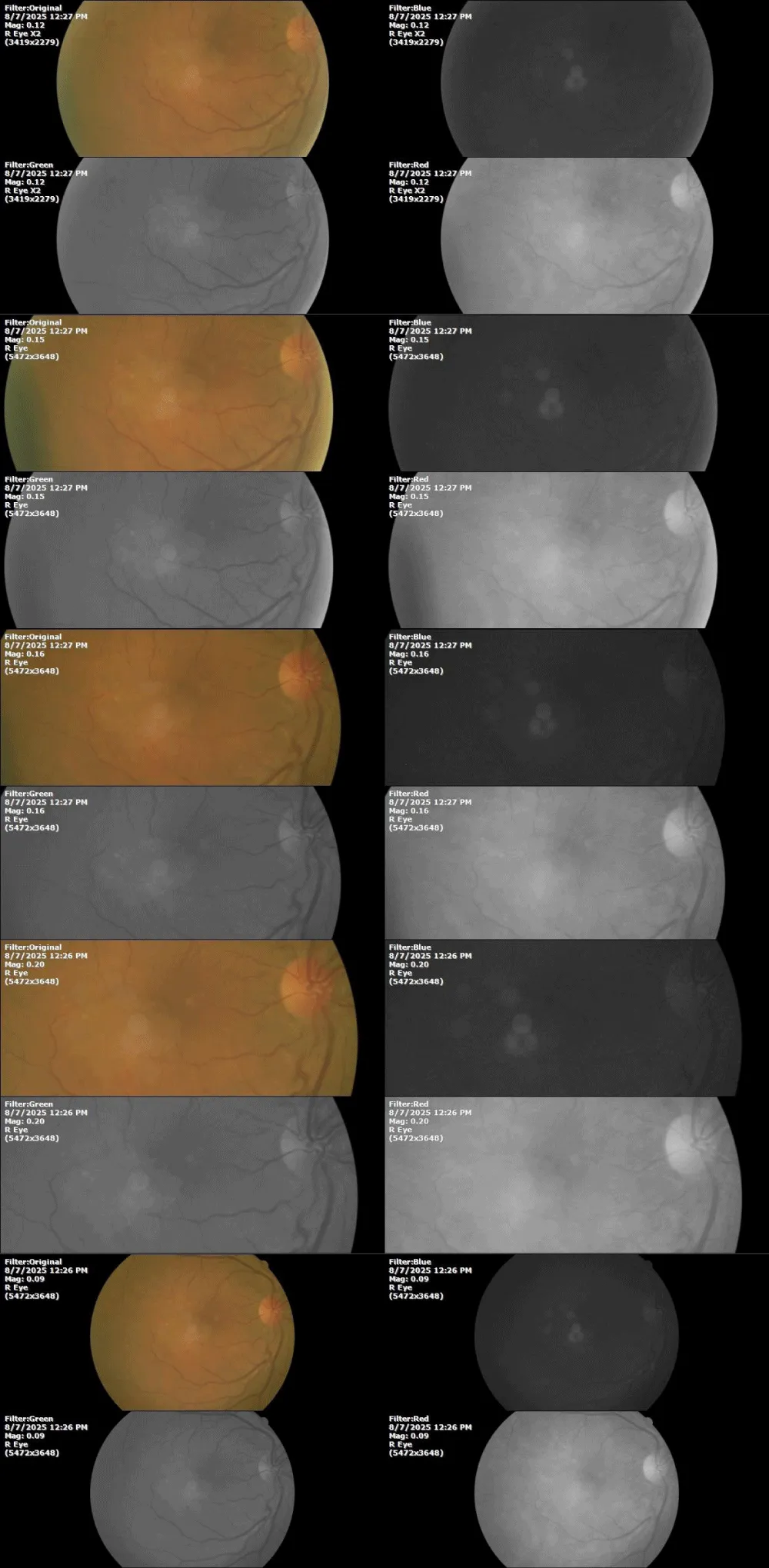
The diagnosis was: Idiopathic preretinal gliosis, in the right eye.
Once the patient explained our therapeutic approach to oxygen balance, and informed consent had been signed, she was prescribed QIAPI 1®, sublingual drops, at the rate of three sublingual drops every hour, for the entire time she was awake.
February 7, 2026.
8 days ago, she felt very bad and anxious; then, the family took her to the clinic.
Figure 10) The prescription of the doctor who treated her, indicating just antibiotics and anti-inflammatories.

During our examination, we found the following clinical parameters:
% SpO2: 94 %
Heartbeat: 71 x´
Objective retinoscopy: -/+
Healthy ocular fundus.
Figure 11) Photograph of the anterior segment of the Right eye. No significant changes were found, beyond nuclear sclerosis of the lens, to a moderate degree.
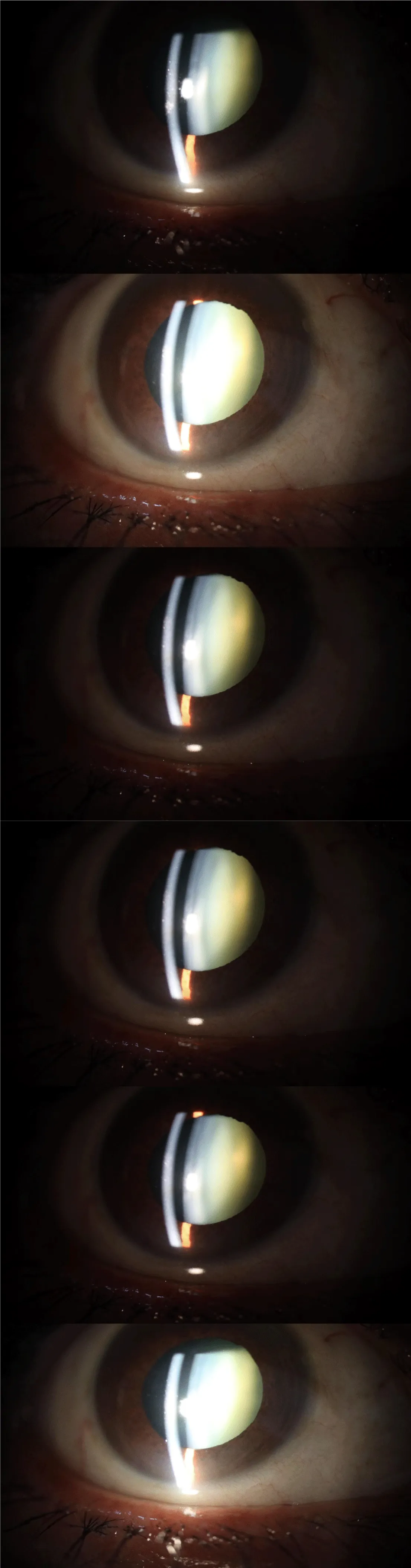
Figure 12) The photographs correspond to the anterior segment of the left eye. Nor does it present significant changes in relation to the previous biomicroscopic examination.
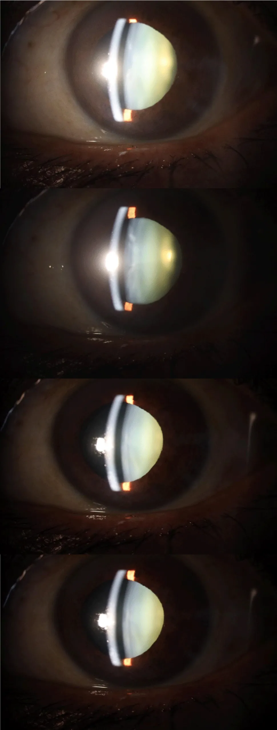
Figure 13) The transparency of the cornea, lens and vitreous body, in this case the left eye, is within normal limits.
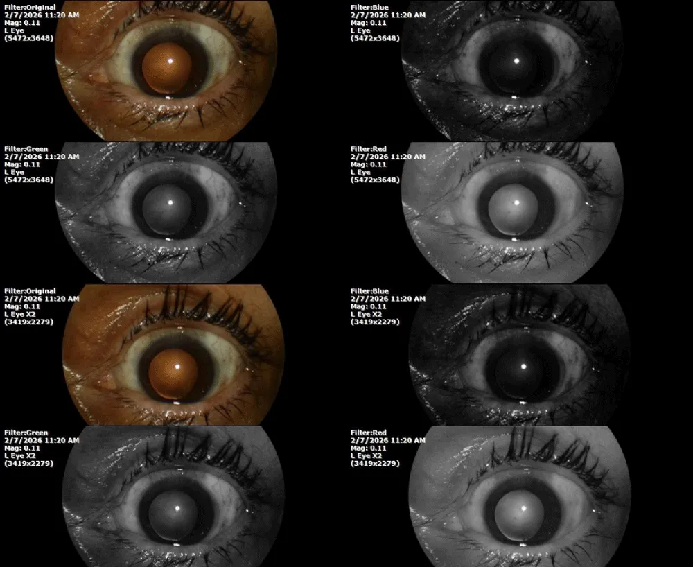
Figure 14) The two photographs correspond to the observation of the vitreous body of the left eye, where a glassy condensation is observed in the lower left quadrant, perhaps a little smaller than in the previous examination.
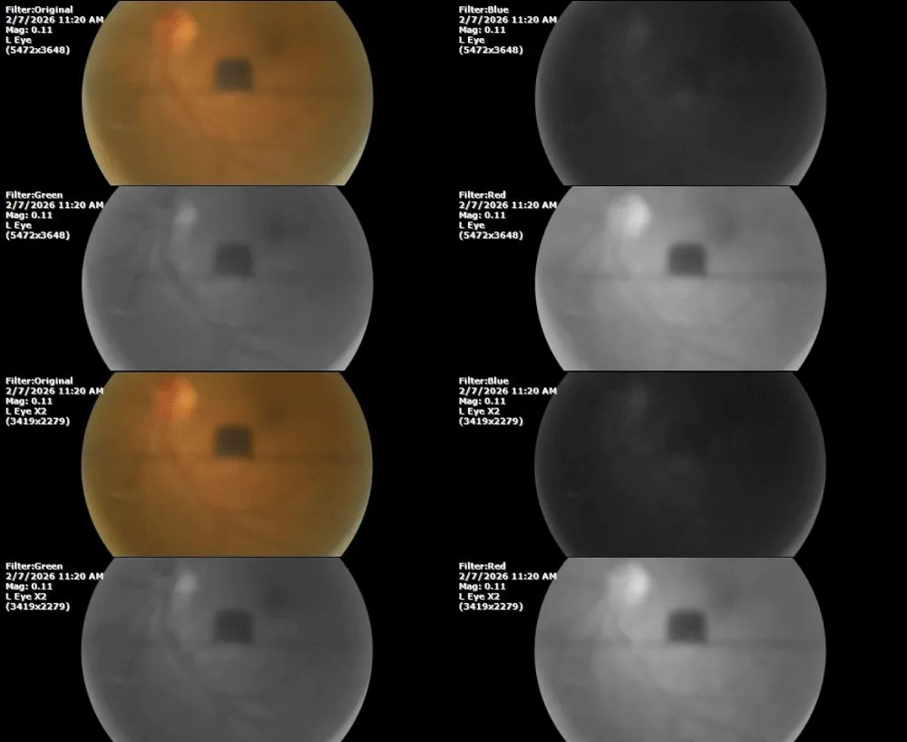
Figure 15) Fundus images of the left eye, taken with different filters, show preserved macula anatomy, physiological excavation (> 30%), choroidal vascularity with good density, and mild to moderate arteriovenous tortuosity. There are no areas of bleeding or exudate.
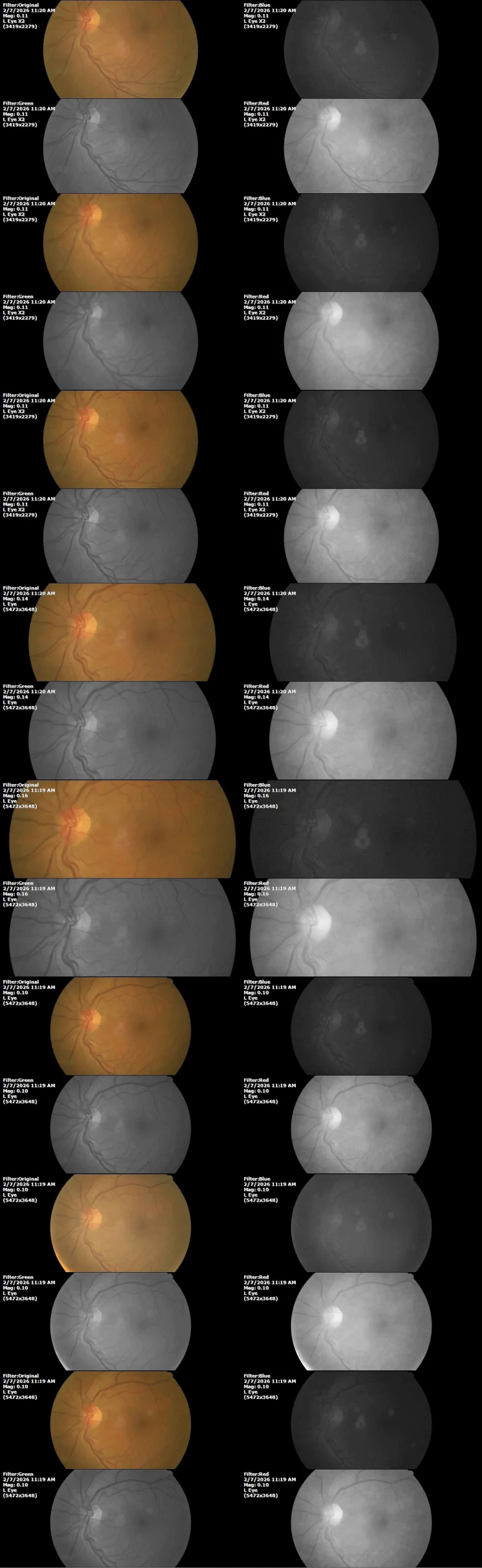
Figure 16) The photographs correspond to the fundus of the eye on the right side, where a clear decrease in the vitreous-retinal condensation zone located in the temporal region of the macula is appreciated, including the temporal region of the clivus and umbo macular, they are recovering their normal morphological characteristics, which results in a better visual function.
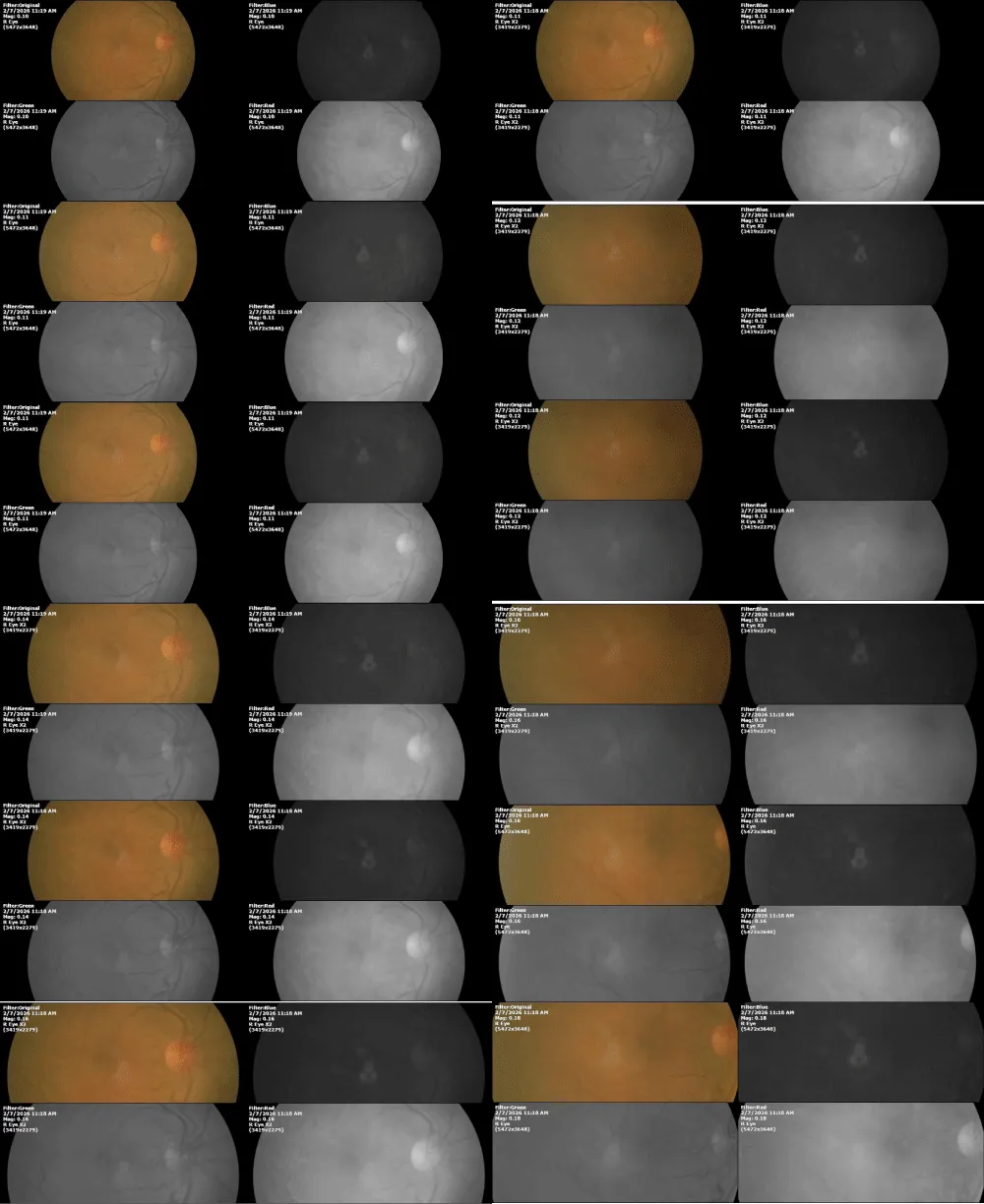
Figure 17) Photographs that correspond to the vitreous body of the right eye. There is no condensation or floating bodies or opercula
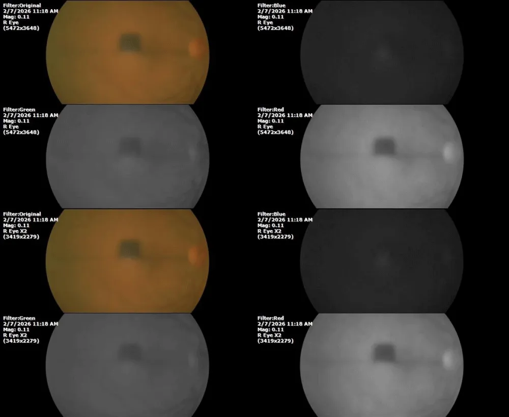
Figure 18) The transparency of the cornea, lens, and vitreous body, dimensioned through specular reflection and recorded through the fundus chamber, is appreciated within acceptable, functional limits.
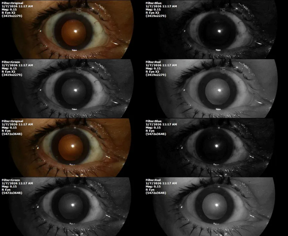
Comment: The diagnosis of this examination was pre-retinal gliosis of the right eye, which is improving according to ophthalmological images. The patient is instructed to maintain the treatment and is scheduled in six months (July 2026).
Date: July 4, 2026.
The patient came in referring to some problems with vision. Sometimes glasses work for her, and sometimes they don’t, because sometimes she sees better without glasses.
On the general examination, the following values were recorded:
% SpO2: 92 %
Heartbeat: 26 x ´
Objective retinoscopy: +- / +-.
Figure 19) Photographs through the bio-microscope of the left eye show no significant changes. Nuclear sclerosis of the lens is mild to moderate.
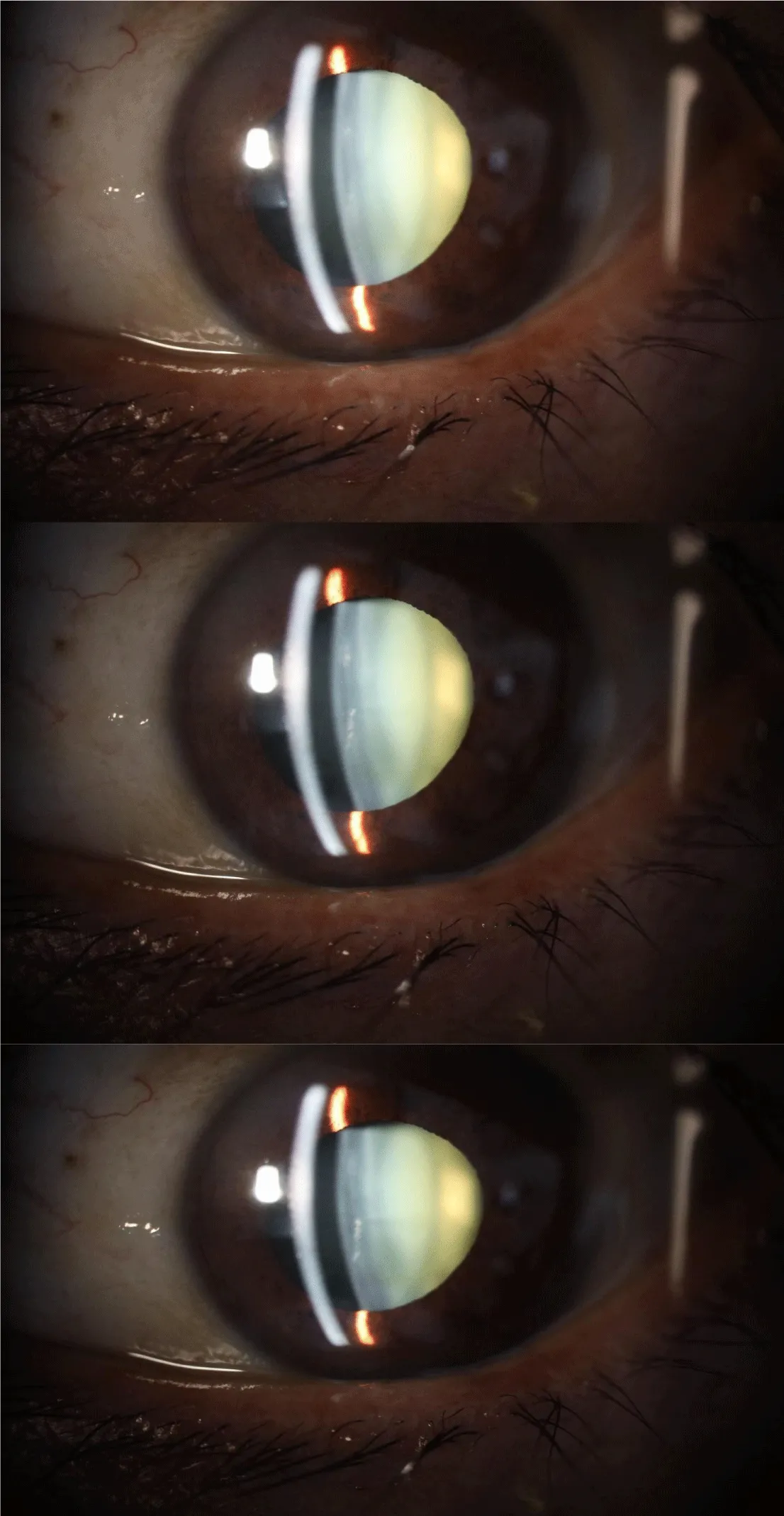
Figure 20) Bio-microscopy of the right eye does not detect major changes. Nuclear sclerosis of the lens is only slightly more pronounced than sclerosis of the left eye.
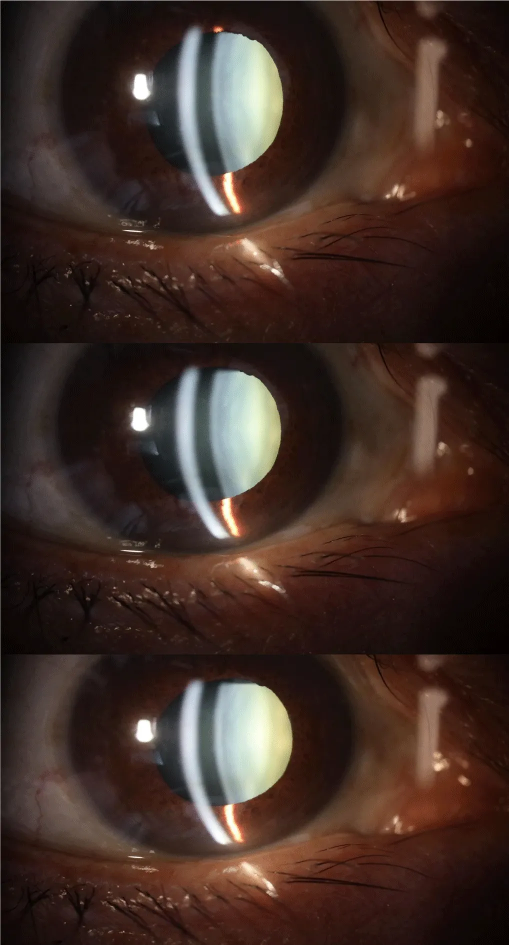
Figure 21) The evaluation of the transparency of the cornea, lens and vitreous body of the left eye, carried out with the fundus camera, shows a very acceptable, very functional transparency.
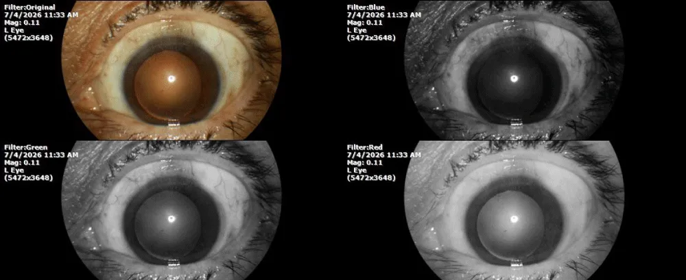
Figure 22) Images of the posterior pole of the right eye show normal macular architecture; the excavation of the optic nerve is within normal (> 30%). Arteriovenous tortuosity of retinal vessels is mild to moderate. The choroidal vascularity is dense, indicating good tissue perfusion.
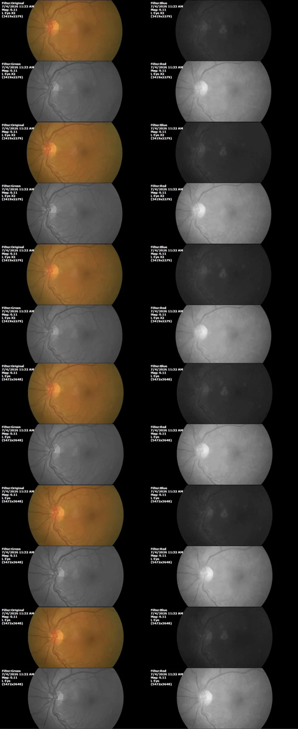
Figure 23) Examination of the posterior pole of the right eye shows a decrease in the vitreoretinal condensation zone, indicating that the contraction processes of the posterior hyaloid membrane continue to improve. The choroidal vascularity is dense.
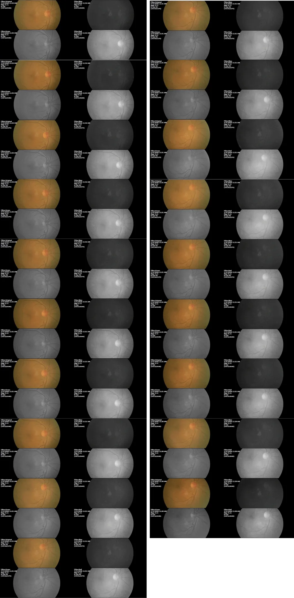
Figure 24) The transparency of the cornea, crystalline lens, and vitreous body of the right eye, sized by means of the fundus camera, demonstrates a very acceptable functional transparency.
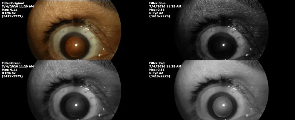
Figure 25) The transparency of the cornea, lens, and vitreous body of the left eye is very acceptable.
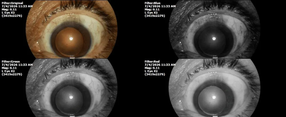
By our standards, the patient's evolution is good, very good, since the vitreoretinal condensation zone of the right eye continues to decrease with the treatment instituted.
The patient tells us that her relatives insisted that she be evaluated by another doctor to evaluate the evolution of the case, and the results of the tests performed in another city are shown below:
Figure 26) The visual fields of the right eye, carried out by computer, show an absolute concentric defect, very peripheral. But it does not suggest any pathology, and may only be an examination artefact, given the relative subjectivity of the study of visual fields.
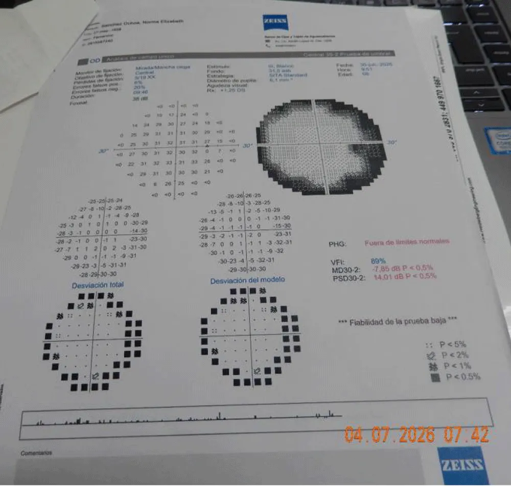
Figure 27) The result of the computerised perimetric study of the visual field of the left eye. Nor does it show data suggestive of pathology, except for the peripheral area, but this can be explained by the fact that the study was carried out without drug mydriasis in both eyes.
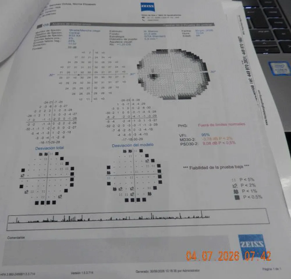
Figure 28) The study of the retinal nerve fibre layer (RNFL) of both eyes was found to be within normal limits, compatible with good visual function.
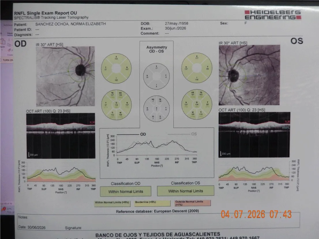
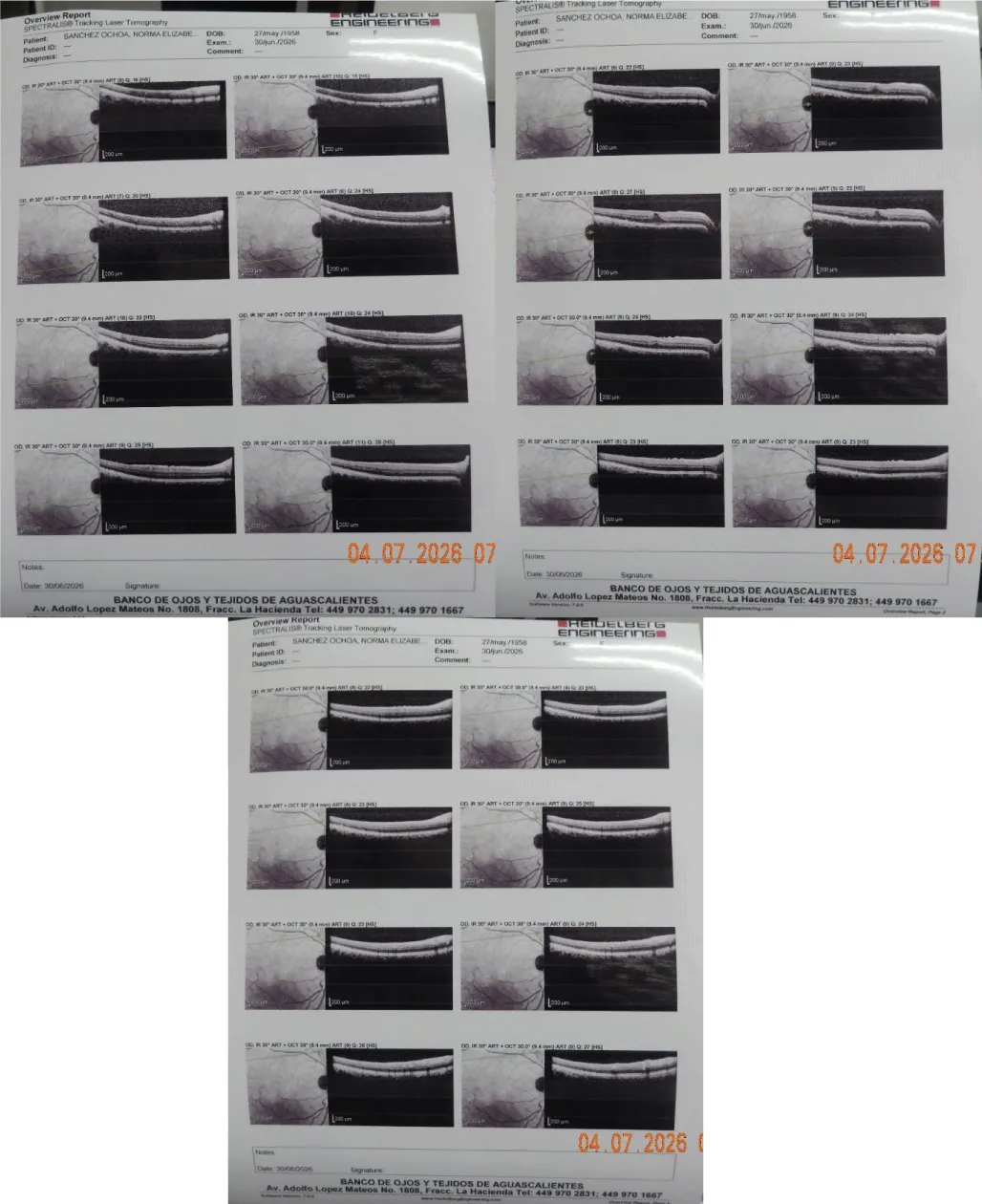
Figure 29) The optical coherence tomography of the macular region of the optic nerve of both eyes is almost normal, although we must consider that they are computer-generated images.
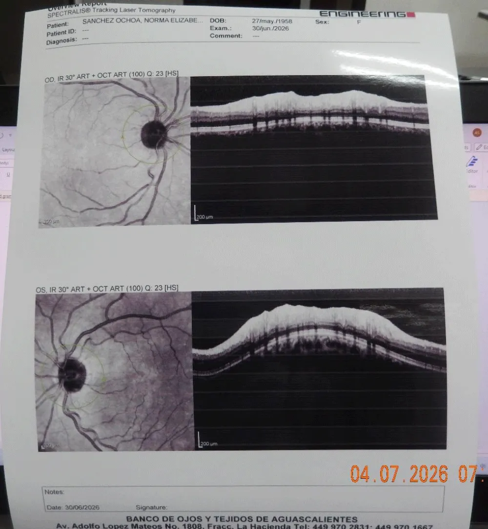
Figure 30) The optical coherence tomography of the right eye, which included the macular umbo, shows a slight elevation, compatible with the traction generated in this tissue by the area of vitreoretinal condensation, but which fortunately has responded to treatment.
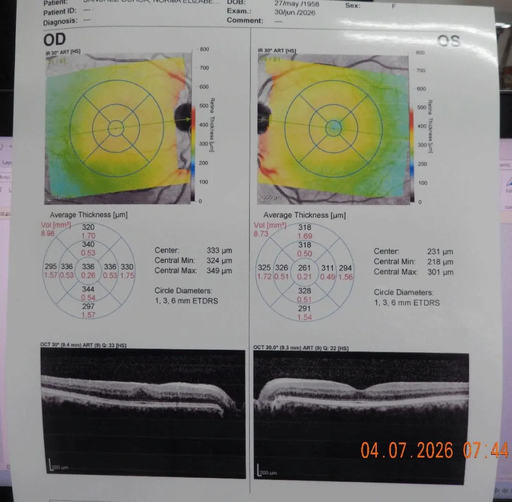
Figure 31) Optical coherence tomography of the macular region of the right eye, which shows minimal traction of the macular umbo, but which retains its internal structure, which is compatible with a good prognosis, given that the condensation zone of the vitreous and retina has been significantly reduced with our therapeutic approach.
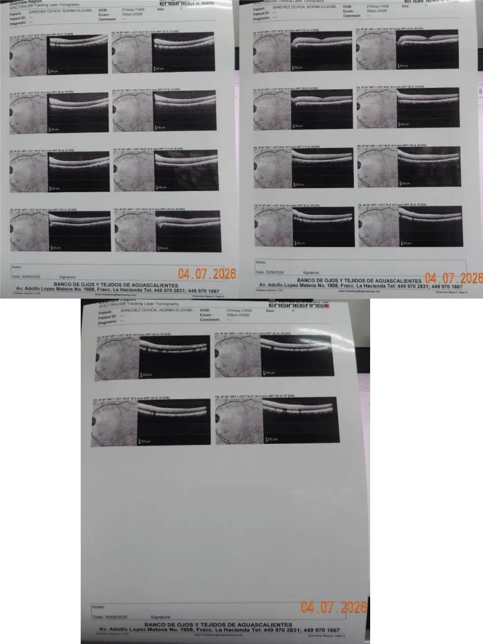
The macular region of the left eye shows an anatomy of the clivus and umbo, which is more conserved in this study, compared to the right eye (Figure 31).
Figure 33) Bilirubin, alkaline phosphatase, and lactic dehydrogenase (DHL), within normal limits.

Figure 34) The white formula and the red formula of blood cytology are within normal limits.

Figure 35) C-reactive protein and rheumatoid factors negative, general urinalysis within normal limits; and slightly elevated cholesterol.
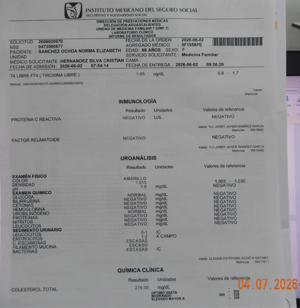
Figure 36) Glucose, triglycerides, serum creatinine, high-density cholesterol, urine albumin, aspartate aminotransferase, and alanine aminotransferase were normal.

Figure 37) The bone densitometry study of the spine was acceptable according to the age of the patient.
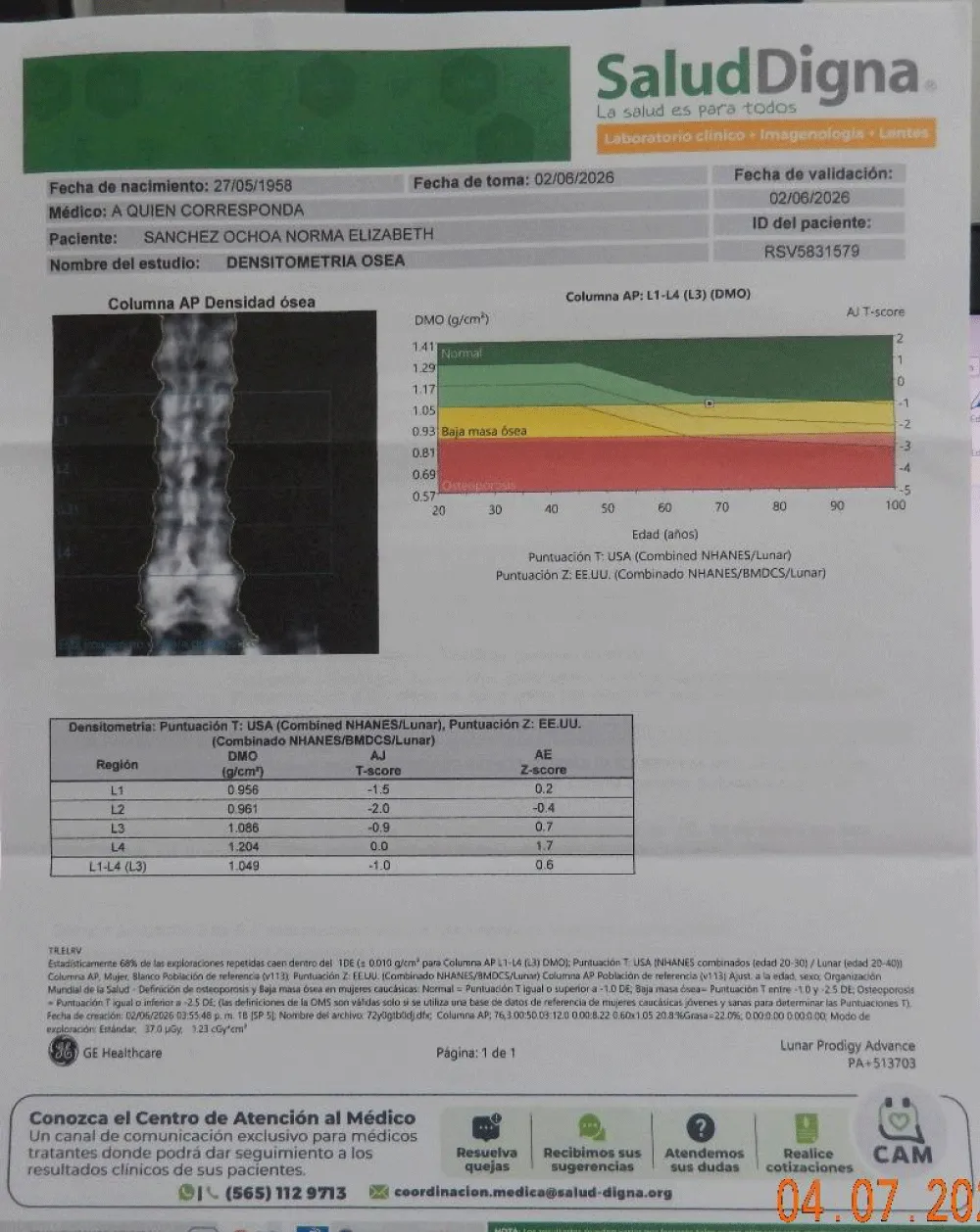
Figure 38) The evaluation of bone densitometry of the coxofemoral joint is within low limits, according to the age of the patient.
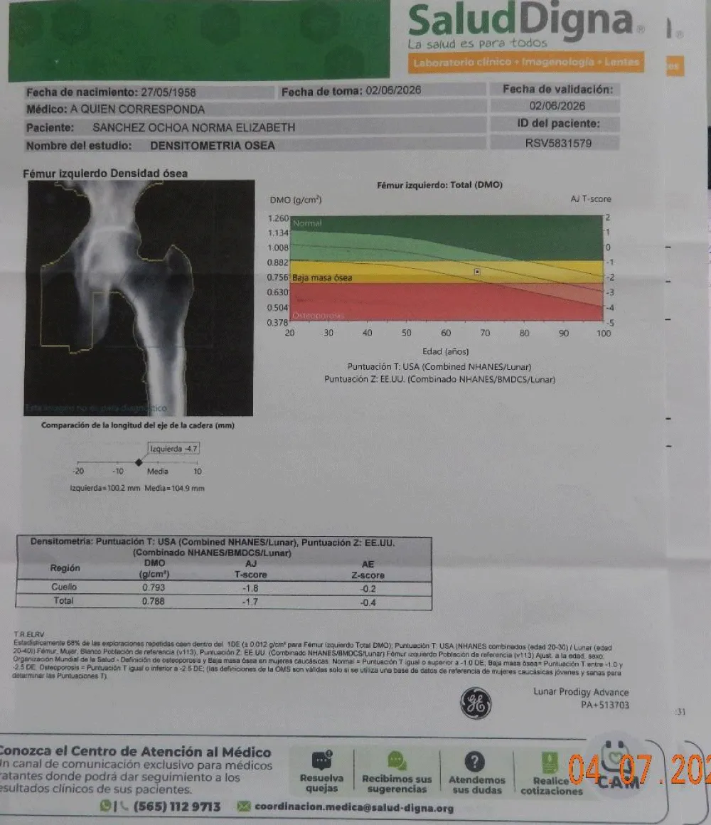
Figure 39) Bone mineral density (BMD) is reported to be low, but it is in accordance with the age of the patient.
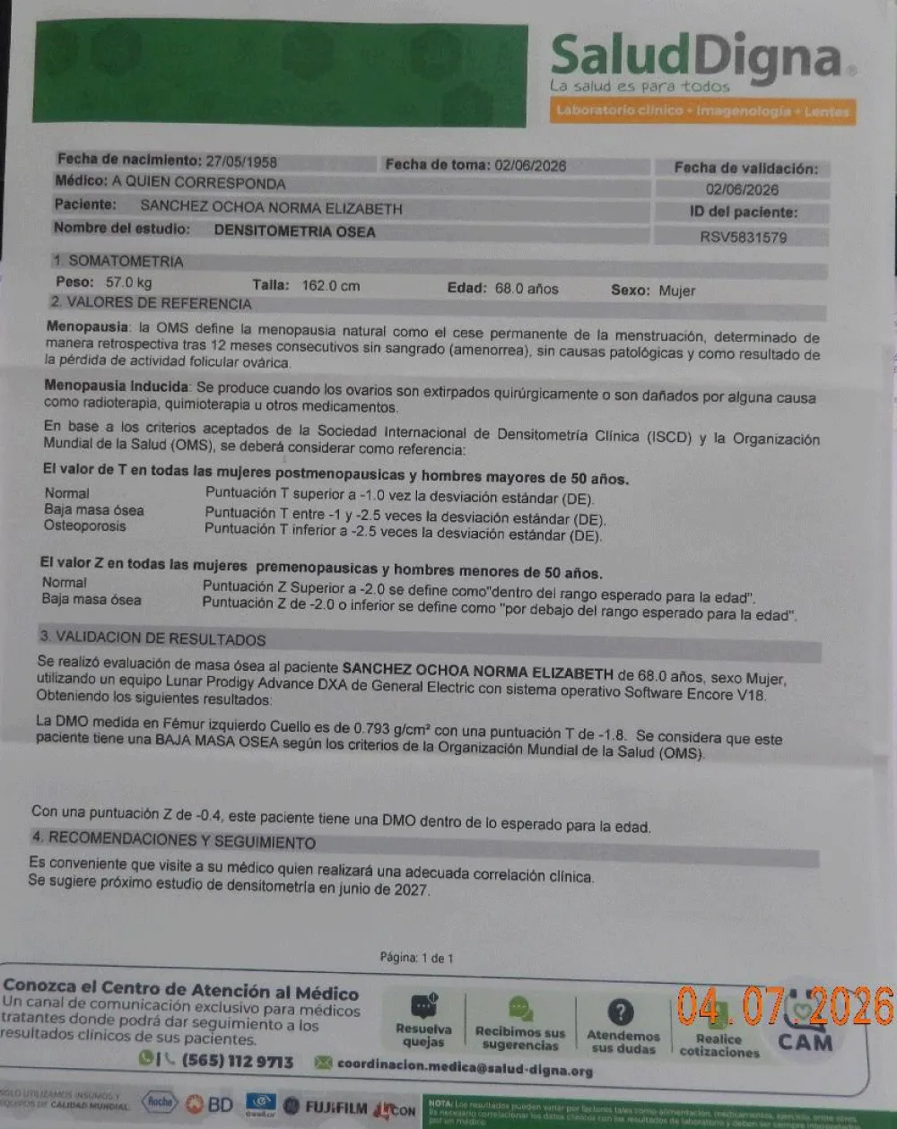
Figure 40) In coagulation studies, partial thromboplastin and prothrombin times were within normal limits.
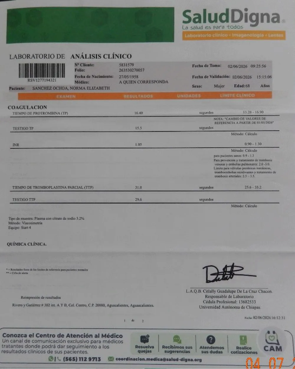
Figure 41) The determination of electrolytes in the blood yielded results within normal parameters.
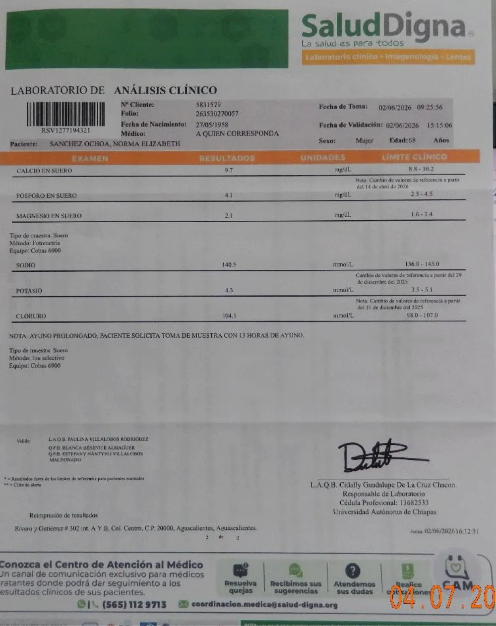
Figure 42) The eye professional's report confirms the patient's condition, and advises continuing with the treatment instituted by us (QIAPI 1®).
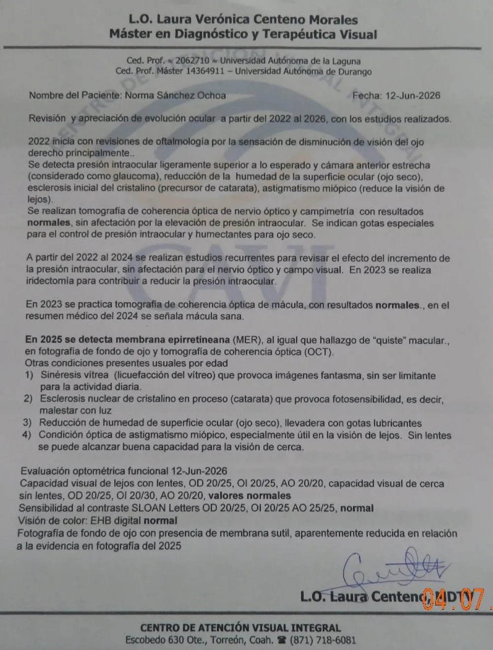
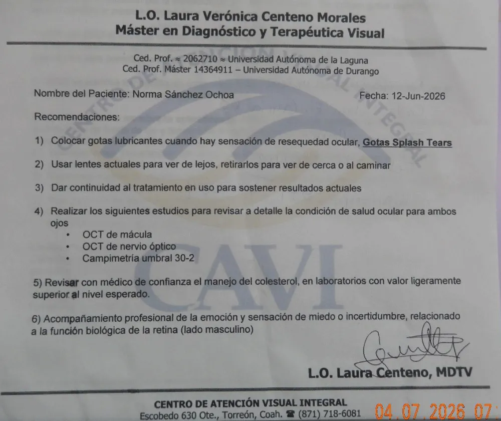
Figure 43) General recommendations of the eye care professional.
Comment: The involution of the epiretinal membrane in this patient is significant and is explained by the re-establishment of the balance between intracellular generation and oxygen consumption, so its effect is sufficiently extensive and profound to restore the complex biochemical and histological balances that make up the normal functionality of a tissue, but all of them depend for the most part on the balance between generation (intracellular) and consumption (intracellular) of oxygen. The part attached between the vitreous and the retina is the one that takes the longest to recover its normal functionality, since the histochemical alterations seem to be more intense in this vitreoretinal junction, normally less strong and extensive in that area, as it is usually only strong enough to preserve the anatomy of the region, but at the same time flexible enough to tolerate the intense normal movement of the eyeballs, with its consequent differential inertia between the different tissues, to allow normal function for decades or even the lifespan of the individual.
Conclusion
Our observation about the unsuspected intrinsic property of human eukaryotic cells to transform the power of sunlight into chemical energy susceptible to be used by the same cell, through the dissociation of the intracellular water molecules, like in plants, constitutes a new paradigm that allows us the development of new treatments for this chronic condition. The observational-analytical research that allowed us to detect that human eukaryotic cells are capable of producing their own oxygen, began in 1990 and ended in 2002, the ophthalmological records of 6000 patients were included, and to date it has gradually gained acceptance, and like all new knowledge, it was very attacked at the beginning, and this has happened with other discoveries throughout history (Galileo Galilei, Copernicus, etc.).
But defining where the oxygen present inside each one of the cells that make us up comes from is a disruptive discovery.
Acknowledgements
This work was supported by an unrestricted grant from Human Photosynthesis® Research Centre, C. S., located in Aguascalientes 20000, México.
Conflict of interest: The finding of the unsuspected intrinsic property of molecules placed inside the human eukaryotic cells, to dissociate the water molecule and the development of QIAPI 1®, was made at our clinical facilities.
- Epiretinal Membranes - Patients - The American Society of Retina Specialists. Retrieved June 2026.
- Ames A III. Energy requirements of CNS cells as related to their function and to their vulnerability to ischemia: a commentary based on studies on retina. Can J Physiol Pharmacol 1992; 70: S158–S164.
- Kaur C, Foulds WS, Ling EA. Hypoxia ischemia and retinal ganglion cell damage. Clin Ophthalmol 2008; 2: 879– 889
- Fortmann SD and Grant MB. Molecular mechanisms of retinal ischemia. Curr Opin Physiol 2019; 7: 41–48.
- Kiel JW. Anatomy. In: The ocular circulation. Morgan & Claypool Life Sciences, https://www.ncbi.nlm.nih.gov/books/NBK53329/
- Luo L, Uehara H, Zhang X, et al. Photoreceptor avascular privilege is shielded by soluble VEGF receptor-1. eLife 2013; 2: e00324.
- Wangsa-Wirawan ND and Linsenmeier RA. Retinal oxygen: fundamental and clinical aspects. Arch Ophthalmol 2003; 121: 547–557
- Santos GSP, Prazeres PHDM, Mintz A, et al. Role of pericytes in the retina. Eye 2018; 32: 483–486.
- Alarcon-Martinez L, Villafranca-Baughman D, Quintero H, et al. Interpericyte tunnelling nanotubes regulate neurovascular coupling. Nature 2020; 585: 91–95.
- Ferland-McCollough D, Slater S, Richard J, et al. Pericytes, an overlooked player in vascular pathobiology. Pharmacol Ther 2017; 171: 30–42.
- Alarcon-Martinez L, Shiga Y, Villafranca Baughman D, et al. Pericyte dysfunction and loss of interpericyte tunnelling nanotubes promote neurovascular deficits in glaucoma. Proc Natl Acad Sci U S A 2022; 119: e2110329119.
- Joyal J-S, Sun Y, Gantner ML, et al. Retinal lipid and glucose metabolism dictates angiogenesis through the lipid sensor Ffar1. Nat Med 2016; 22: 439–445.
- Fortmann SD and Grant MB. Molecular mechanisms of retinal ischemia. Curr Opin Physiol 2019; 7: 41–48.
- Lejoyeux R, Benillouche J, Ong J, et al. Choriocapillaris: fundamentals and advancements. Prog Retin Eye Res 2022; 87: 100997.
- Herrera, AS., and Arias Esparza, Md., 2025. The unsuspected ability of the human cell to oxygenate itself. Applications in cell biology. Medical Research Archives, [online] 13(3). Available from: https://doi.org/10.18103/mra.v13i3.6344
- Kaur C, Sivakumar V and Foulds WS. Early response of neurons and glial cells to hypoxia in the retina. Invest Ophthalmol Vis Sci 2006; 47: 1126–1141.
- Phipps JA, Dixon MA, Jobling AI, et al. The renin-angiotensin system and the retinal neurovascular unit: a role in vascular regulation and disease. Exp Eye Res 2019; 187: 107753.
- Iyer NV, Kotch LE, Agani F, et al. Cellular and developmental control of O2 homeostasis by hypoxia-inducible factor 1α. Genes Dev 1998; 12: 149–162.
- Country MW. Retinal metabolism: a comparative look at energetics in the retina. Brain Res 2017; 1672: 50–57.
- Kaur C, Sivakumar V and Foulds WS. Early response of neurons and glial cells to hypoxia in the retina. Invest Ophthalmol Vis Sci 2006; 47: 1126–1141
- Marmor MF. Mechanisms of fluid accumulation in retinal oedema. Doc Ophthalmol 1999; 97: 239–249.
- Mrugacz M, Bryl A and Zorena K. Retinal vascular endothelial cell dysfunction and neuroretinal degeneration in diabetic patients. Journal of Clinical Medicine 2021; 10: 458.
- Petersson J, Glenny RW. Gas Exchange in the Lung. Seminars in Respiratory and Critical Care Medicine. 2023 Oct;44(5):555-568. Available from: https://dx.doi.org/10.1055/s-0043-1770060
- Occhipinti R, Boron WF. Role of Carbonic Anhydrases and Inhibitors in Acid-Base Physiology: Insights from Mathematical Modelling. Int J Mol Sci. 2019 Aug 6;20(15):3841. Available from: https://dx.doi.org/10.3390/ijms20153841
- Subczynski WK, Widomska J, Stein N, Swartz HM. Factors determining barrier properties to oxygen transport across model and cell plasma membranes based on EPR spin-label oximetry. Appl Magn Reson. 2021 Oct;52(10):1237-1260. Available from: https://dx.doi.org/10.1007/s00723-021-01412-4
- Subczynski WK and Swartz HM, in Vol. 23. Biomedical ESR – Part A: Free Radicals, Metals, Medicine, and Physiology, edited by Eaton SS, Eaton GR, and Berliner LJ (Kluwer, Boston, 2005), pp. 229–282
- Subczynski WK, Pasenkiewicz-Gierula M, • Widomska Justyna, Mainali L, and • Raguz Marija, Cell Biochemistry and Biophysics 75, 369 (2017)
- Krogh A, Krogh M. On the rate of diffusion of carbonic oxide into the lungs of man. Skandinavisches Archiv Für Physiologie. 1910;23:236–247.
- Haldane J, Smith JL. The absorption of oxygen by the lungs. J Physiol (London) 1897;22:231–258. Available from: https://dx.doi.org/10.1113/jphysiol.1897.sp000689.



 Save to Mendeley
Article Alerts
Subscribe to our articles alerts and stay tuned.
 This work is licensed under a Creative Commons Attribution 4.0 International License.