Imaging Journal of Clinical and Medical Sciences
Leucocrit-The white face of acute myeloid leukemia
Tomas V Karajan*
Cite this as
Karajan TV (2021) Leucocrit-The white face of acute myeloid leukemia. Imaging J Clin Medical Sci 8(1): 002-002. DOI: 10.17352/2455-8702.000132CASE / Clinical Image
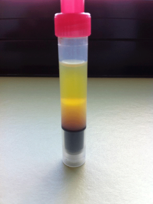
Of a 72-year-old patient who was brought to our emergency room with fever and worsening condition for several weeks. Initial blood count showed a hemoglobin of 48g/L with a hematocrit of 0.14. White blood count was 350G/L and showed severe leukocytosis. Additionally we found thrombocytopenia at 49G/L. As you can see in the image (her blood, after staying 30 minutes in a edta tube) there is a massive widening to 0.32 (normally up to 0.01) of the «leukocrite» (the white part of the pillar) after sedimentation .
The patient was later diagnosed with acute myeloid leukemia and unfortunately couldn`t be saved.
Article Alerts
Subscribe to our articles alerts and stay tuned.
 This work is licensed under a Creative Commons Attribution 4.0 International License.


 Save to Mendeley
