Imaging Journal of Clinical and Medical Sciences
Leiomyomatosis Peritonealis Dissiminata
Hanne Christensen*
Cite this as
Christensen H (2019) Leiomyomatosis Peritonealis Dissiminata. Imaging J Clin Medical Sci 6(1): 086-086. DOI: 10.17352/2455-8702.000125The leiomyomatosis peritonealis dissiminata (LPD) a histologically benign condition, but it can residivere and be metastatic. Since the macroscopically picture imitates diffuse peritoneal carsinosis, the risk of unnecessary drastic precautions are nearby.
Clinical Picture
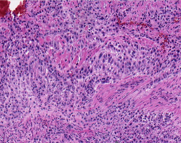
LPP is a multifocal tumor with a macroscopic image of multiple fibroids and subperitoneal nodules varying in size from 0.1 to 0.3 cm, histologically constructed of smooth muscle cells with fibroblasts. It is believed that Noduli is formed by metaplasic transformation of submesothelial stroma derived from the mulleric ductuli, induced and activated by changes in the body's oestrogen and/or progesteron ballance.
The LDP is usually subclinical and discovered incidentally in women, even in pregnancy and rarely in postmenopausal patients. The diagnosis is made peroperatively unexpected in laparascopic examinations, hysterectomies, sectio casesarean or by autopsy. Surgical treatment of LPD is indicated only by subjective symptoms as most fibroids of LPD will disappear by themselves.
Metastatic properties with the proliferation of uterine fibroids via the lymphatic and vascular system have been discussed, but are contradicted by the fact that LPD is noted in women who are hysterectomated earlier. Malignancies development is never described.
Conclusion
Dissiminated peritoneal leiomyomatosis is usually discovered incidentally and symptoms are unusual. The lesions are completely benign and apparently undergo spontaneus regression, therefore minimal or no treatment is indicated after the diagnosis is confirmed by biopsy (Figure 1).
Article Alerts
Subscribe to our articles alerts and stay tuned.
 This work is licensed under a Creative Commons Attribution 4.0 International License.


 Save to Mendeley
