Imaging Journal of Clinical and Medical Sciences
Evaluation of the diagnostic efficacies of Doppler Ultrasonography, time-resolved contrasted Magnetic Resonance Angiography and Digital Subtraction Angiography in comparison to Rotational Digital Subtraction Angiography in Carotid Artery Stenosis
Aysegul Altunkas*
Cite this as
Altunkas A (2018) Evaluation of the diagnostic efficacies of Doppler Ultrasonography, time-resolved contrasted Magnetic Resonance Angiography and Digital Subtraction Angiography in comparison to Rotational Digital Subtraction Angiography in Carotid Artery Stenosis. Imaging J Clin Medical Sci 5(1): 016-022. DOI: 10.17352/2455-8702.000039Purpose: In this study, rotational (3 dimentional) digital subtraction angiography being taken as a reference, the sensitivity, specificity, positive and negative predictive values, and diagnostic accuracy of Doppler ultrasonography, and time-resolved imaging of contrast kinetics MR angiography, and lateral plane digital subtraction angiography were used in the detection of internal carotid artery stenosis.
Materials and Methods: With Doppler US, TRICKS MRA and DSA, stenosis rates were evaluated in 39 internal carotid arteries of 22 patients. Stenosis measurements were performed by two independent radiologists in TRICKS MRA and DSA. Stenosis grades were calculated both in the DSA and TRICKS MRA by both lateral and sagittal plan and area measurements both subjectively and objectively. In Doppler US, the rate of stenosis according to velocity, diameter and area was measured by another radiologist.
Results: Sensitivity, specificity, and diagnostic accuracy rates according to the velocity were respectively 100%, 96.4%, 97.4%, according to diamater respectively 72%, 92%, 87.2%, and according to the area, resoectively 90.9%, 85.7%, and 87.2% in the detection of stenosis in Doppler US. In the lateral plan DSA, 72.7%, 100% and 92.3%, respectively, were found. Sagittal and areal TRICKS MRA measurements were 72.7% and 81.8%, 96.4% and 92.8%, 89.7% and 89.7%, respectively.
Conclusion: Doppler US is the most important diagnostic modality of PSV and it is a reliable parameter to evaluate carotid artery stenosis. TRICKS MRA can be used as a safe alternative diagnostic method to DSA by evaluating all segments of carotid arteries in noninvasive and nonionizing form with high diagnostic accuracy rates in all parameters in carotid stenosis.
Introduction
Atherosclerosis accounts for about 90% of the cerebral thromboembolic cases, which are the 3rd leading cause of stroke after cardiac diseases and cancer in developed countries, among all causes of death [1]. Atherosclerosis is a multifactorial disease affecting the carotid, femoral, and coronary arteries primarily. Among these, the prevalence of carotid atherosclerosis is reported to be 41-59% in various studies [2]. In addition, it is reported that 22-40% of all stroke cases are associated with the diseases of large arteries [3]. Although conventional cerebral angiography is accepted to be the “golden standard” in evaluating the carotid arteries and vertebrobasilar system, it bears around a 1-4% risk for neurological deficits and mortality, as well as, being invasive and expensive. Because the technique is far from being a screening method for those reasons, non-invasive or minimally invasive imaging techniques like Doppler ultrasonography (US), magnetic resonance angiography (MRA), and computed tomography angiography (CTA) are preferred more commonly [4].
Looking from this point of view and accepting the rotational 3 dimensional (3D) DSA images on the axial plane as the golden standard, the diagnostic efficacy of Doppler US (stenosis rates according to velocity, diameter, and areameasurements), TRICKS MRA (area and diameter), and the conventional DSA (lateral plane) were evaluated with the NASCET method in making the diagnosis of carotid artery stenosis in patients with the pre-diagnosis of carotid artery stenosis and cerebrovascular disease.
Material and Methods
Following the approval of the ethics committee (GOU.0.01.00.00/183), 15 females and 7 males with a pre-diagnosis of carotid stenosis and cerebrovascular disease, making a total of 22 patients, tested consecutively with Doppler US, TRICKS MRA, and DSA with relatively shorter intervals in between, were included in the study to be evaluated retrospectively. In five patients who had previously undergone unilateral ICA stenting, the ICAs with stents were excluded from the study. Using a linear 10 MHz probe (A.A.), Doppler US (Logic 7, GE Medical Systems and Milwaukee-USA) was performed in all study patients by the same radiologist. The area and diameter measurements were taken from the narrowest segment of ICA and the PSV (Peak Systolic Velocity) data were obtained from the points immediately distal to this segment. Then, for adaptation into North American Symptomatic Carotid Endarterectomy Trial (NASCET), the diameter and the areadata were collected from the most distal segments of the internal carotid arteries, provided that the segment does not contain any lesions. The rate of stenosis measured in Doppler US was determined based on the 2003 criteria established by the Consensus Panel of the Society of Radiologists in Ultrasound [5].
The MRA investigations of all study patients were performed with the 1.5 T device (Signa excite HD; GE Healthcare, Milwaukee, WI, USA, 2005). After injecting the contrast agent, the coronal plane images covering the area extending from the aortic arch to the circle of Willis were obtained with a 3-dimensional (3D) TRICKS sequence (TR: 4.1, TE: 1.6, NEX: 0.75, FA: 35°, effective sectional thickness 1 mm, FOV: 28x20 cm, matrix: 320x224, bandwidth: 62.5, voxel volume: 1.1x1.3x1.0 mm). In this investigation, the slab thickness was adjusted to completely cover the carotid and vertebral arteries, so that the number of slices ranged from 60to 70 while the imaging time ranged from 1.10 to 1.45 seconds. The elliptic-centric method was used for K-space encoding. After injecting a 0.1 mmol/kg contrast agent (Omniscan-GE Healthcare) into the antecubital vein with an automatic injector (Nemoto Sonic Shot, Tokyo- Japan) and a 22 G cannula and at a velocity of 1.5 ml/s, the catheter was cleansed with physiologic saline solution of 20 ml. Approximately 20 seconds after the start of the sequencing, 0.1 mmol/kg contrast agent was administered in approximately 26.6 seconds using an automatic injector. 3D images were reconstructed with the maximum intensity projection (MIP) algorithm using the “Volume Viewer” software in “GE Advantage Windows Workstation 4.2”. Following the examination of the segments with stenosis, the segments with the highest rate of stenosis were identified and the 3D images were reconstructed with a sufficient magnification rate in the sagittal and axial projections without missing any levels in order to allow visualization of the internal carotid artery bifurcation level. Peak arterial phase images showing the highest contrast resolution with no venous contamination were selected for stenosis measurement.
In performing the DSA (GE Innova 3100, Milwaukee-USA) examination, selective ICA imaging was performed using a 4F and vertebral Simmons catheter after performing the femoral artery puncture with Seldinger’s method. During the posteroanterior and lateral projections, 10 ml of iodinated contrast agent (Omnipaque, 350 mg of iodine per milliliter; GE) was administered at a velocity 5 ml/s into each carotid artery. In order to perform the rotational DSA, a total of 20 ml of iodinated contrast agent was given at a velocity of 5 ml/s into each of the carotid arteries. DSA images were obtained with 1000x1000 and 750x750 matrices. “GE Advantage Windows Workstation 4.3” was used for Stenosis measurements.
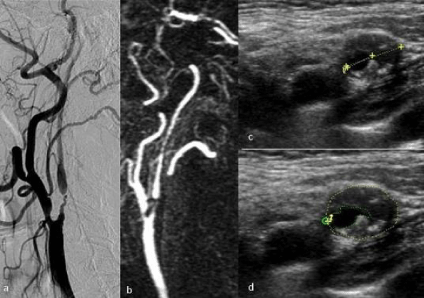
TRICKS MRA and DSA images of the cases were evaluated by two radiologists experienced in vascular radiology (E.G., B.A.). Observers evaluated the MRA images first and then they evaluated DSA images at different times and independently while the images from the other technique were classified and each observer was uninformed of the other observer’s findings. In both examinations, the stenosis measurements were made in the narrowest segment of the ICA lumen. Stenosis rates were determined separately according to the area and diameter, using the NASCET method. The subjective evaluations were performed by examining all other projections except the axial planes in TRICKS MRA and DSA. On the other hand, the objective evaluations were performed on the sagittal plane in TRICKS MIP, on the axial area in TRICKS, on the lateral plane in conventional DSA, and on the axial plane in 3D DSA, according to the area, by marking with a cursor at the narrowest points of the stenotic segments and by manual drawings in area measurements. In TRICKS MRA investigation, the stenosis rate was accepted to be 99% if the flow was identified immediately distal to the ICA segment where no flow was observed (Figure 1). The axial plane images obtained with rotational DSA was considered as the “golden standard” in evaluating the narrowness. As regards to the areameasurements of the stenoses, the oblique MIP images were converted to axial planes by the radiologists who performed the measurements in order to obtain the full axial plane view of the narrowest segment of the ICA.
Statistically, the agreement between the two observers in terms of DSA, MRA, and Doppler US measurements was examined by the significance test of the difference between two pairs and the agreement was determined between the two observers. The averages of the values measured by the two evaluators were taken and the measurement results were coded according to their placements in the following ranges defined as 0-29%, 30-49%, 50-69%, 70-99%, 100%. The agreement between the methods were evaluated using these values. The Kappa Coefficient (κ) was used to assess the diagnostic agreement between the methods. In order to investigate the diagnostic efficiency of Doppler US, TRICKS MRA, and conventional DSA in comparison to the axial planes from rotational DSA; the sensitivity, specificity, positive predictive value (PPV), negative predictive value (NPV), and diagnostic accuracy rate were calculated, accepting the stenosis levels <70% as intact and the stenosis levels ≥70% as pathologic. The continuous variables were presented in mean and standard deviation (SD) values. The categorical variables were presented in numbers (n) and percentages (%). When the calculated p values were below 0.05, it was accepted to be statistically significant. Calculations were done using a statistical software (IBM SPSS Statistics 19, SPSS Inc., an IBM Co., Somers, NY).
Results
The mean age of the patients was 69.8 (53-82) years. Indicating a high level of concordance for each of the groups, the kappa coefficients were found to be 65.4%, 72.1%, 62.7%, and 63.7%, respectively; for the subjective evaluation of the carotid artery stenoses with TRICKS MRA and conventional DSA, for the objective evaluation of the carotid artery stenosis with 3D MIP TRICKS MRA on the sagittal plane and with conventional DSA on the lateral plane, for the objective evaluation of the areas of carotid artery stenoses with 3D DSA on the axial plane and 3D TRICKS MRA, and finally for the objective evaluation of the carotid artery stenoses according to velocity in Doppler US and in the 3D DSA on the axial plane. In the objective evaluation of carotid artery stenoses according to the area and diameter in Doppler US, the Kappa coefficient was 59.8% between the two tests, indicating a sufficient concordance.
According to the NASCET classification, the stenosis rates determined in TRICKS MRA and DSA subjectively; on the sagittal plane in 3D MIP TRICKS MRA and on the conventional lateral plane DSA; on the axial plane in 3D DSA and in 3D TRICKS MRA; on the axial plane in 3D DSA and in Doppler US according to velocity; and in Doppler US according to the area and diameter are given in tables 1-4 respectively.
The rates of sensitivity and specificity, the PPV and NPV, and the rate of diagnostic accuracy, which are obtained by comparing the regional stenosis rates in DSA in the patients with carotid artery stenoses to the stenosis rates measured in Doppler US according to the velocity, diameter, and area; to the stenosis rates measured in TRICKS MRA subjectively on the sagittal plane according to the area; and in DSA on the sagittal and lateral planes, are presented in table 5.
Discussion
DSA, which is accepted as the golden standard method in the diagnosis of supraaortic occlusive arterial diseases, has a therapeutic value in imaging the communication between the intracerebral arteries dynamically. However, it is an invasive procedure with established morbidity (0.5-4%) and mortality (0.01%) risks. Furthermore, it bears other disadvantages as well, including that it emits ionizing radiation, the contrast agent used in the tests has a nephrotoxic effect, the patients need some recovery time to return to their daily lives, and the cost is higher compared to the MRA [6-8]. On the other hand, Doppler US is the most common radiologic imaging method in carotid artery disease because it not only bears any of these disadvantages but allows the evaluation of the structure of the atherosclerotic plaques as well. In relation to this, it is increasingly used in the preoperative evaluation of carotid stenosis cases. In the Consensus Conference of the Society of Radiologists in Ultrasound in 2003, panelists reported that 80% of the patients who underwent carotid endarterectomy in the United States were applied only Doppler US before the surgery [5].
In calculating the stenosis rates in the Doppler US examination; the diameter, area, and flow velocities are measured as well besides the usually utilized PSV values. In addition, “B flow” studies have been on the rise recently with study reports noting high concordance with DSA findings in stenosis measurements [9,10]. In our study, too, the diagnostic accuracy rate (97.4%) of PSV in Doppler US was higher compared to the other modalities in determining the severity of stenosis. Furthermore, our diagnostic accuracy rates are quite high (87.2%) in determining the stenoses on the basis of the diameter and area measurements in Doppler US. On the other hand, they are well-known facts that US test results depend on the personal skills and experience and that stenosis measurements are influenced by several technical parameters and patient characteristics [10]. MRA, which is not an individual-based imaging method, represents a favorable option as a diagnostic test with its superior features like being minimally invasive and rapid. Over the last 15 years, a number studies have been made available in the literature evaluating the comparison of MRA versus DSA in examining the arteries of the head and neck. In most of these studies, contrast MRA and TOF MRA were used. With the TOF technique, which is based on obtaining contrasting images due to the flow, several disadvantages have been presented including reductions in signal intensity depending on the saturation rates, a relatively smaller area of examination, a higher signal intensity loss in stenotic areas, a longer duration of the procedure (5-6 min), and the risk of obtaining poor-quality images due to the increased saturation effect associated with the decreases in the thicknesses of the sections. 3D post-contrast MRA techniques with high temporal resolution allow performing one or multiple arterial-phase MR angiograms in each patient regardless of the venous return rates [11]. Accordingly, the time-resolved 3D MRA sequences have eliminated the need for precise timing for the contrast agent injections. In this technique, imaging starts simultaneously with the contrast agent injection, allowing to obtain 40-60 sequential images with a rate of 1 image per second. A significant advantage of the technique is that it has eliminated the user dependency in timing [11].
The comparative studies report that DSA has the established risk for several complications, whereas MRI and MRA do not present with any risks. Contrast-enhanced MRA, on the other hand, bears a very low risk for potential complications. In addition, an important advantage of MRA over DSA in imaging the carotid bifurcation is the necessity of at least two injections to obtain standard biplanar images with DSA, unlike MRA which allows obtaining several images after a single injection of the contrast agent. The major advantages of the contrast-enhanced MRA technique are the reduction of artefacts due to the flow and patient movements, favorable spatial resolution, and the sufficient capacity to scan a wide area extending from the aortic arch to the circle of Willis. These advantages allow us to estimate the carotid artery stenosis rates accurately, to distinguish between the occlusion or pseudo-occlusion which are very important factors clinically and therapeutically, and to identify the consecutive stenoses in a row [12]. Remonda et al. study in 2002, evaluating 240 ICAs in terms of stenosis, reported the specificity rate as 96%, sensitivity rate 98%, PPV as 95%, and NPV as 98% after comparing MRA to DSA according to NASCET method based on the MIP images in MRA [13]. There are other studies available in the literature in which more than two methods were investigated. A meta-analysis, conducted by Wardlaw et al. in 2006, investigated the studies comparing US, MRA, CTA, and contrast-enhanced MRA to DSA, and reported that contrast-enhanced MRA was superior to all other noninvasive techniques in determining the rates of carotidarterystenosis. The same study determined a 94% sensitivity and 93% specificity in carotid stenoses with the extent ranging from 70% to 99%, whereas, a 77% sensitivity and 97% specificity were determined in carotid stenosis ranging from 50% to 69%. On the other hand, a 96% rate for both sensitivity and specificity were determined in stenoses <50% [14]. In 2003 Borisch et al. evaluated 71 vessel segments with Doppler US and contrast-enhanced MRA in 39 patients with carotid stenosis. Both modalities were compared with DSA findings. It was determined that the sensitivity and specificity of the contrast-enhanced MRA were 94.9% and 79.1%, respectively, in carotid stenoses, with stenosis rates above 70%. For Doppler US, sensitivity was determined to be 92.9% and specificity was determined to be 81.9%. As both tests were concordant with each other, the mean sensitivity of both tests is 100% and the specificity is 81.4% [15]. Anzalone et al. evaluated 98 carotid segments with Doppler US in 49 patients with carotid stenosis. In their study, the 3D TOF MRA, contrast-enhanced MRA, DSA, and rotational angiography techniques were compared according to NASCET method in carotid artery stenoses. The sensitivity and specificity rates were 100% and 90%, respectively, in contrast-enhanced MRA. These rates were 95.5% and 87.2% respectively for 3D TOF MRA and 88.6% and 100% respectively for DSA. Taking all groups into consideration, the highest concordance was determined between the contrast-enhanced MRA and rotational angiography; and between DSA and rotational angiography. On the other hand, the lowest concordance was between DSA and contrast-enhanced MRA. The study reported that the stenosis area was evaluated in several projections in MRA, allowing the calculation of the rate of stenosis in the narrowest lumen area. Therefore, it was reported that the stenosis rates might have been higher compared to those obtained with DSA [16].
In our study, when the limit for the stenosis rate was accepted to be 70%, 4 cases fell into different stenosis intervals according to the results of the evaluations of the stenotic area in TRICKS MRA compared to those found in rotational DSA. All stenoses falling into different intervals of stenosis in TRICKS MRA fell into the intervals with highest rates of stenosis in DSA. This may be due to the fact that the spatial resolution in MRA is lower than that of DSA and may be due to a lower signal to noise ratio. In Doppler US, 5 of the stenoses detected by area measurement are within different stenosis intervals. In Doppler US, all vessel segments located in different stenosis intervals are within the upper interval limits of stenosis. When the stenosis percentages were calculated in Doppler US, area and diameter measurements were taken from the distal ICA segment in order to adjust for NASCET criteria. It was considered that the percentages of stenosis were high because Doppler US might not have allowed the imaging of an ICA segment at a location distal enough.
U-King-Im JM et al., conducted a study comparing contrast-enhanced MRA and DSA and reported that one of the limitations in their study was to define a cut-off point like a serious limit level of 70% according to NASCET and ECST methods, leading to variations in the statistical results although rates like 67% would not create a significant difference clinically [17]. In the area measurements in our study, 3 vessel segments falling into the 70-99% interval in TRICKS MRA fell into the 50-69% interval in rotational DSA as the measurements yielded the following percentages of 68%, 68%, and 67% consecutively. Secondly, 2 vessel segments falling into the 70-99% interval in Doppler US fell into the 50-69% interval in rotational DSA because the measured stenosis rates were 67% and 65% consecutively. Therefore, the data falling into different interval groups had an impact on the statistical results.
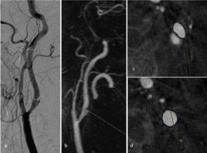
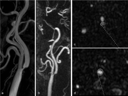
One of the major advantages of our study was the performance of the stenosis measurements in both the sagittal and axial planes in TRICKS MRA and DSA [18,19]. the stenosis segments could not be evaluated in some patients optimally as a result of the superpositioning especially in DSA in the sagittal plane measurements. However, the 3D images obtained from DSA allowed reformatting and evaluating the stenosis in a 3D format. The area measurements are considered to improve the sensitivity and the diagnostic accuracy in DSA and TRICKS MRA in the presence of irregular plaques. For instance, in the area measurements, 3 patients falling into the stenosis interval of 50-69% were found to be in the 70-99% interval and 2 patients falling into the 30-49% interval were found to be in the 50-69% interval (Figure 2). 2 patients with stenosis rates of 51% and 69% on the sagittal plane were evaluated at higher levels of stenosis according to the results of area measurements in TRICKS MRA (77% and 85%) (Figure 3).
In our study, the measurements were performed manually by marking with a cursor in the lateral projections of the DSA images and in the TRICKS MRA MIP images on the sagittal plane. As each of the observers preferred different stenotic segments and different points of distal vessel segments for NASCET method in case of longer sized plaques, this caused millimetric discordances in the measured values yielding to different stenosis rates. We are of the opinion that this is one of the major factors leading to the differences between the classifications.
In litarature there is a higher incidence of the disease in men than in women. However, in our study, the number of female patients was higher than male patients.
The most important limitation of our study was the small number of patients. It will be appropriate to increase the number of patients in future studies.
Conclusion
We determined the PSV as the modality with the highest diagnostic efficacy in Doppler US, providing a reliable parameter to evaluate the carotid artery stenoses. TRICKS MRA can be used as a reliable and safe diagnostic technique alternative to DSA as it provided high rates of diagnostic accuracy in all parameters and it allowed the evaluation of all carotid artery segments with a non-invasive and non-ionizing method. Although the area measurements with rotational DSA are the golden standards in the evaluation of stenoses, performing this technique is not required in each patient because the objective and subjective stenosis evaluations on the lateral and on other planes provided high diagnostic accuracy rates. However, the decision to perform rotational DSA can be made during the procedure especially if superpositions are present on the lateral planes in DSA or if the narrowness of stenosis requires optimal evaluation by area measurements in cases with irregularly ulcerated plaques.
- Osborn A (1993) Cerebral vasculature: Normal anatomy and pathology, in Diagnostic Neuroradiology, D.L.W., Editör. Mosby. 330-341.
- Rubba P, Riccardi G, Pauciullo P, Vaccaro O, Carbone L, et al. (1989) Different localization of early arterial lesions in insulin-dependent diabetes mellitus and familial hypercholesterolemia. Metabolism 38: 962-966. Link: https://tinyurl.com/ya6rorsy
- Jeng JS, Chung MY, Yip PK, Hwang BS, Chang YC (1994) Extracranial carotid atherosclerosis and vascular risk factors in different types of ischemic stroke in Taiwan. Stroke 25: 1989-1993. Link: https://tinyurl.com/ydc63jgm
- Adla T, Adlova R (2015) Multimodality Imaging of Carotid Stenosis. Int J Angiol 24: 179-184. Link: https://tinyurl.com/ycdqnvc6
- Grant EG. Benson CB, Moneta GL, Alexandrov AV, Baker JD, et al. (2003) Carotid artery stenosis: gray-scale and Doppler US diagnosis- Society of Radiologists in Ultrasound Consensus Conference. Radiology 229: 340-346. Link: https://tinyurl.com/yb5mvyf6
- Heiserman JE, Dean BL, Hodak JA, Flom RA, Bird CR, et al. (1994) Neurologic complications of cerebral angiography. AJNR Am J Neuroradiol 15: 1401-1407. Link: https://tinyurl.com/yagoq3wc
- Waugh JR, Sacharias N (1992) Arteriographic complications in the DSA era. Radiology 182: 243-246. Link: https://tinyurl.com/ybgjqzgh
- Willinsky RA, Taylor SM, TerBrugge K, Farb RI, Tomlinson G, et al. (2003) Neurologic complications of cerebral angiography: prospective analysis of 2,899 procedures and review of the literature. Radiology 227: 522-528. Link: https://tinyurl.com/yaar5eru
- Zwiebel WJ, Pellerito JS (2006) Introduction to Vascular Ultrasound. Mihmanli I (Translated) 1st Edition, Istanbul. İstanbul Medical Publishing 586-609.
- Yurdakul, M, Tola M, Cumhur T (2004) B-flow imaging of internal carotid artery stenosis: Comparison with power Doppler imaging and digital subtraction angiography. J Clin Ultrasound 32: 243-248. Link: https://tinyurl.com/y7fuhhu7
- Binkert CA, Baker PD, Petersen BD, Szumowski J, Kaufman JA (2004) Peripheral vascular disease: blinded study of dedicated calf MR angiography versus standard bolus-chase MR angiography and film hard-copy angiography. Radiology 232: 860-866. Link: https://tinyurl.com/ya9hcyes
- Korosec FR, Turski PA, Carroll TJ, Mistretta CA, Grist TM (1999) Contrast-enhanced MR angiography of the carotid bifurcation. J Magn Reson Imaging 10: 317-325. Link: https://tinyurl.com/y7m232l2
- Remonda L, Senn P, Barth A, Arnold M, Lövblad KO, et al. (2002) Contrast-enhanced 3D MR angiography of the carotid artery: comparison with conventional digital subtraction angiography. AJNR Am J Neuroradiol 23: 213-219. Link: https://tinyurl.com/y8oceu3x
- Wardlaw JM, Chappell FM, Best JJ, Wartolowska K, Berry E (2006) Non-invasive imaging compared with intra-arterial angiography in the diagnosis of symptomatic carotid stenosis: a meta-analysis. Lancet 367: 1503-1512. Link: https://tinyurl.com/y8m79292
- Borisch I, Horn M, Butz B, Zorger N, Draganski B, et al. (2003) Preoperative evaluation of carotid artery stenosis: comparison of contrast-enhanced MR angiography and duplex sonography with digital subtraction angiography. AJNR Am J Neuroradiol 24: 1117-1122. Link: https://tinyurl.com/y9cnnapl
- Anzalone N, Scomazzoni F, Castellano R, Strada L, Righi C, et al. (2005) Carotid artery stenosis: intraindividual correlations of 3D time-of-flight MR angiography, contrast-enhanced MR angiography, conventional DSA, and rotational angiography for detection and grading. Radiology 236: 204-213. Link: https://tinyurl.com/ybkmnvuz
- U-King-Im JM, Graves MJ, Cross JJ, Higgins NJ, Wat J, et al. (2007) Internal carotid artery stenosis: accuracy of subjective visual impression for evaluation with digital subtraction angiography and contrast-enhanced MR angiography. Radiology 244: 213-222. Link: https://tinyurl.com/yamdtya6
- Razek AA, Gaballa G, Megahed AS, Elmogy E (2013) Time resolved imaging of contrast kinetics (TRICKS) MR angiography of arteriovenous malformations of head and neck. Eur J Radiol 82: 1885-1891. Link: https://tinyurl.com/y94nzbfb
- Abdel Razek A, Ashmalla G, Samir S (2017) Clinical value of classification of venous malformations with contrast enhanced MR angiography. Phlebology 32: 628–633. Link: https://tinyurl.com/ya26p3zr
Article Alerts
Subscribe to our articles alerts and stay tuned.
 This work is licensed under a Creative Commons Attribution 4.0 International License.


 Save to Mendeley
