Imaging Journal of Clinical and Medical Sciences
Severe Congestive Dysmenorrhea in an Adolescent
Swaramya Chandrasekaran*, Haritha Sagili and Sunitha V Chakkalakkoombil
Cite this as
Swaramya C, Haritha S Sunitha VC (2017) Severe Congestive Dysmenorrhea in an Adolescent. Imaging J Clin Medical Sci 4(1): 011-012. DOI: 10.17352/2455-8702.000035Case Notes
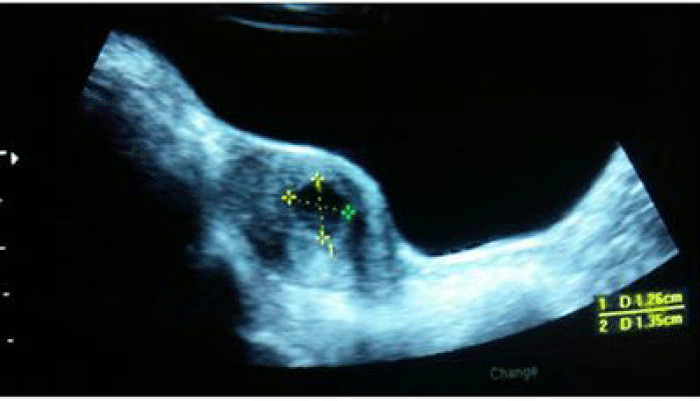
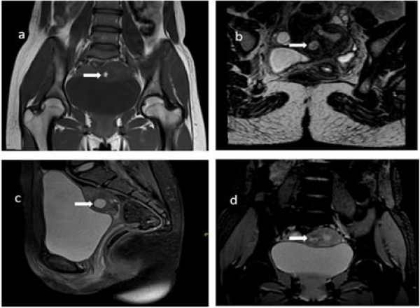
A 16 year old girl presented with severe congestive dysmenorrhea shortly after the onset of menarche at 14 years of age. Menstrual cycles were otherwise regular with normal flow pattern. She was prescribed multiple NSAIDS, which provided minimal relief. She was subsequently referred from a secondary health centre with an ultrasound report of bicornuate uterus. Clinical examination did not reveal any abnormality. Pelvic ultrasound showed a normal sized uterus with cystic lesion in the anterior wall of the uterus raising the possibly of a degenerated fibroid (Figure 1). Endometrium appeared thin and both ovaries were normal. In view of uncertainty in diagnosis, MRI study of pelvis was done and a diagnosis of juvenile cystic adenomyoma of uterus was made based on findings of a well-defined uterine intramural lesion with signal characteristics of blood products within (Figure 2).
Single line condensation: Juvenile cystic adenomyoma diagnosed on MRI in an adolescent girl presenting with severe congestive dysmenorrhea.
Comment
Adenomyomas occur in multiparous women usually over 40 years of age. An uncommon variant is the cystic type and is especially rare amongst adolescents. Juvenile form of cystic adenomyoma is enountered in girls between 13-20 years of age [1]. It is believed to be the result of repeated focal haemorrhages within the ectopic endometrial glands in myometrium resulting in cystic spaces filled with altered blood products. It often poses a diagnostic challenge, as on initial ultrasound evaluation it may mimic congenital malformations of the uterus such as a non- communicating horn with hematometra, fibroid with hemorrhagic degeneration or congenital uterine cysts [2]. Further imaging with MRI is essential for appropriate diagnosis as it may have implications on the future reproductive health. MRI is the most sensitive and specific imaging modality for this condition and will demonstrate cystic regions with variable signal on both T1 and T2 weighted images, correlating with the presence of blood products. Juvenile cystic adenomyoma often presents with secondary dysmenorrhea refractory to first line management with NSAIDs as in our patient. The girl is currently on continuous pharmacological menstrual suppression with oral contraceptive pills.
- Ho ML, Ratts V, Merritt D (2009) Adenomyotic cyst in an adolescent girl. J Pediatr Adolesc Gynecol 22: e33-38. Link: https://goo.gl/4r3KkN
- Branquinho MM, Marques AL, Leite HB, Silva IS (2012) Juvenile cystic adenomyoma. BMJ Case Rep. Link: https://goo.gl/uA9Ewq
Article Alerts
Subscribe to our articles alerts and stay tuned.
 This work is licensed under a Creative Commons Attribution 4.0 International License.


 Save to Mendeley
