Imaging Journal of Clinical and Medical Sciences
Correlation Ultrasound, Clinical and Biological in the Management of Chronic Renal Failure at the Medical Imaging, Exploration and Diagnostics Cabinet (CIMED) and at the National Hemodialysis Center of Donka
1Medical Imaging Exploration and Diagnosis Office, Guinea
2Luxembourg” Mother-Child Hospital Center, Luxemburg
3National Social Security Fund of Conakry, Guinea
4Faculty of Medicine of the Gamal Abdel Nasser University of Conakry, Guinea
Author and article information
Cite this as
Mamoudou C, Issa C, Siré N, Ibrahima B, Sory DI, Mamadou D, et al. Correlation Ultrasound, Clinical and Biological in the Management of Chronic Renal Failure at the Medical Imaging, Exploration and Diagnostics Cabinet (CIMED) and at the National Hemodialysis Center of Donka. Imaging J Clin Medical Sci. 2024; 11(1): 001-010. Available from: 10.17352/2455-8702.000142
Copyright License
© 2024 Mamoudou C, et al. This is an open-access article distributed under the terms of the Creative Commons Attribution License, which permits unrestricted use, distribution, and reproduction in any medium, provided the original author and source are credited.Introduction: This study aimed to describe the clinical, ultrasound, and biological correlation in the diagnosis of chronic renal failure at CIMED and the Donka National Hemodialysis Center.
Materials and methods: This was a descriptive cross-sectional study lasting 16 months from November 1, 2021, to March 31, 2023. Study variables were sociodemographic, clinical, and paraclinical.
Results: During the study period, 6943 ultrasound scans were performed, 48 of which showed CKD (0.7%). There was a predominance of males, with a sex ratio of 1.08. The mean age was 52, with extremes of 17 and 78. Blue-collar workers were the most common socio-professional group (33.3%). The edematous syndrome was the most frequent reason for consultation (33.3%). Severe CKD was the most common in our study (56.3%). Ultrasound stage III was the most frequent, accounting for 52.1% of cases. There was a correlation between a decrease in creatinine clearance and an increase in the ultrasound stage of CKD and an increase in the severity of CKD with an increase in the ultrasound stage.
Conclusion: Our study made it possible to cross-reference clinical, ultrasound, and biological data of chronic renal failure, allowing us to observe significant trends by location which suggests a correlation between these different aspects in the diagnosis of chronic renal failure within the practice of medical imaging for exploration and diagnostics (CIMED) and at the Donka National Hemodialysis Center in Conakry.
Chronic Kidney Failure (CKD) is a common and constantly increasing pathology worldwide [1]. It is defined as a progressive and irreversible reduction in Glomerular Filtration Rate (GFR) below 60 ml/min per 1.73 m2, persisting for 3 months or more [2].
Chronic kidney failure (CKD) affects between 1.74 and 2.5 million people in France and end-stage chronic kidney disease (ESRD) is around 45,000 [3]. The incidence of this pathology is around 100 new patients per million inhabitants per year. It is around 95/million inhabitants in Sweden but goes up to 218/million inhabitants in the United States. The epidemiology of CKD varies considerably from one country to another and access to treatments depends on the socio-economic level of the country [4].
The aging of the population and the progression of vascular and diabetic nephropathy lead to a steady increase in the prevalence of chronic kidney failure [5].
Patients suffering from CKD are subjected to replacement treatment by dialysis or by kidney transplantation, hemodialysis is a technique used for more than 40 years to replace the main functions of healthy kidneys: filtering the blood (eliminating its wastes), and balancing fluid levels by removing wastes from the blood and excess water. Hemodialysis also has the role of removing excess accumulated fluid. During dialysis treatment, this excess liquid can be removed by creating pressure in the filter [6].
Renal imaging is based on three main types of exploration: ultrasound, CT, and nuclear magnetic resonance. Ultrasound is an anatomical and functional imaging technique, which communicates information on the morphology and vascularization of the kidney. It makes it possible to obtain a renal analysis at the four stages of the exploration with the obtaining of angiographic and urographic equivalents [7].
In the United States, in 2010, the estimated prevalence of all stages of chronic kidney disease was close to 13% and affected nearly 20 million Americans. Unlike what happens in certain low-income developing countries where inaccessibility to replacement treatments remains the major difficulty encountered [8].
In France, in 2015, according to the Epidemiological and Information Network in Nephrology (REIN), the overall incidence of so-called end-stage renal failure was 166 per million inhabitants. Half of the incident cases were over 70.5 years old. Associated comorbidities were frequent, in particular, diabetes (45% of incident cases) and cardiovascular comorbidities (58%), the frequency of which increased with age [9].
In Africa, several studies have been carried out to establish an epidemiological and clinical profile of CKD in hospitals:
In Ivory Coast, in 2011, the incidence of end-stage chronic renal failure (ESRD) in the internal medicine department of Treichville University Hospital was 7.5% [10].
In Mali, several studies have been carried out on the subject; already in 1990, there was a frequency of 1.9% in the internal medicine department of the Point G University Hospital [11]. B. Djanka in 2004 reported a frequency of 12.35% in the nephrology department of Point G University Hospital [12].
In Guinea, in 2015, a study carried out in the nephrology department of the Donka National Hospital revealed that 579 (49%) patients presented with CKD [13]. The absence of previous studies on the subject, the increasing number of kidney diseases in Guinea, and the existence of adequate ultrasound equipment are the reasons that allowed us to choose this subject with the following objectives:
- Describe the sociodemographic characteristics of patients with chronic renal failure.
- Determine the clinical, ultrasound, and biological aspects of chronic renal failure.
- Compare clinical, biological, and ultrasound data in the diagnosis of chronic renal failure to establish a possible correlation.
Methods
Study framework
Two centers served as a framework for us of study namely:
a) The medical imaging exploration and diagnosis office (CIMED)
b) The DONKA National Hemodialysis Center (CNHD)
Material
Served as study support:
- The patient consultation register
- Patient medical records
- The renal ultrasound report
- The investigation sheet established for this purpose
Methods
Type and duration of study: This was a descriptive cross-sectional study lasting 16 months from November 1, 2021, to March 31, 2023.
Target population: These are patients of all ages and both sexes who received a renal ultrasound at CIMED and then were treated at the DONKA National Hemodialysis Center during the study period.
Selection criteria:
Inclusion criteria:
Were included in our study:
- Patients with IRC, clearance ≤ 60 ml/1.73 m2 who had a renal ultrasound at CIMED during the study period.
- Patients treated at the DONKA National Hemodialysis Center who have had a renal ultrasound at CIMED.
Non-inclusion criterion:
Not included in our study:
All patients without clinical or biological reports and renal ultrasound.
Method of recruitment
Recruitment was precise and involved 48 patients who had consulted the Donka hemodialysis center and had a renal ultrasound performed at the CIMED.
Sampling and sample size
- We recruited 48 patients meeting the inclusion criteria and our study period.
- After applying our selection criteria, the final size was 48 patients.
Operational definition of variables
Our variables were divided into:
- Sociodemographic data
- Clinical data
- Paraclinical data
Socio-demographic data
- Age: the elapsed period, in completed years, from the patient's birth to the day of their inclusion in our study. Patients will be classified by age group with a range of 10 years. Then the average age and standard deviation will be calculated.
Sex: the set of morphological characteristics that differentiate the male sex from the female sex in search of predominance. The sex ratio will be calculated.
- Marital Situation: determines the patient's marital situation about the law during our study period distributed as follows:
o Single: an unmarried person.
o Married: person united to another by marriage according to the civil code.
o Divorced: Legal dissolution of civil marriage pronounced by a court during the lifetime of the spouses, at the request of one or both spouses according to the forms determined by law.
Widower: a person whose spouse is dead
- Socio-professional layer: the primary activity carried out by patients to earn their living. We will distinguish patients according to the following professional status:
o Housewife: the woman who has no other activity outside the home.
o Civil servant: Public or private agent appointed to a permanent job and tenured at a grade in the administrative hierarchy.
o Pupil/Student: a person who receives an education in a general or university establishment.
o Worker: the person who, possibly for a salary, performs generally manual work for an employer.
o Trader/Merchant: a person who makes a profession of buying and selling.
Unemployed: a person not exercising a profession.
o Other profession to specify
- Origin: the place of residence of the respondents. We will consider the 5 communes of Conakry (Ratoma, Kaloum, Matoto, Matam, Dixinn) and other origins outside Conakry (Boké, Kindia, Mamou, Labé, Faranah, Kankan, N’Zérékoré).
- Level of education: This is the index that locates the level of a living being from the point of view of literacy. It was distributed as follows:
o Out of school: a person who has not been to school;
o Primary: a person whose level of study is limited to the 1st to 6th elementary year.
o Secondary: the person whose level of study is limited from the 7th year to the final year.
o Higher: the person whose level has reached higher education.
Clinical data:
The clinical data were distributed:
- Functional signs: are symptoms that motivated the patient to consult our department. These were:
* Physical asthenia: Physical asthenia refers to general weakness or physical fatigue, characterized by decreased muscle strength and a persistent feeling of fatigue.
* Hiccups: Hiccups are an involuntary contraction of the diaphragm followed by rapid closure of the glottis.
* Insomnia: Insomnia is a sleep disorder characterized by difficulty falling asleep, staying asleep, or getting quality sleep, which can lead to daytime fatigue and other problems.
* Pruritus: Pruritus is an itchy sensation of the skin that can be due to a variety of causes, including allergic reactions, skin conditions, or liver problems.
* Drowsiness: Drowsiness is a state of strong desire to sleep or fall asleep, often associated with decreased alertness and responsiveness.
* Edematous syndrome: Edematous syndrome is characterized by excessive accumulation of fluid in the tissues, leading to edema. This may be due to heart, kidney, liver, or other problems.
* Abdominal pain: Abdominal pain is an unpleasant or painful sensation felt in the abdomen area.
Vomiting: Vomiting is the forced expulsion of stomach contents through the mouth.
- Physical signs: All the signs observed by the doctor in the patient during the physical examination. These were:
* Pallor: Pallor refers to a skin tone that is lighter than normal due to decreased blood flow or decreased pigmentation.
* Agitation: Agitation is a state of physical and mental excitement or restlessness, often characterized by intense activity, nervous behavior, or an inability to calm down.
OAP: Acute pulmonary edema is a serious medical condition in which the lungs fill with fluid. This may occur due to congestive heart failure or other lung problems, causing severe difficulty breathing.
- Medical history: pathologies developed by patients already cured or undergoing treatment before the consultation. These were:
* Phytotherapy
* Excl. VAT
* Diabetes
* IV
* Sickle cell anemia
* Recurrent urinary tract infection
* Pulmonary tuberculosis
* Cataract
Paraclinical data:
Blood
The serum creatinine is used in the calculation of clearance using the simplified MDRD formula:
Stage 1: clearance ≥120 ml/min (No renal failure)
Stage 2: clearance 60-89 ml/min (mild renal impairment)
Stage 3: clearance 30-59 ml/min (moderate renal impairment)
Stage 4: clearance 15-29 ml/min (severe renal failure)
Stage 5: clearance < 15 ml/min; End-stage renal failure.
Urea: normal is between 2.7 - 7.5 mmol/l.
Blood count (CBC): allowed us to make the diagnosis of anemia and define its characteristics. We will speak of anemia when the Hb level < 13 g/dl in men and 12 g/dl in women.
The blood ionogram:
- Serum calcium: normal is between 2.2 - 2.6 mmol/l
- Serum sodium: normal: 135 - 145 mmol/l
- Serum potassium: normal: 3.5 - 5.5 mmol/l
Urine
- Reactive urine strip (ComboStik 10): presenting dry chemistry reactive zones allowed us to search in urine for the quantitative and/or semi-quantitative presence of different parameters such as leukocytes, nitrites, proteins, and erythrocytes.
- Proteinuria: demonstration of albumin thanks to the color change of a pH indicator: Negative = absence of protein; Trace = trace of protein; + = 30 mg/dl or 0.3 g/l; ++ = 100 mg/dl or 1 g/l; +++ = 300 mg/dl or 3 g/l; ++++ = 1000 mg/dl or 10 g/l.
Hematuria: demonstration of erythrocytes and myoglobin by peroxidase activity and the change of an indicator; Negative = absence of blood; Trace = trace of blood; + = 25 red blood cells/µl; ++ = 80 red blood cells/µl; +++ = 200 red blood cells/µl.
- Leukocyturia: demonstration of the activity of esterases secreted by granular leukocytes; Negative = absence of leukocytes; Trace = trace of leukocytes; + = 70 leukocytes/µl; ++ = 125 leukocytes/µl; +++ = 500 leukocytes/µl.
- Nitrite: demonstration of nitrites obtained by the activity of nitrate reductases of certain germs; Negative = absence of nitrite; Trace = trace of nitrite; Positive = presence of nitrite.
24-hour proteinuria: the 24-hour proteinuria assay allows quantification of proteinuria:
- Minimal: < 1g/24h.
- Average: 1 to 3g/24.
- Massive: > 3g/24h Cytological and bacteriological examination of urine (ECBU): ECBU looking for leukocyturia, hematuria, and germs.
Ø Imaging
- Abdominopelvic ultrasound: carried out by a radiologist with an SIEMENS HEALTHINEERS ultrasound machine using a deep 5C1 frequency probe. This examination made it possible to assess the size of the kidneys:
Decreased, normal, or increased (it is considered reduced when the long axis is < 9 cm); their echogenicities; their differentiations; their contours and symmetries; to look for dilation of the excretory pathways, to assess the bladder, the possible presence of lithiasis, cysts, and other aspects; it allows you to explore other organs (liver, spleen, pancreas, gallbladder)
Ultrasound stage of CKD
Stage 0 (normal kidney): hypo-echoic renal cortex compared to the liver and spleen, well-differentiated
Stage I: renal cortex iso-echoic concerning the liver and spleen,
Stage II: renal cortex hyperechoic concerning the liver and spleen, but hypo echogenic in the renal sinus with preservation of cortico-medullary differentiation and loss of cortico-sinus differentiation,
Stage III: renal cortex hyperechoic to the liver, and iso-echoic to the renal sinus with the absence of cortico-medullary and cortico-sinus differentiation
- The fundus view: Allows the detection of hypertensive retinopathy (Kir Kendall classification) and/or diabetic retinopathy depending on their evolution.
Kir Kendall classification
Stage I: severe and disseminated arterial narrowing,
Stage II: in addition, the presence of retinal hemorrhage, dry exudates and cottony nodules,
Stage III: in addition to stages I and II, the presence of papilledema.
- The electrocardiogram: allows you to look for ventricular hypertrophy, signs of necrosis or ischemia, rhythm, and cardiac conduction disorders.
- Front chest x-ray: used to look for cardiomegaly using the calculation of the cardiothoracic index (ICT > 0.50) and other abnormalities such as pleurisy, acute lung edema, pneumopathy, or any chest tumor.
Type of initial nephropathy: In the absence of a renal biopsy, the semiological classification was adopted to identify the type of initial nephropathy. Thus we found the following initial nephropathies:
- Vascular nephropathy: considered in the presence of severe and long-standing hypertension; low proteinuria (< 1.5 g/l); the absence of hematuria and leukocyturia; hypertensive retinopathy at the back of the eye; left ventricular hypertrophy.
- Glomerular nephropathy: retained in the face of proteinuria > 2 g/l; hypertension; a recurrent edematous syndrome; microscopic hematuria; kidneys that are small, poorly differentiated, symmetrical, and regular.
- Diabetic nephropathy: considered in the presence of diabetes that has been present for at least 5 years; proteinuria > 500 mg/24h or micro albuminemia > 300 mg/24h; the absence of hematuria; kidneys preserved in their size or enlarged, dedifferentiated; diabetic retinopathy at the back of the eye. Tubulointerstitial nephropathy: retained in the face of absent or moderate hypertension; polyuria; the existence of recurrent urinary infection and/or herbal medicine; low proteinuria (< 1.5 g/l); asymmetrical renal atrophy, irregular and bumpy contours on ultrasound.
- Nephropathy of undetermined origins: retained in the face of cortico-sinus dedifferentiation whose clinical and paraclinical anomalies do not allow us to relate them to one of the above-mentioned categories.
- Mixed nephropathy: association of diabetic nephropathy and vascular nephropathy in the same patient.
Data processing and analysis:
Our data was entered using the Word Excel software from Office Pack 2019 and analyzed by SPSS V21 software.
Ethical and regulatory consideration
Our data was collected anonymously and the information obtained was used for purely scientific purposes.
Limitations and difficulties
During our study, our limitations and difficulties focused on the lack of certain medical information in certain medical records and the difficulty of associating the results of ultrasound examinations with the medical record of the same patient due to poor record keeping.
Results
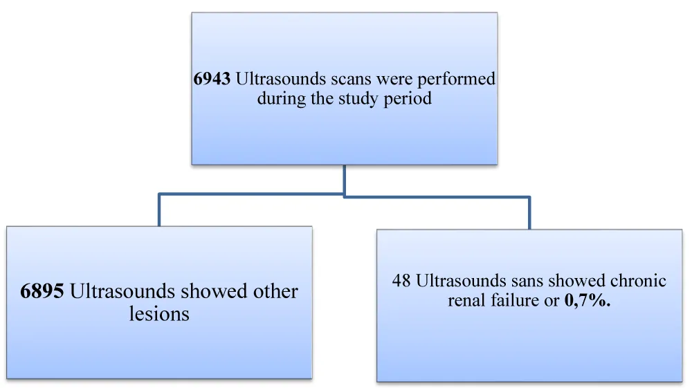
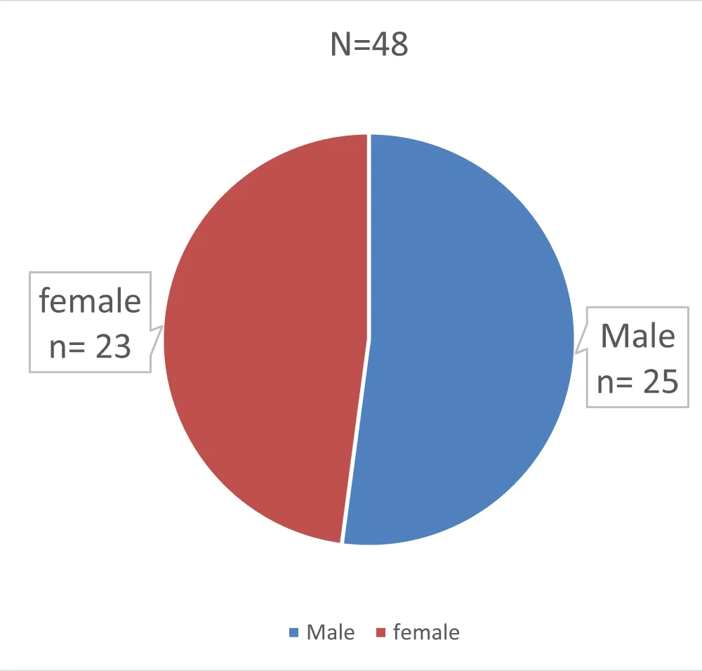
Out of 6943 ultrasounds performed during the study period, 48 ultrasounds showed CKD or 0.7% (Figure 1). The average age of our patients was 52 years, workers were the most predominant, i.e. 33.3%, and more than half of the patients were married, i.e. 83.3% (Table 1). The sex ratio was 1.08 in favor of men (Figure 2). Edematous syndrome was the most frequent functional sign, i.e. 33.3%, herbal medicine was the most common antecedent, i.e. 60.4%, and antibiotic therapy was the most common previous treatment, i.e. 70.8% (Table 2). In biology, the most encountered serum creatinine was 301-450 µmol/L, serum potassium, and serum calcium were mostly reduced, i.e. 41.7% and 83.3%, serum phosphorus was increased in 62.5%, the rate hemoglobin was normal in 66.7%, serum sodium was normal in 87.5% and proteinuria was massive in 12.5% (Table 3).
The glomerular filtration rate was 56.3% between 15-29 ml/min belonging to severe chronic renal failure (Table 4). The initial glomerular and indeterminate nephropathy was the most frequent, i.e. 22.9% and 25% (Table 5).
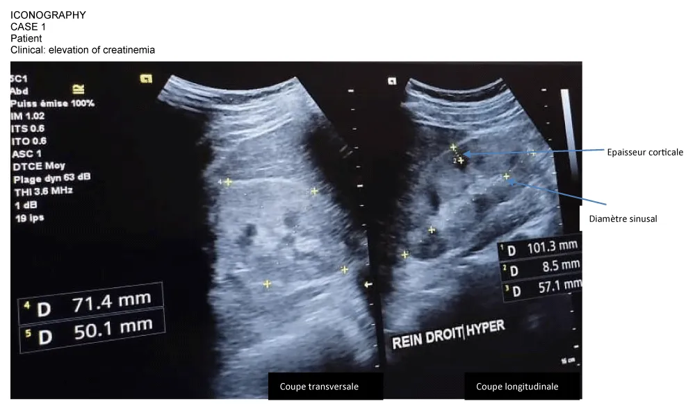
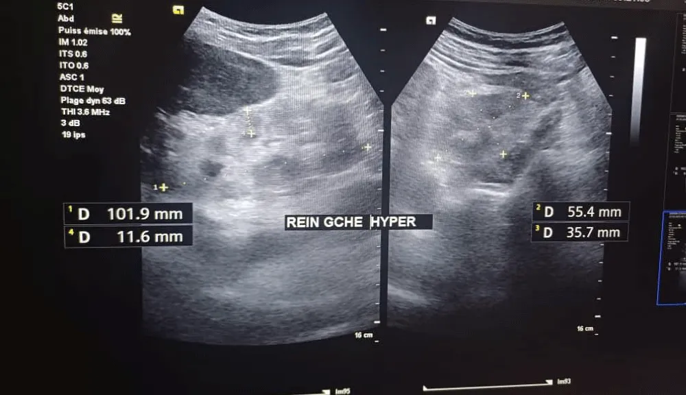
On ultrasound, the size of the kidneys was normal in 58.3% (Table 6). The cortical index was increased in both kidneys, 47.9% on the right and 56.3% on the left (Table 6). Renal ultrasound stage III was the most frequent, i.e. 52.1% (Table 7). All ultrasound stage I patients were classified as stage 4 of CKD and mainly present a serum creatinine between 150-300 µmol/L. For ultrasound stage II, the majority of patients had a serum creatinine between 301-450 µmol/L. Among them, most are classified as stage 4 of CKD, with a few cases at stage 3 and stage 5. For ultrasound stage III, patients mainly present with serum creatinine between 451-600 µmol/L. Most patients in this group were classified as stage 5 CKD. Overall, there was an increase in CKD severity with increasing ultrasound stage, which is consistent with clinical expectations (Table 8) (Figures 3,4).
Discussion
During the 16 months of our study, 6943 ultrasound scans were performed at CIMED, including 48 cases of CKD from national hemodialysis centers, representing a frequency of 0.7%. Our results are lower than those found in Ivory Coast in 2011 where the hospital prevalence of CKD was 7.5% in the internal medicine department of the Treichville University Hospital [10]. This difference could be explained by the fact that the study was carried out in an office.
The most represented age group was 30-44 years and 45-59 years, i.e. 29.2% for each of these intervals, with extremes ranging from 17 to 78 years and an average age of 52 years. This result is different from that of Amekoudi [14] where the average age was 37.3 ± 15.4 years and Kessler et al [15] who found an average age of 62.8 ± 16 years. This could indicate that ESRD is not limited to a specific age category, but rather affects a diverse range of age groups.
The sex ratio was 1.08 in favor of men, or 52%. This is comparable to the result of Chaabouni, et al. [16] who found a sex ratio of 1.27 and 1.4 respectively. This could be due to a difference in susceptibility between the sexes or specific risk factors in men.
Workers and housewives, with respective percentages of 33.3% and 27.1%, were the professions most represented in our study.
The most common functional sign was edematous syndrome, representing 33.3% of cases. The two reasons for hospitalization most cited by Amekoudi [14] were renal failure and severe hypertension with 40.3% and 23.1% respectively. Our result highlights the importance of clinical assessment of edema in patients, as this could be an early sign of renal failure.
Herbal medicine and hypertension were the most common antecedents with respective frequencies of 60.4% and 47.9%. In the literature, hypertension has been found as a pathological antecedent of patients with CKD [12]. Its association with diabetes further jeopardizes the prognosis of diabetic patients. This association highlights the close relationship between hypertension and CKD. In our study, the most observed physical sign was pallor, i.e. 33.3% of patients. Amekoudi [14] found morning vomiting at 65.7% and asthenia at 64.7%. Lengani A. [17] in Burkina, reported asthenia and vomiting in 78.2% and 63.2% of cases respectively. The polymorphism of these manifestations could be explained by the late treatment of patients who mostly arrive at the terminal stage.
56.3% of patients had a serum creatinine between 451-600 µmol/l with an average of 403.5625 µmol/l. Akinsola et al in Nigeria [18], Sidikath in Burkina [19], and Ramiltiana in Madagascar [20] reported respective mean serum creatinine levels of 1130 ± 576 µmol/l, 1134 ± 857.4 µmol/l, and 911.3 µmol/l. The observed differences could be attributable to differences in awareness, and early detection.
Initial glomerular and vascular nephropathy were the most common causes of CKD in our study with 25% of patients. This could be explained by the fact that hypertension is an important risk factor in the diagnosis of CKD.
After analysis, the majority of patients, i.e. 56.3%, presented severe CKD (GFR between 15-29 ml/min) while Amekoudi [14] reported that 68% of patients had a clearance less than 15. This difference could be linked to a dissimilarity in the patient selection criteria or to a late consultation of patients in Amekoudi's study [14]. GFR is a key measure of kidney health, and its decline in the majority of patients highlights the severity of chronic kidney disease. Appropriate management, including consideration of dialysis or kidney transplantation, may be necessary to optimize patient quality of life and survival.
Renal clearance was statistically associated with patient age with p value = 0.023. A decrease in renal clearance with age is consistent with the general understanding of aging and its impact on renal function.
At ECBU, 14.6% of patients had leukocyturia and 16.7% had hematuria. Our results are lower than those of Amekoudi [14] who reported 43.1% and 50% leukocyturia respectively. The presence of leukocyturia and hematuria in some patients may indicate infections highlighting the importance of regular screening in patients with CKD.
The systematic performance of ultrasound in all patients allowed us to identify ultrasound stages of CKD. This therefore proves that ultrasound is a key and crucial examination in the diagnosis of CKD.
The ultrasound stage most represented in our study was stage III with 52.1% of cases. This indicates advanced kidney disease and highlights the importance of proactive management to slow the progression of kidney failure.
The ultrasound stage of CKD was very significantly associated with age with a p-value = 0.02 and poorly associated with clearance with p p-value = 0.09. This association may suggest that age may be an influential factor in the progression of CKD with a higher risk of stage III in older patients. Furthermore, it was not associated with sex (p value = 0.332), previous traditional treatment (p value = 0.354), and 24-hour proteinuria (p value = 0.24). These results suggest that the result of 24-hour proteinuria and the use of traditional treatment before admission does not seem to have a significant impact on the ultrasound stage of CKD.
Kidney size was not statistically associated with age (p value = 0.10), sex (p value = 0.807), 24-hour proteinuria (p value = 0.80), and renal pain (p value = 0.16).
The distribution of patients received at CIMED and Donka University Hospital according to the ultrasound, clinical and biological stages of Chronic Kidney Failure (CKD), with an evaluation of serum creatinine indicated an absence of statistical association with p-value = 0 .09. This suggests that there are no significant associations between ultrasound stages, serum creatinine, and CKD type.
Conclusion
Our study made it possible to cross-reference clinical, ultrasound, and biological data of chronic renal failure, allowing us to observe significant trends by location which suggests a correlation between these different aspects in the diagnosis of chronic renal failure within the practice of medical imaging for exploration and diagnostics (CIMED) and at the Donka National Hemodialysis Center in Conakry.
Adult patients aged 30-44 and 45-59 years were most affected by CKD. Analysis of the study population suggests that CKD is not limited to a specific group.
The most common functional sign was edematous syndrome. The high prevalence of herbal medicine and hypertension in medical histories highlights the complexity of risk factors associated with CKD.
The prevalence of elevated serum creatinine and decreased renal clearance reflects the chronicity of renal failure at the time of diagnosis.
The systematic use of ultrasound has highlighted the crucial importance of this examination in the early diagnosis of CKD.
These results reinforce the importance of a holistic and integrated approach in the management of this complex pathology, highlighting the need for close collaboration between healthcare professionals to optimize care for patients with kidney failure.
Author contributions
All authors contributed to the acquisition, analysis, and interpretation of the data, as well as the writing of the article.
- Berlaud Y, Bertand D. Nephrology for interns. edscimed. Tome 1. Paris, 1998.
- Levey AS, Eckardt K-U, Tsukamoto Y, Levin A, Coresh J, Rossert J, et al. Definition and classification of chronic kidney disease: a position statement from kidney disease: Improving Global Outcomes (KDIGO). Kidney Int. 2005;67(5):2089–2100. Available from: https://doi.org/10.1111/j.1523-1755.2005.00365.x
- Diagnosis of CKD in adults [online]. Available from: http://www.anaes.fr.
- Jungers P, Robino C, Choukroun G. Evolution of the epidemiology of chronic renal failure and forecast of replacement dialysis needs in France. Néphrologie. 2001. Available from: https://pubmed.ncbi.nlm.nih.gov/11436669/
- Kossi AS, Befa NKK, Eyram YMA, Weu MT, Vigan J, Ibrahim H, et al. Acute Kidney Injury during Malaria in Togolese Children. Open J Nephrol. 2018;8. Available from: https://www.scirp.org/journal/paperinformation?paperid=88437
- Jungers P, Joly D, Man N. Chronic renal failure: prevention and treatment. Librairie Lavoisier. (accessed 24 February 2023). Available from: https://www.lavoisier.fr/livre/medecine/l-insufficiency-renale-chronique-4-ed/jungers/descriptif-9782257204301
- Renard-Penna R, Marcy P-Y, Lacout A, Thariat J. Imaging of the kidney. Bull Cancer. 2012;99(3):251–262. Available from: https://doi.org/10.1684/bdc.2011.1487
- Collins AJ, Foley RN, Gilbertson DT, Chen SC. United States Renal Data System public health surveillance of chronic kidney disease and end-stage renal disease. Kidney Int Suppl. 2015;5(1):2. Available from: https://doi.org/10.1038/kisup.2015.2
- R.E.I.N. (Epidemiological and Information Network in Nephrology). Biomedicine Agency. (accessed 24 February 2023). Available from: https://www.agence-biomedecine.fr/R-E-I-N-Reseau-Epidemiologique-et-Information-en-Nephrologie
- Ouattara B, Kra O, Yao H, Kadjo K, Niamkey EK. Particularities of chronic renal failure in black adult patients hospitalized in the internal medicine department of the Treichville University Hospital. Nephrol Ther. 2011;7(6):531–534. Available from: https://doi.org/10.1016/j.nephro.2011.03.009
- Cissé I. Epidemiological aspect of CKD in the internal medicine department of Point G Hospital. 1990.
- Djanka B. Epidemiology of chronic renal failure in the nephrology department of Point G. 2003.
- Bah AO, Lamine C, Balde MC, Mamadou Lamine Yaya Bah, Lionel Rostaing. Epidemiology of chronic kidney diseases in the Republic of Guinea; future dialysis needs. J Nephropathol. 2015;4(3):127–133. Available from: https://doi.org/10.12860/jnp.2015.24
- Amekoudi EYM. Epidemiological-clinical profile of chronic renal failure in the nephrology and hemodialysis department of CHU point G. (accessed 2 January 2024). Available from: https://www.bibliosante.ml/handle/123456789/1527
- Kessler M, Frimat L, Panescu V, Briançon S. Impact of nephrology referral on early and midterm outcomes in ESRD: EPidémiologie de l'Déficience REnale chronique terminale en Lorraine (EPIREL): results of a 2-year, prospective, community-based study. Am J Kidney Dis. 2003;42(3):474–485. Available from: https://doi.org/10.1016/s0272-6386(03)00805-9
- Chaabouni Y, Yaich S, Khedhiri A, Zayen MA, Kharrat M, Kammoun K, et al. Epidemiological profile of end-stage chronic kidney disease in the Sfax region. Pan Afr Med J. 2018;29:1–9. Available from: https://doi.org/10.11604/pamj.2018.29.64.12159
- Lengani A. Epidemiology of severe chronic renal failure in Burkina. Health Notebook. 1997;7:379. Available from: https://www.scirp.org/reference/referencespapers?referenceid=2998469
- Akinsola A, Durosinmi MO, Akinola NO. The hematological profile of Nigerians with chronic renal failure. Afr J Med Med Sci. 2000;29(1):13–16. Available from: https://pubmed.ncbi.nlm.nih.gov/11379460/
- Sidikath S. Biological profile of chronic kidney failure (CKD) in the internal medicine department of the Yalgado Ouédraogo National Hospital Center (CHN-YO) in Ouagadougou: Thesis Med. Ouagadougou, 2003; No. 27.
- Ramilitiana B, Ranivoharisoa EM, Dodo M, Razafimandimby E, Randriamarotia WF. A retrospective study on the incidence of chronic renal failure in the Internal Medicine and Nephrology department of the University Hospital Center of Antananarivo. Pan Afr Med J. 2016;23:141. Available from: https://doi.org/10.11604/pamj.2016.23.141.8874
Article Alerts
Subscribe to our articles alerts and stay tuned.
 This work is licensed under a Creative Commons Attribution 4.0 International License.


 Save to Mendeley
