Imaging Journal of Clinical and Medical Sciences
Gigant Laryngocele Airway Management
1Hospital General Universitario de Ciudad Real, Anesthesiology Department, Spain
2Hospital General Universitario de Ciudad Real, Ear, Nose and Throat Department, Spain
Author and article information
Cite this as
Pascual-Ramirez J, del Hierro JC (2014) Gigant Laryngocele Airway Management. Imaging J Clin Medical Sci. 2014; 1(2): 18-19. Available from: 10.17352/2455-8702.000012
Copyright License
© 2014 Pascual-Ramirez J, et al. This is an open-access article distributed under the terms of the Creative Commons Attribution License, which permits unrestricted use, distribution, and reproduction in any medium, provided the original author and source are credited.Emergent airway obstruction is a dreaded emergency among anesthesiologists. Classically has been managed with awake options, particularly fibreoptic intubation. Laryngoceles, if size and accessibility allows for it, can be evacuated by needle aspiration, postponing definitive management.
Emergent airway obstruction is a dreaded emergency among anesthesiologists. Classically has been managed with awake options, particularly fibreoptic intubation. Laryngoceles, if size and accessibility allows for it, can be evacuated by needle aspiration, postponing definitive management.
Case Report
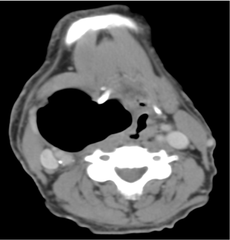
A 69 years-old male presented at the emergency room with hoarseness, fever and a right-sided cervical mass. He had a previous history significant for cigarette smoking and chronic obstructive pulmonary disease. He used to be a glass-blower. The mass was already detected 10 years ago, but the patient neglected medical care at that point in time. It was noticed to suddenly increase its size over the last 15 days. He was admitted to the ward for observation. The computed tomography scan revealed a gigantic cyst (9.5 cm lateral x 7 cm anterior-posterior diameters) of air-density in close relation to the Morgagni ventricle, placed in the anatomic cervical areas II and III. It displaced the submaxilar gland anteriorly, the sterno-cleido-mastoid muscle and carotid artery posteriorly and, very significantly, displaced laterally the larynx lumen, epiglottis and hyoid bone. The diagnosis was made for a giant laryngocele. Three days later the patient complained about sudden respiratory difficulty, cervical pain and difficulty to swallow. Thirty mL of air were obtained by ultrasound-guided percutaneous needle aspiration and a drain was left in place to avoid refilling of the space. The clinical picture improved significantly. He was scheduled for surgery ten days later. Direct endotracheal intubation was achieved at its first attempt, although with moderate difficulty due to having a Cormack-Lehane grade III, but no significant lateral displacement was noticed. A tracheostomy and cyst excision under general anesthesia was completed afterwards without problems.
Discussion
Laryngocele is a rare benign laryngeal disease [1,2]. Laryngoceles can be congenital or acquired. It was first described during Napoleon's Egyptian campaign. Often-times are asymptomatic and that is the reason real incidence is unknown, although it has been estimated to be close to 2.5 cases per million population. It grows in size due to a valvular air-entrapping mechanism [3] until incidental discovery or symptoms appear, due to infection (pyocele formation), aspiration with subsequent bronchitis and pneumonia or upper airway obstruction [4]. Malignant degeneration is extremely rare but has been described [5]. Laryngoceles may expand medially, causing a reduction of the supraglottic space; and laterally, exiting through the thyroid membrane near the internal branch of the upper laryngeal nerve, causing neck bulging. Sometimes it shows mixed medial and lateral growth as our case did. Differential diagnosis includes: saccular cyst, ranchial cyst, neck abscess and lympho-adenopathy. These kind of cysts do not communicate with the laryngeal lumen and are usually filled with fluid. Medial expansion of the laryngocele usually represents an upper airway menace. Management frequently includes awake options, either fibreoptic intubation [6] or, more classically, tracheostomy under local anesthesia to preserve a patent airway. The ultrasound-guided [7] transcutaneous needle aspiration approach that we used has been already described as a safe strategy for urgent airway management. Definite surgical excision can be safely completed later on.
- Pennings RJ, van den Hoogen FJ, Marres HA (2001) Giant laryngoceles: a cause of upper airway obstruction. Eur Arch Otorhinolaryngol 258: 137-140 .
- Soler EM, Vecina VMZ, Vintro XL, Agusti MQ, Vila JB, et al. (1995) Laryngoceles: clinical and therapeutic study of 60 cases. Acta Otorrinolaring Esp 46: 279-286.
- Drozd M, Szuber K, Szuber D (1996) The significance of the valve mechanism in pathology of laryngocele. Otolaryngol Pol 50:17-20.
- De Paula Felix JA, Felix F, Pires de Mello LF (2008) Laryngocele: a cause of upper airway obstruction. Rev Bras Otorrinolaringol 74: 143-146.
- Akbas Y, Unal M, Pata YS (2004) Asymptomatic bilateral mixedtype laryngocele and laryngeal carcinoma. Eur Arch Otorhinolaryngol 261: 307-309.
- Leong SCL, Badran K, McCormick MS (2007) Laryngocoele presenting as acute airway obstruction. Singapore Med J 48: e84.
- Mace AT, Ravichandran S, Dewar G, Picozzi GL (2009) Laryngopyocoele: simple management of an acute airway crisis. J Laryngol Otol 123: 248-249.


 Save to Mendeley
Article Alerts
Subscribe to our articles alerts and stay tuned.
 This work is licensed under a Creative Commons Attribution 4.0 International License.

