Global Journal of Anesthesiology
A Challenging Extubation Caused by the Inability to Deflate the Cuff of an Endotracheal Tube
V Abraham1, Ria2* and M Singh3
2Resident, Department of Anaesthesia and Critical Care, Christian Medical College and Hospital, Ludhiana, Punjab, India
3Professor, and HOD, Department of Anaesthesia and Critical Care, Christian Medical College and Hospital, Ludhiana, Punjab, India
Cite this as
Abraham V, Ria, Singh M. A Challenging Extubation Caused by the Inability to Deflate the Cuff of an Endotracheal Tube. Glob J Anesth. 2024;11(1):003-006. Available from: 10.17352/2455-3476.000057Copyright
© 2024 Abraham V, et al. This is an open-access article distributed under the terms of the Creative Commons Attribution License, which permits unrestricted use, distribution, and reproduction in any medium, provided the original author and source are credited.Background: Extubation is considered a critical event in general anesthesia. A smooth endotracheal intubation or an uneventful intra-operative period can still lead to a difficult extubation. This is a case report of an eventful and unanticipated difficult elective extubation in a post coronary artery bypass grafting patient.
Observations: A knot was found on the pilot balloon tubing during extubation of the patient which made deflation of the endotracheal tube cuff almost impossible. After several minutes of troubleshooting, we were able to extubate the patient with stable hemodynamics without any evidence of sore throat.
Significance: Rare cases and difficulties like these, challenge a healthcare provider to think beyond common knowledge and this serves as an example for other anaesthesiologists that how minute malfunctions in our common devices/instruments can lead to disastrous outcomes and hence vigilance is important.
Introduction
Challenges faced during extubation have been well-known to all anesthesiologists. Sometimes, even a seemingly easy airway can lead to unanticipated outcomes. The objective of this case report is to advocate the importance of a thorough examination of the devices/instruments being used on a patient to avert disastrous consequences.
Case report
A 62-year-old male, with a known case of hypertension and diabetes mellitus-type 2 with a history of anterior wall myocardial infarction, and coronary angiography showing triple vessel disease, was taken up for coronary artery bypass grafting after pre-operative optimization of the cardiovascular status. On examination, vitals were stable and on airway examination, all parameters were within normal limits like a Mallampati score of 2 (a score to predict the ease of endotracheal intubation by visually assessing the distance from tongue base to roof of the mouth, the higher the score, higher the chances of a difficult intubation) and normal neck mobility. All blood investigations were within normal limits, cross-matching was done and blood products according to anticipated blood loss were arranged.
On the day of surgery, the baseline hemodynamic parameters were normal. The patient was pre-oxygenated for three minutes, and induced with intravenous fentanyl 100 mcg, propofol 150 mg, and vecuronium 6 mg. After three minutes of mask holding and manual ventilation, tracheal intubation was accomplished smoothly with an 8 mm cuffed PVC endotracheal tube. Bilateral chest air entry was confirmed and the tube was fixed at 22 mm on the lip. Cormack Lahane’s grade was 2a (difficulty of intubation when assessed during direct laryngoscopy and view of the glottis, higher the grade, higher the chances of a difficult intubation). Anaesthesia was maintained with isoflurane, oxygen, and air. A central venous catheter in the right internal jugular vein and right radial arterial cannulation was performed for central venous pressure and invasive blood pressure monitoring, respectively. Inotropic support infusions like Nor-adrenaline, dopamine, and adrenaline were prepared and attached to the central venous catheter for maintaining the target intra-operative blood pressure. The intra-operative period including aortic cross-clamping, cardioplegia, re-perfusion, etc was uneventful. The surgery lasted for about four hours and the patient was shifted to CTVS ICU unextubated and an elective extubation was planned after 12 hours.
Following the procedure, in the ICU, the patient was hemodynamically stable. He was kept on a low dose of infusion fentanyl for post-operative analgesia. All sedative medications were discontinued one hour before extubation. After some time, the patient was awake and started responding to verbal commands. Ventilatory settings showed adequate tidal volumes generated by the patient. During the process of attempting to remove the Endotracheal Tube (ETT), there was unexpected resistance encountered while deflating the pilot balloon. Despite efforts to forcefully deflate the balloon and subsequent collapse, the tube could not be extracted due to ongoing resistance. Examination of the pilot balloon tubing did not reveal any visible kinks or malposition issues. After several minutes of troubleshooting without success, it was determined that the valve within the pilot balloon might have malfunctioned. The decision was made to separate the valve from the tubing using scissors, yet the ETT still could not be withdrawn.
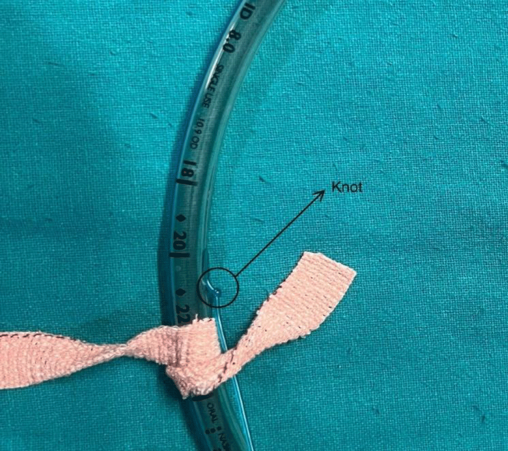
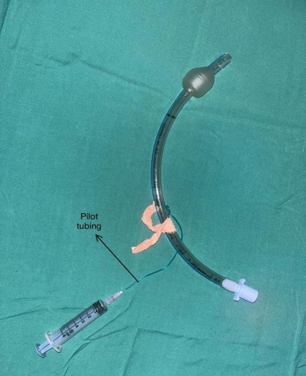
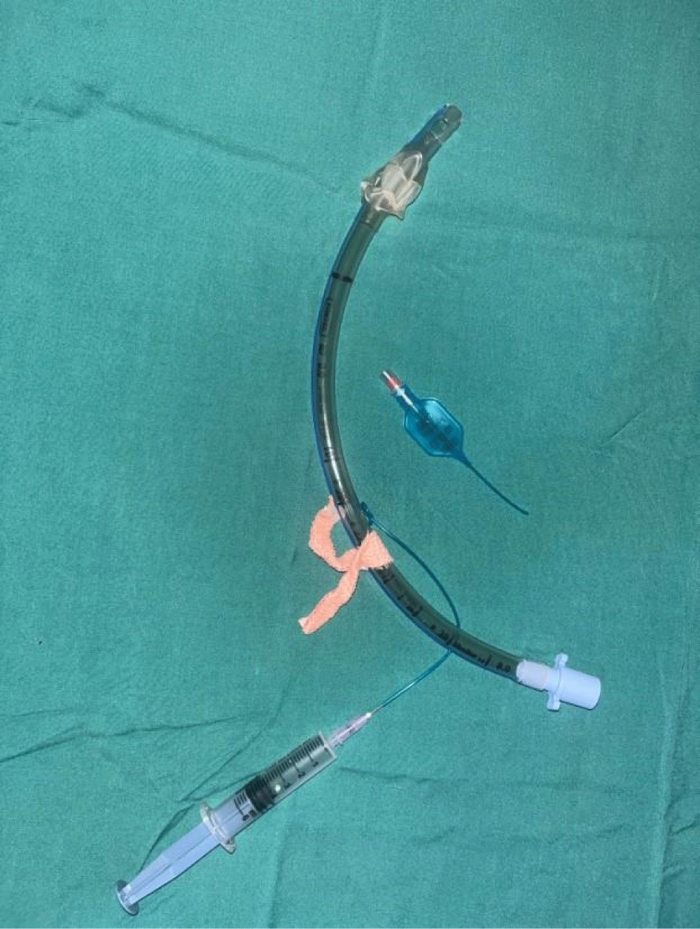
As the patient began showing signs of restlessness, sedation was administered again, and direct laryngoscopy was performed. During this examination, it was discovered that there was a knot in the pilot tubing near its attachment to the ET tube (Figure 1), which had impeded the deflation process. Carefully, the tube was partially extracted, and the knot was gently loosened using artery forceps (Figure 2). Subsequently, a 5cc syringe with a hypodermic needle was carefully inserted into the tubing, and the cuff was meticulously deflated (Figure 3) under direct laryngoscopic visualization. Once the cuff was confirmed to be completely deflated, the patient regained consciousness a few minutes later, and the extubation proceeded smoothly.
Following extubation, the patient was hemodynamically stable, and recovered without any complications such as a sore throat, indicating an uneventful post-procedural course.
For ethical purposes, written informed consent for publication was taken from the patient.
Discussion
Extubation failure is defined as the need for reinstitution of ventilatory support within 24 hours - 72 hours of planned endotracheal removal [1].
Common causes of extubation failure include factors leading to airway obstruction like upper airway edema, laryngospasm, tracheal collapse, tracheomalacia, bleeding, secretions pooling in the upper airway, etc [2]. Complications from extubation can range from minor to major. Minor complications are often transient and do not typically require reintubation, which may lead to underreporting. These minor issues include temporary increases in blood pressure, rapid heart rate, coughing, restlessness, and agitation. However, these physiological disturbances can potentially lead to more serious complications in specific patient groups. Individuals with significant heart or cerebrovascular conditions, elevated intracranial or intraocular pressure, and those who have undergone neck, neurosurgical, or certain plastic surgery procedures are particularly at risk for severe complications [3]. All India Difficult Airway Association (AIDAA) in 2016, showed how an anticipated difficult extubation can be managed and all complications associated with it can be minimized. The use of Airway Exchange Catheters (AEC) and fiber optic bronchoscopes etc, has led to an additional advantage in achieving an uneventful extubation [4].
Rarely, a difficult extubation due to difficult ‘decannulation of the airway’ has also been observed. This mostly depends upon mechanical factors related to the patient, surgery, or anesthesia.
For instance, conditions related to the patient could involve undetected subglottic stenosis or significant swelling that physically obstructs the removal of the Endotracheal Tube (ETT). A surgery-related factor could include a misplaced surgical stitch inadvertently anchoring the ETT to the tracheal wall. Anesthesia-related causes might include incomplete deflation of the ETT cuff due to cuff malfunction or oversight [2,3]. Forcibly pulling out the tube with a fully inflated cuff can result in laryngeal trauma, vocal cord edema, and dislocation of the arytenoid cartilage.
Common reasons for this complication include difficulties in deflating the Endotracheal Tube (ET) cuff, a cuff that is too large to obstruct the vocal cords, and the ET adhering to the tracheal wall due to insufficient lubrication or being secured by a suture or wire. A case reported by N.G Lall, et al. had difficulty extubating a patient due to a fold in the distal attachment of the cuff caused by the repetitive boiling of the tube, leading to loss of elasticity and folding of the cuff on itself, increasing its external diameter [5].
Sriramka, et al. also faced a similar problem due to unusual kinking of the inflation tube due to the ET tube adhesive tape [6].
The majority of challenging extubations result from problems with cuff deflation, which can stem from issues such as compression of the pilot balloon tubing, malfunctioning of the inflation valve, accidental detachment of the inflation port from the ET, and the unadvised practice of forcefully pulling the pilot balloon tubing from the ET to speed up cuff deflation [7].
The Difficult Airway Society (DAS) guidelines for tracheal extubation in adult patients stress the importance of planning, preparation, performance, and post-extubation care [8]. A kink in the pilot balloon tubing leading to failure of deflation has also been reported multiple times, mostly due to improper fixation of ETT with the adhesive tapes [9,10].
Our case had a knot on the pilot balloon tubing, leading to a failure of deflation of the cuff. And since pulling the ETT tube while the cuff is fully or partially inflated is known to cause injury to vocal cords amongst other complications [11], deflating the cuff under direct vision via a 5cc syringe after loosening the knot, appeared to be a better idea. Hence our patient could be extubated easily and post-extubation sore throat and other tissue injuries were prevented.
Airway extubation is typically a planned procedure that should be performed under ideal conditions. It requires thoughtful consideration tailored to each patient and specific surgical context [12].
Conclusion
In this case, a knot found on the pilot balloon tubing was untied using an artery forceps and extubation could be done.
Looking back, it’s clear that equipment malfunction can result in severe and irreversible consequences. Therefore, it’s crucial to meticulously inspect such devices and maintain vigilance throughout every stage of their use, like a knot, kink, etc. on the pilot balloon tubing, as seen in this case. A seemingly uncomplicated airway can lead to an unanticipated difficult extubation, hence all possibilities should be considered, and eventually, one solution will prevail.
- Rothaar RC, Epstein SK. Extubation failure: magnitude of the problem, impact on outcomes, and prevention. Curr Opin Crit Care. 2003;9(1):59-66. Available from: https://doi.org/10.1097/00075198-200302000-00011
- Cavallone LF, Vannucci A. Extubation of the difficult airway and extubation failure. Anesth Analg. 2013;116(2):368-383. Available from: https://doi.org/10.1213/ane.0b013e31827ab572
- Parotto M, Cooper RM, Behringer EC. Extubation of the challenging or difficult airway. Curr Anesthesiol Rep. 2020;10(4):334-340. Available from: https://doi.org/10.1007/s40140-020-00416-3
- Kundra P, Garg R, Patwa A, Ahmed SM, Ramkumar V, Shah A, et al. All India Difficult Airway Association 2016 guidelines for the management of anticipated difficult extubation. Indian J Anaesth. 2016;60(12):915-921. Available from: https://doi.org/10.4103%2F0019-5049.195484
- Lall NG. Difficult extubation: A fold in the endotracheal cuff. Anaesthesia. 1980;35(5):500-501. Available from: https://doi.org/10.1111/j.1365-2044.1980.tb03829.x
- Sriramka B, Natarajan A, Sharma K, Zubair S. Failed endotracheal tube cuff deflation due to unusual kinking of inflation tube. J Dent Anesth Pain Med. 2023;23(4):241-243. Available from: https://doi.org/10.17245%2Fjdapm.2023.23.4.241
- Suman S, Ganjoo P, Tandon MS. Difficult extubation due to failure of an endotracheal tube cuff deflation. J Anaesthesiol Clin Pharmacol. 2011;27(1):141-142. Available from: https://www.ncbi.nlm.nih.gov/pmc/articles/PMC3146146/
- Weatherall AD, Burton RD, Cooper MG, Humphreys SR. Developing an extubation strategy for the difficult pediatric airway—Who, when, why, where, and how? Paediatr Anaesth. 2022 May;32(5):592-599. Available from: https://doi.org/10.1111/pan.14411
- Nag S, Samaddar DP. Inappropriate fixation of an endotracheal tube causing cuff malfunction resulting in difficult extubation. J Anaesthesiol Clin Pharmacol. 2013;29(4):571-572. Available from: https://doi.org/10.1016/j.bjane.2013.04.009
- Bradley DB, Sprung J. An unusual cause of difficult tracheal extubation. J Clin Anesth. 2003;17(2):279-280. Available from: https://doi.org/10.1053/jcan.2003.64
- Zenga J, Galaiya D, Choumanova I, Richmon JD. Endotracheal tube obstruction caused by cuff hyperinflation. Anesthesiology. 2018;129(3):581. Available from: https://doi.org/10.1097/aln.0000000000002233
- Li LT, Gjuzi K, He P, Daly Guris RJ. Airway extubation: A narrative review. J Oral Maxillofac Anesth. 2024;24(3). Available from: https://joma.amegroups.org/article/view/6544/html

Article Alerts
Subscribe to our articles alerts and stay tuned.
 This work is licensed under a Creative Commons Attribution 4.0 International License.


 Save to Mendeley
