Archives of Hematology Case Reports and Reviews
Thalassemia-Beta major-Case report
Vesna Ambarkova1*, Tina Krmzova2 and Zoran Nonkulovski2
2Resident on Specialization at the Department of Pediatric and Preventive Dentistry, Faculty of Dental Medicine University St. Cyril and Methodius, Skopje, Republic of North Macedonia
Cite this as
Ambarkova V, Krmzova T, Nonkulovski Z (2021) Thalassemia-Beta major-Case report. Arch Hematol Case Rep Rev 6(1): 021-025. DOI: 10.17352/ahcrr.000034Beta thalassemia (β thalassemia) is a group of inherited blood disorders. Case report A 17-year old boy accompanied by medical support staff visited our Department for preventive and pediatric dentistry within the University Dental Center Ss.Pantelejmon in Skopje, due to a dental pain which comes from the first molar tooth(36) of the low jaw from the left side. From the accompanying documentation we saw that the boy has congenital beta thalassemia major and was admitted to the Department for Hematology within University State Hospital Mother Theresa a month ago. The boy was hospitalized in an agreed term for realization of allogeneic bone marrow transplantation, from a related donor (sister). Dental problems in patients with thalassemia are common and should not be neglected. With the introduction of stem cell transplantation from a related donor, a bright future has been opened for children with thalassemia in the Republic of North Macedonia.
Introduction
Hereditary anemias are genetically conditioned conditions that result in insufficient hemoglobin synthesis or abnormal hemoglobin synthesis in erythrocytes. Hemoglobin is made up of two alpha (α) and two beta (β) globin chains, so depending on which genes are affected, α-thalassemia, reduced or absent production of α-globin chains, and β-thalassemia, reduced or absent production, are distinguished. of β-globin chains.
The first descriptions of the clinical picture of β-thalassemia homozygotes are given by TB Cooley, et al. in Detroit 1925 and 1927. They reported several patients with anemia, an enlarged spleen, a strange Mongoloid appearance on the face, and increased osmotic resistance of erythrocytes [1].
A selective role in the spread of the thalassemia gene is thought to have played by malaria. The reason for this is the increased resistance of thalassemia heterozygotes to malaria infection [2].
The analyzes showed that the average prevalence of β-thalassemia in Macedonia is 2.6%, of α-thalassemia is 1.5%, of delta-β-thalassemia is 0.2%, while the prevalence of the Swiss type of Hereditary Fetal Hemoglobin Persistence (HPFH) is 0.3% [3].
Certain types of thalassemia are more common in certain parts of the world. β-thalassemia is much more common in Mediterranean countries such as Greece, Italy, and Spain. Many Mediterranean islands, such as Cyprus, Sardinia and Malta, have a significantly higher incidence of severe β-thalassemia, which poses a major health problem in these regions. β-thalassemia is also quite common in North Africa, the Middle East and Eastern Europe. In contrast, α-thalassemia is common in Southeast Asia, the Middle East, and Africa [4].
Β-thalassemia syndrome includes β-thalassemia heterozygotes in which there is mild, clinically insignificant anemia (thalassemia minor), and β-thalassemia homozygotes (thalassemija major and thalassemija intermedija) in which defects in both globin genes occur due to defects in both globin genes. Homozygotes for β-thalassemia are divided into two groups based on transfusion needs.
Patients with thalassemia major (Cooley’s anemia) are completely dependent on erythrocyte transfusions, in contrast to patients with thalassemia major, where the hemoglobin level is maintained above 6 g / dl without transfusions. This hemoglobin level is sufficient for relatively normal development and growth, and most of these individuals reach adulthood.
Thalassemia major
At birth, all patients with thalassemia major have normal haematological parameters because the synthesis of γ-globin chains is not affected, ie there is an adequate level of Hb F. However, during the transition to hemoglobin synthesis (from Hb F to Hb A), β- globin becomes evident. Between 6 and 9 months of age, there is marked anemia accompanied by paleness, stunted growth, inadequate nutrition, and significant hepatosplenomegaly. Hemoglobin ranges between 3 and 6 g/dl, and typical changes in erythrocytes in terms of microcytosis, hypochromia, and fragmentation are seen in the peripheral smear.
Herein, we describe a case of a 17 year old boy with ß-thalassemia major who underwent a stem cell transplant from his younger sister(donor) and who had dental problems shortly before the intervention. The patient and their parents gave consent to the publication of this report.
Case report
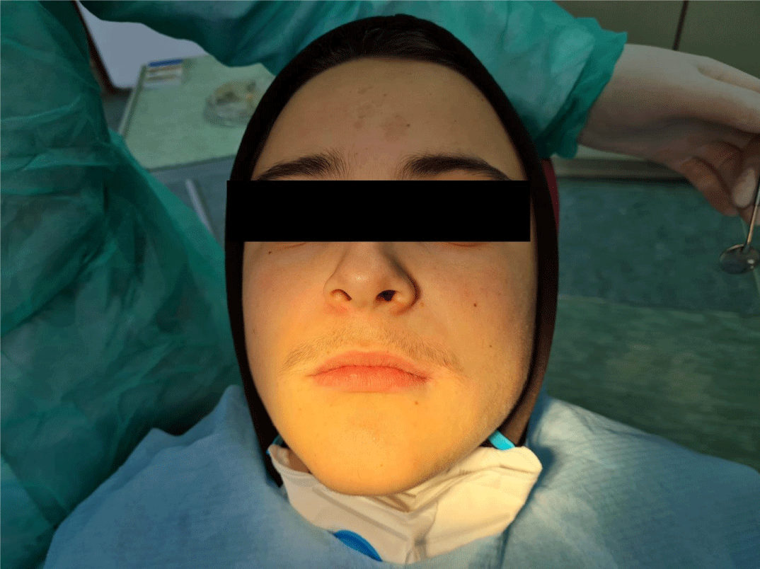
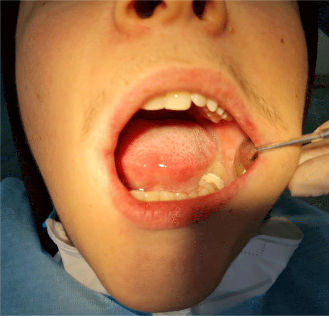
A 17-year old boy accompanied by medical support staff visited our Department for preventive and pediatric dentistry within the University Dental Center Ss.Pantelejmon in Skopje, due to a dental pain which comes from the first molar tooth (36) of the low jaw from the left side (Figure 1). Case report was approved by Ethics Committee of University Ss. Cyril & Methodius.
From the accompanying documentation we saw that the boy has congenital beta thalassemia major and was admitted to the Hematology Clinic a month ago. Due to the covid 19 pandemic, the patient was tested for the SARS-CoV-2, covid-19 virus prior to hospitalization and negative results were obtained. He was previously treated and examined at the University Clinic for Pediatric Diseases with combined transfusion regimen and chelation therapy with Ferriprox. Hepatic biopsy was performed in support of degeneratively altered liver tissue with a mild degree of hemosiderosis.
During magnetic resonance imaging (MRI) of the heart and small pelvis, the estimated value of the deposited iron (Fe) concentration in the myocardium is 0.598 mg/g dry matter, and in the liver parenchyma 2.404 mg/g dry matter. The patient has a sister, which is HLA identical.
The family history indicates that mother and father heterozygotes for thalassemia. Past diseases are status post appendectomiam, status post splenectomiam, tonsilitis chronica, degeneratio parenchymatosis hepatitis (low grade haemosyderosis).
Pharmacological history Tbl. Ospen a 500 mg, Tab. Exjadea 500 mg 2 X1, Tab. Imuran a 50 mg 3X1. Cps. Hydrea a 500 mg 2X1.
Objective finding on admission Afebrile, conscious, contactable, time, space and person oriented. Takes an active position on the bed. Nutrition and osteo-muscular structure are neat. Turgor and elasticity are neatly expressed. Leaves the impression of a moderately ill patient. Skin and visible mucous membranes icterically colored, with no signs of hemorrhagic syndrome. Lymph nodes in areas accessible for palpation - do not palpate.
Head and neck prominent frontal bone, prominent zygomatic bones, neck centrally positioned, cylindrical, symmetrical, movable in all directions. Thyroid not palpable, enlarged. Language centrally placed, moist, uncoated. Normosthenic chest was observed. Weakened vesicular respiration is heard on an auscultatory basis. Oxygen saturation 97%. Ictus cordis invisible and palpable. Cardiac action rhythmic, heart tones are clear, without the presence of pathological noises. Beats Per Minute (BPM) BPM=87/min, blood pressure TA=120/80 mmHg. The urogenital tract confirmed the male phenotype, according to age. Succusio renalis was сmutually negative.
The patient live in the Vladievci village, which is located in the vicinity of Strumiva city in the southeastern region of the Republic of Northern Macedonia, which is known for the increased presence of this hereditary hematological disease.
Before a dentist can perform any dental treatment, he must know some important information about type and treatment of thalassemia, the degree of iron load and systemic complications. In all invasive interventions, antibiotic prophylaxis is necessary. Hepatic tests and coagulation tests should always be considered.
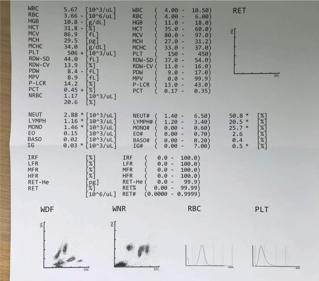
It is planned to perform allogeneic hematopoietic stem cell transplantation from a related donor-sister. Immediately before the dental intervention, a Complete Blood Count (CBC) was made (Figure 2).
At the first visit to our clinic, the patient was diagnosed with gangrene of the dental pulp, trepanation of the tooth 36 was preformed and application of the medication against tooth pain (sol. Chlumsky) and we left the tooth open. In the meantime, we asked for a blood test and for the next visit of the patient to be covered with antibiotic therapy. His platelet count was 406,000platelets per micro liter of blood.
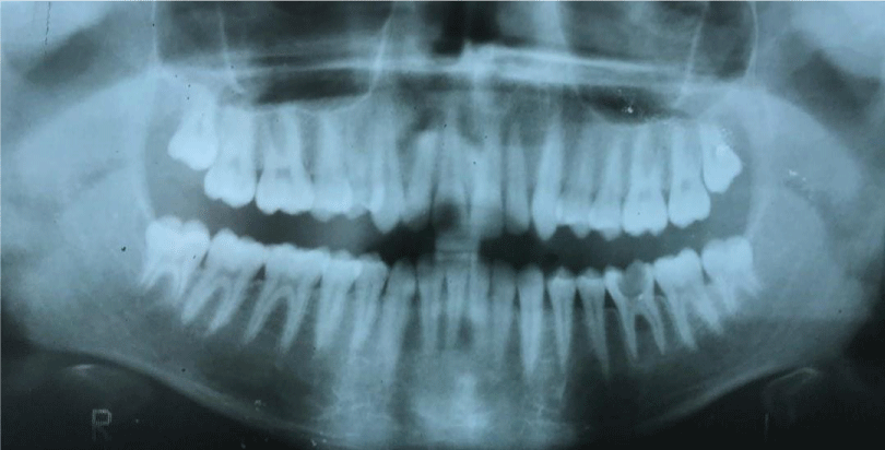
After conducting orthopantomography X-ray imaging (Figure 3) we decided to treat the tooth and in the next session we performed extirpation of the root canals and filled them with pasta (solution Clumsky and iodoform powder) and closed it temporarily (Figure 4). After a week of our intervention the patient received stem cells from his 12-year-old sister.
In the case of a bone marrow transplant, Hematopoietic Stem Cells (HSCs) are removed from a large bone in the donor, usually from the pelvis, using a large needle that reaches the center of the bone. This technique is called bone marrow collection and is performed under general anesthesia.
An intervention was performed in a surgical room, for a collection of bone marrow with a regular flow, at the pediatric surgery clinic. The period after the intervention passes without significant side effects. The girl donor is discharged in good general condition.After the intervention she receives therapy that includes Amp.Lendacin a 2mg, Amp. Niflam, Amp. Apotel, Amp.Famotidine, Amp. Granocyte.
The patient with thalassemia major has blood group B Rh +, while his sister (donor) has the same blood group B Rh +. After the intervention, she received transfusions of Frozen Serum plasma (FSA) and decanted erythrocytes 2 units. Upon discharge from the hospital he received a recommendation to continue with tablet therapy: tab. Pancef ȧ 400mg 1x1 / 7 days, tab.Brufen ȧ 400 mg 2x1 / as needed and topical dressings. An adequate hygienic-diet regime is required according to the previously given recommendation, rest.
The Macedonian Hematopoietic Stem Cell Transplantation Team has been a member of the EBMT (European Society for Blood and Marrow Transplantation) since 2003.
Disscusion
Efremov’s study, which covers a period of 40 years in the former Yugoslavia and involved about 37,000 school-age children and about 1,600 adults, found an average incidence of β-thalassemia of 1.2%. The highest incidence was registered in Macedonia (2.9%), and the lowest in Croatia (0.8%). A screening study of 12,680 newborns showed a 1.5% incidence of α-thalassemia [5].
Within the Balkan’s cuntries, after Greece, most cases are registered in the Republic of North Macedonia, about 2%. Most cases are β-thalassemia heterozygotes, HB Lepore in relatively small number, followed by δβ-thalassemia heterozygotes and HPFH, while other forms of thalassemia are present in very small numbers [6].
Children suffering from β-thalassemia major should be placed on regular transfusion programs (every 3-4 weeks) so that the hemoglobin level can be maintained at over 10g/dl, allowing for relatively normal growth and development. To prevent or delay the onset of haemochromatosis, excess iron should be removed by chelation. Splenectomy can reduce the need for transfusion in a condition that occurs when your spleen becomes enlarged (splenomegaly) [7]. Allogeneic stem cell transplantation has been shown to be a successful treatment, but efficacy has been reduced due to necessary histocompatibility with the stem cell donor as well as lifelong immunosuppression.
Significant success has been achieved with allogeneic bone marrow transplantation in patients with homozygous β-thalassemia who have had an HLA-identical donor. The mortality from the procedure in these patients is quite high (30%), due to the complications of iron overload, but it is significantly lower (below 10%) if the intervention is performed in early childhood [8].
The orofacial manifestations in people with thalassemia are numerous and intense. They are mainly the result of bone changes due to inefficient erythropoiesis. A major problem is malocclusion, which results from maxillary protrusion [9-12].
Patients with β-thalassaemia major reported in this study is at increased risk of developing dental caries and periodontal disease. It is worrying that he was not suggested to undergo dental treatment by his dentist before coming to hospital for stem cell transplantation. Especially because he was diagnosed damage to the liver with a degeneratively altered tissue by biopsy due to discontinuity in the application of chelation therapy.
To ensure safe and effective treatment, it is necessary for the dentist to have as much information about the disease as possible before starting the dental treatment of a person with thalassemia. The type and treatment of thalassemia, the degree of iron load, systemic complications, are important information to plan dental treatment. In all invasive interventions, antibiotic prophylaxis is necessary. Hepatic tests and coagulation tests should always be considered. Sedation / anesthesia is an important aspect. It should be carefully planned to reduce intra and post-intervention complications. Interventions should be minimally aggressive and as less traumatic as possible for patients [13]. Bayrak S, et al. recently evaluate the osseous changes preku radiomorphometric indexes and fractal dimension in dental panoramic radiography of 59 thalassemia patients and 59 healthy control subjects. They conclude that results of dental panoramic radiography can be used to refer these patients to appropriate medical investigation [14]. Rani ST, et al. also evaluate and compare the body mass index (BMI), dental age, salivary alkaline phosphatase levels, malocclusion, and treatment needs in 50 children with β thalassemia major and 50 healthy controls. The high OHI-S index scores, low gingival index scores, low BMI, salivary alkaline phosphatase levels and higher prevalence of malocclusion are find within tallasemia children. They concluded that greater dental treatment needs in children with thalassemia [15].
In a study conducted by Hatab, et al. involving 54 patients with Thalassemia major aged 5–18 years, clinical and radiological features were analyzed. In addition, poor oral hygiene was registered in 60% and gingivitis in 43%. More than half had a frontal anomaly, saddle nose and maximal protrusion. Dental discoloration and pallor of the oral mucosa were present in 44% and 39%, respectively. Dental pain was observed in 40%, and headache in 29% [16]. Hazza, et al. evaluate the dental development of patients within 44 patients with thalassemia major in comparison with unaffected children, using the Demirjian method for dental age estimation. He analyse panoramic radiographs taken before the age of 16 years. The delay in dental development slow varied according to the patient’s age within patients with thalasemia major [17].
Conclusion
The dental problem that occurred in our patient just before the transplantаtion procedure can compromise the planned therapy. Therefore, timely dental care and preventive care of every patient with ß-thalassemia is necessary to be preformed.
With the introduction of stem cell transplantation from a related donor, a bright future has been opened for children with thalassemia in the Republic of Northern Macedonia. We hope that the allergenic stem cell transplant performed in the presented case although was not committed during early childhood will be succesful and will improve the quality of life of the patient.
Author’s contributions
AV contributed to conception, drafting, and critical revision of the manuscript; TA and ZN searched the literature relevant to the work. All authors gave final approval and agree to be accountable for all aspects of the work.
- Smith CH (1948) Detection of mild types of Mediterranean(Cooley’s) anemia. Am J Dis Child 75: 505–527. Link: https://bit.ly/3jDy2J6
- Modell B, Darlison M, Birgens H, Cario H, Faustino P, et al. (2007) Epidemiology of haemoglobindisorders in Europe: an overview. Scand J Clin Lab Invest 67: 39-69. Link:https://bit.ly/3lYSdnX
- Efremov GD (2007) Dominantly Inherited beta-Thalassemia. Hemoglobin 31: 193-207. Link: https://bit.ly/3fOCoMD
- De Sanctis V, Kattamis C, Canatan D, Soliman AT, Elsedfy H, et al. (2017) β-Thalassemia Distribution in the Old World: an Ancient Disease Seen from a Historical Standpoint. Mediterr J Hematol Infect Dis 9: e2017018. Link: https://bit.ly/3yHfE8w
- Efremov GD (2007) Thalassemias and other hemoglobinopathies in the Republic of Macedonia. Hemoglobin 31: 1-15. Link:https://bit.ly/3iEW8nP
- Efremov GD (1992) Hemoglobinopathies in Yugoslavia: an update. Hemoglobin 16: 531-544. Link: https://bit.ly/3AzwoiM
- Jankulovski N, Antovic S, Kuzmanovska B, Mitevski A (2014) Splenectomy forhaematological disorders. Prilozi 35: 181-187. Link:https://bit.ly/3xHSZYl
- Ansari-Moghaddam A, Adineh HA, Zareban I, Mohammadi M, Maghsoodlu M (2018) The survival rate of patients with beta-thalassemia major and intermedia and its trends in recent years in Iran. Epidemiol Health 40: e2018048. Link:https://bit.ly/3Cw97Qq
- Ambarkova V (2018) Dental Aspects of Thalassemias. EC Dental Science 17: 638-639. Link:https://bit.ly/3fPGQdN
- Hattab FN, Yassin OM (2011) Dental arch dimensions in subjects with beta-thalassemia major. J Contemp Dent Pract 12: 429-433. Link:https://bit.ly/37AeP5K
- Einy S, Hazan-Molina H, Ben-Barak A, Aizenbud D (2016) Orthodontic Consideration in Patients with Beta-Thalassemia Major: Case Report and Literature Review. J Clin Pediatr Dent 40: 241-246. Link:https://bit.ly/3CDo1nX
- Bouguila J, Besbes G, Khochtali H (2015) Skeletal facial deformity in patients with β thalassemia major: Report of one Tunisian case and a review of the literature. Int J Pediatr Otorhinolaryngol 79: 1955-1958. Link:https://bit.ly/37yFjV9
- Sinokapovska S, Dimova C (2015) The importance of dental care in patients with thalassemia. Vox Dentarii 10: 42-43.
- Bayrak S, Göller Bulut D, Orhan K, Sinanoğlu EA, Kurşun Çakmak EŞ, et al. (2020) Evaluation of osseous changes in dental panoramic radiography of thalassemia patients using mandibular indexes and fractal size analysis. Oral Radiol 36: 18-24. Link:https://bit.ly/3skMI49
- Rani ST, Reddy ER, Kiranmai M, Mudusu SP, Srikanth S, et al. (2019) Comparative Evaluation of BMI, Dental Age, Salivary Alkaline Phosphatase Levels, and Oral Health Status in Children with β Thalassemia Major. Int J Clin Pediatr Dent 12: 303-306. Link:https://bit.ly/37FErOl
- Hattab FN (2013) Patterns of physical growth and dental development in Jordanian children and adolescents with thalassemia major. J Oral Sci 55: 71-77. Link:https://bit.ly/3lUimnU
- Hazza'a AM, Al-Jamal G (2006) Dental development in subjects with thalassemia major. J Contemp Dent Pract 7: 63-70. Link: https://bit.ly/2VKH3s6
Article Alerts
Subscribe to our articles alerts and stay tuned.
 This work is licensed under a Creative Commons Attribution 4.0 International License.
 Save to Mendeley
