Archives of Hematology Case Reports and Reviews
Follicular B-Cell Lymphoma Embedded in a Pulmonary Chondroid Hamartoma: A Case Report
Lara Mannelli1*, Sofia Kovalchuk1, Benedetta Puccini1, Gemma Benelli1, Catia Dini2, Alberto Bosi1 and Luigi Rigacci1
2Department of Radiodiagnostic, AOU Careggi, General and Academic Hospital, Florence, Italy
Cite this as
Mannelli L, Kovalchuk S, Puccini B, Benelli G, Dini C, et al. (2017) Follicular B-Cell Lymphoma Embedded in a Pulmonary Chondroid Hamartoma: A Case Report. Arch Hematol Case Rep Rev 2(1): 005-007. DOI: 10.17352/ahcrr.000005Background: Here we report a rare case of concomitant pulmonary chondroid hamartoma and follicular non Hodgkin lymphoma in a 65-year-old woman with a history of 4-month nonspecific symptoms like fever, sweating and weight loss.
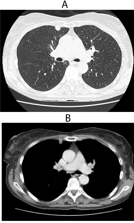
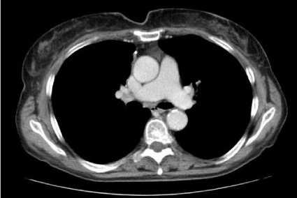
Methods: Chest CT scan revealed a 12-mm nodule with relatively regular margins in the right middle lung field, adhering to the chest wall, with no involvement of hilar or mediastinal lymph nodes. The patient underwent thoracoscopy and resection of the lesion. Following histological exam and immunohistochemical staining, the patient was diagnosed as having follicular b-cell lymphoma embedded in a pulmonary chondroid hamartoma. FISH analysis was performed and showed the presence of typical follicular lymphoma chromosomal translocation t (14;18) (q32;q21).
Conclusions: Following the final diagnosis, we decided not to refer the patient to chemotherapy and she enter watch and wait regimen with imaging control every 6 months.
After 18 months, the chest X-ray and abdominal ultrasound are still negative with no evidence of recurrence.
Introduction
Pulmonary chondroid hamartoma (PCH) is the most common benign neoplasm of the lung with an incidence of 0.25% [1], and accounts for 7-14% of all solitary lung nodules [2].
PCH are usually well circumscribed from the lung parenchyma or protude into the bronchial lumen when located in the bronchus [3].
Primary pulmonary lymphomas (PPL) are extremely rare, representing less than 1% of primary malignant lung cancers, fewer than 1% of malignant lymphomas, and accounting for only 3.6% of extranodal lymphomas [4].
Here we report a rare case of pulmonary chondroid hamartoma containing grade I-II follicular lymphoma in a 65-yr-old female patient.
Case Description
A 65 year-old woman presented with dry cough and progressive dyspnea. She experienced 10 kg of weight loss during 4 months and reported nocturnal fever and sweating.
The patient was a chain-smokers with prior history of asthma.
She was admitted to a hospital and received broad-spectrum antibiotics with no improvement. In her second hospital admission, no focal findings were noted on examination, especially no palpable lymphnodes and no hepatosplenomegaly. A complete blood test showed a white cell count of 6.21x10⁹ cells/l and the percentage of neutrophil was 54.9%. The renal and hepatic function and serum lactate dehydrogenase levels were unremarkable.
Chest CT scan revealed a 12-mm nodule with regular margins in the right middle lung field, adhering to the chest wall. No hilar or mediastinal lymph nodes were significantly visible (Figures 1a,b,2).
According to thoracic surgeon, the nodular lesion was enucleated, with the rest of the lung being considered normal and not resected.
Histologically, the lesion was composed mainly of mature cartilage, fatty tissue and normal bronchial epithelium, containing lymphoid infiltrate of small lymphocytes, organized into follicles with expansion of marginal areas.
Immunohistochemical staining resulted positive for CD20, bcl6, bcl1-2 and negative for CD3, CD10, IRF4, with MIB-1 index <10%. FISH analysis showed the presence of chromosomal traslocation t(14;18)(q32;q21).
Therefore, the patient was diagnosed with chondroid hamartoma containing primary pulmonary follicular lymphoma grade I-II.
Bone marrow trephine biopsy and a complete enhanced CT scan and FDG-PET/CT showed no further abnormalities.
The absence of any detectable sign of disease after surgery led us to perform no further therapy, while keeping the patient under strict clinical control.
After 18 months, the chest X-ray and abdominal ultrasound are still negative with no evidence of recurrence.
Conclusions
Pulmonary chondroid hamartoma was first described by Albrecht in 1904 [5].
The onset of the tumor is generally in the adulthood (peak age incidence in the sixth decade) (2) with a male: female ratio being 2:1 to 3:1 [5].
Chondroid hamartomas are usually solitary and well circumscribed, with dimensions usually ranging from 1 to 4 cm [5]. Rarely, they may be larger and multiple. Based on the localization, PCH may be distinguished into two types, peripheral parenchymal type and central endobronchial type. The former comprises over 90% of all chondroid hamartomas, arises from small bronchi and is generally asymptomatic. The latter arises from large bronchi, is less frequent but is often symptomatic [5].
PCHs are frequently discovered on routine X-ray chest and they appear as solitary coin lesions. Less commonly, they may show up as multiple coin lesions [2] or as cavitary lesions in case of cystic PCH.
Some cases show popcorn pattern of calcification. Trans-thoracic FNAC can be used to establish the diagnosis [5]. The cytologic diagnosis is based on the recognition of mesenchymal components with fibromyxoid and chondroid elements.
PCH histologically consists of islands of cartilage, fat, fibromyxoid stroma, and narrow spaces lined by respiratory epithelium. [5].
Recently, cytogenetic studies have demonstrated an abnormal karyotype in several instances. Thus, it was proposed that PCH represents a true benign mesenchymal neoplasm instead of a developmental abnormality [1].
Recurrence of chondroid hamartoma is extremely unusual [5] and malignant transformation is exceptional.
Primary pulmonary lymphomas are extremely rare and represent <1% of primary malignant lung tumors, <1% of lymphomas, and only 3%–4% of extranodal lymphomas [6]
PPL is defined as clonal lymphoid proliferation affecting one or both lungs [7], lobes or primary bronchus, with or without mediastinal involvement, and no evidence of extrathoracic lymphoma at the time of diagnosis or for 3 months thereafter.
The most common PPL type is mucosa-associated lymphoid tissue (MALT) lymphoma,
An extranodal marginal zone lymphoma that accounts for 80%–90% of PPL cases.
DLBCL is the second most common type of PPL [6] followed by follicular B-cell lymphoma and Hodgkin lymphoma.
Because of the small number of PPL cases, it is very difficult to draw definitive conclusions about its clinical features and treatment. The radiological aspect (in 50%–90% of PPL cases) is a localized alveolar opacity [6], less than 5 cm-diameter and blurred or well-defined contours (according to the series); PPL is associated in nearly 50% of cases with an air bronchogram [6], peribronchial disease with a halo of ground-glass shadowing and proximal bronchiectasis.
PPL has low FDG involvement, so the benefit of FDG-PET in the clinical assessment of this kind of lymphoma is still unclear [6].
Although the diagnostic procedure of this disease has not been well defined, open thoracotomy or chest CT‑guided percutaneous biopsy are the preferred methods used [8].
The positive value of open thoracotomy biopsy is high, but it is associated with high medical cost and clinical risk [8].
By contrast, the diagnostic value of chest CT-guided percutaneous biopsy has a low medical cost and less serious complications. However, the procedure is limited for the diagnosis of masses that are in or adjacent to the hilum or mediastinum [8].
Bronchoscopy may be effective in endobronchial lymphoma only (10%).
Although there is no consensus on the treatment of PPL, surgical resection is generally performed in cases of localized tumors. Because the rate of local recurrence after complete resection is reported to be high, chemotherapy or radiation therapy should be considered for non-indolent lymphomas [6].
In the present case, a nodular opacity with regular margins was detected by CT-scan in a patient symptomatic for dyspnea, cough, chest pain, as well as fever and weight loss.
The pathologist confirms the presence of follicular b-cell lymphoma embedded in a chondroid hamartoma, and we didn’t find a similar cases in the previous reported studies.
The patient was considered a surgical candidate because of the neoplasm localization.
Following the final diagnosis, we decided not to refer the patient to chemotherapy and she enter watch and wait regimen with imaging control every 6 months.
After 18 months, the chest X-ray and abdominal ultrasound are still negative with no evidence of recurrence.
- Ozbudak IH, Dertsiz L, Bassorgun CI, Ozbilim G (2008) Giant cystic chondroid hamartoma of the lung. J Pediatr Surg 43: 1909-1911. Link: https://goo.gl/eziATX
- Kim GY, Han J, Kim DH, Kim J, Lee KS (2005) Giant cystic chondroid hamartoma. J Korean Med Sci 20: 509-511. Link: https://goo.gl/1GWzp5
- Minami Y, Iijima T, Yamamoto T, Morishita Y, Terashima H, et al. (2005) Diffuse pulmonary hamartoma: a case report. Pathol Res Pract 200: 813-816. Link: https://goo.gl/zhKj4e
- Graham BB, Mathisen DJ, Mark EJ, Takvorian RW, (2005) Primary pulmonary lymphoma. Ann ThoracSurg 80: 1248-1253. Link: https://goo.gl/sTZr58
- Majeed FA, Nafees M, Rehman A (2010) Chondroid hamartoma presenting as an uncalcified mass. Pakistan Armed Forces MJ 60: 4 Link: https://goo.gl/UR5Ypl
- Yoshino N, Hirata T, Takeuchi C, Usuda J, Hosone M, et al. (2015) A case of primary pulmonary diffuse large B-cell lymphoma diagnosed by transbronchial biopsy. Ann Thorac Cardiovasc Surg 21: 396-398. Link: https://goo.gl/EFWelE
- Zhu J, Wang Y, Gong L, Huang G (2014) Diagnosis of primary pulmonary T cell/histiocyte rich large B-cell lymphoma with tissue eosinophilia via clinicopathological observation and molecular assay. Diagn.Pathol 9: 188. Link: https://goo.gl/KGuA6l
- Jiang AG, Gao XY, Lu HY (2014) Diagnosis and management of a patient with primary pulmonary diffuse large B-cell lymphoma: a case report and review of the literature. Experimental and Therapeutic Medicine 8: 797-800. Link: https://goo.gl/pMmY2c
Article Alerts
Subscribe to our articles alerts and stay tuned.
 This work is licensed under a Creative Commons Attribution 4.0 International License.


 Save to Mendeley
