Archives of Hematology Case Reports and Reviews
Dual Complication of Visceral Leishmaniasis-Hemophagocytic Lymphohistiocytosis and Myocarditis–A Case Report
1Internal Medicine Department, St. Paul’s Hospital Millennium Medical College, Addis Ababa, Ethiopia
2Internal Medicine Department, Saglan Wajee General Hospital, Holeta, Oromia, Ethiopia
3Assistant Professor of Anatomic Pathology, Dermatopathologist Fellow, Armauer Hansen Research Institute, Addis Ababa, Ethiopia
4Hematologist, Internal Medicine Department, St. Peter Hospital, Addis Ababa, Ethiopia
Author and article information
Cite this as
Teshome EL, et al. Dual Complication of Visceral Leishmaniasis-Hemophagocytic Lymphohistiocytosis and Myocarditis–A Case Report. Arch Hematol Case Rep Rev. 2026; 11(1): 1-5. Available from: 10.17352/ahcrr.000052
Copyright License
© 2026 Teshome EL, et al. This is an open-access article distributed under the terms of the Creative Commons Attribution License, which permits unrestricted use, distribution, and reproduction in any medium, provided the original author and source are credited.Visceral Leishmaniosis is a life-threatening protozoal disease that can affect multiple organs. It typically presents with fever, hepatosplenomegaly, and cytopenia, but can sometimes manifest with rare complications. This case report discusses a 20-year-old male who presented with intermittent low-grade fever, night sweats, loss of appetite, vomiting of ingested matter, and a 6 kg weight loss for the past three months. He was acutely and chronically ill with a blood pressure of 80/50 mmHg, a heart rate of 120 beats per minute, a respiratory rate of 28 breaths per minute, and a temperature ranging from 38.3°C to 39.0°C. He had mild pleural effusion and hepatomegaly with liver extending 6 cm below the right costal margin and splenomegaly, with the spleen palpable 4 cm below the left costal margin. The patient also exhibited pedal edema. Initial investigation showed pancytopenia (WBC: 980, Hemoglobin/Hematocrit: 7 and 18.9, and platelets of 80000). He was managed as a case of sepsis of the chest and Gastrointestinal focus for eight days with broad-spectrum antibiotics. He was transfused several times, and his hemoglobin was starting to rise until he developed type I respiratory failure secondary to cardiogenic shock with pulmonary edema secondary to acute myocarditis. For this, he was managed at the ICU with vasopressor and diuresis, and when the cell count further dropped, bone marrow aspiration was done, which suggested secondary Hemophagocytic Lymphohistiocytosis. Other additional serologic tests confirmed the diagnosis (RK39 was positive, Serum Triglyceride was 418, and Serum Ferritin was 1500). The patient was treated according to the 2004 HLH protocol and discharged improved. This complication of leishmaniosis is due to immune system hyperactivation in response to the protozoa in the bone marrow. Additionally, the cardiovascular symptoms can be possibly due to the direct involvement of the protozoa in the heart, which is usually managed supportively. Both conditions are uncommon, and this report highlights the importance of maintaining a high level of suspicion for these complications and initiating early treatment.
Leishmania is a protozoan disease transmitted by the female sandfly. It exists in three main forms: visceral, cutaneous, and mucocutaneous. In Ethiopia, both visceral and cutaneous leishmaniasis are present, with an overall prevalence of 9.13% [1]. Visceral leishmaniasis (kala-azar) is fatal if untreated and typically presents with fever, weight loss, anemia, and hepatosplenomegaly. However, it can occasionally present with rare complications. This case presents two such complications in one patient: visceral leishmaniasis causing secondary Hemophagocytic Lymphohistiocytosis and cardiac involvement. Secondary HLH in leishmaniasis results from immunologic hyperactivation towards the protozoa, leading to the destruction of normal blood cells. Leishmaniasis can also involve the heart, manifesting in various cardiac symptoms.
Case
A 20-year-old male presented with progressively worsening symptoms over three months, including easy fatigability, headaches, and tinnitus. He also reported intermittent low-grade fever, night sweats, loss of appetite, and a 6 kg weight loss during this period. Before seeking medical care, he experienced a dry cough, pleuritic chest pain, and shortness of breath with minimal exertion, along with multiple episodes of vomiting. The patient, a daily laborer, had recently traveled to the northwestern part of Ethiopia and border cities in Sudan.
Upon examination, he appeared acutely and chronically ill. His vital signs were as follows: blood pressure of 80/50 mmHg, heart rate of 120 beats per minute, respiratory rate of 28 breaths per minute, and a temperature ranging from 38.3°C to 39.0°C. He had pale conjunctiva and non-icteric sclera, with no enlarged lymph nodes in accessible areas. Initial chest examination showed signs of minimal pleural fluid collection. Cardiovascular examination revealed a flat Jugular Venous Pressure (JVP) with well-heard S1 and S2 heart sounds. Abdominal examination showed hepatomegaly, with the liver extending 6 cm below the right costal margin, and splenomegaly, with the spleen palpable 4 cm below the left costal margin. The patient also exhibited pedal edema. He was kept at the emergency ward and investigated with a complete blood count, which showed pancytopenia with a WBC count of 970 and a differential of 57% neutrophil and 35 % lymphocyte, Hemoglobin and hematocrit of 7 and 18.8, respectively and platelet count of 80000. With the assessment of septic shock of GI and chest focus and pancytopenia secondary to sepsis to rule out megaloblastic anemia, the patient was given 2 units of intravenous normal saline, started on cefepime, vancomycin, and metronidazole, and transfused with two units of cross-matched packed RBC on the first day. His blood pressure stabilized. Peripheral morphology revealed anisochromic anisopokiliocytic anemia, leukopenia, and thrombocytopenia. Serum folate and Cobalamine levels were in the normal range, 524 and 8.58 respectively. Abdominal Ultrasound showed hepatomegaly measuring 17.8cm and splenomegaly measuring 15.2cm with bilateral pleural effusion moderate ascites. Blood and urine culture did not show any growth. Pleural Fluid analysis showed WBC 20, Neutrophil: 85%, Lymphocyte:14% , Gram stain, AFB, and Gene expert were negative. Organ function tests were Cr:0.82, Urea: 20.3, SGOT:85, SGPT:24, ALP:580 , Total Bilirubin: 1.06 , Direct Bilirubin:0.66, Pt: 17.2 ,PTT:52 , INR:1.59, Albumin: 1.5, and an LDH:536.
He continued to receive the antibiotics and transfusion; his fever, shortness of breath, and anemia symptoms were improving for the first 7 days of admission at the emergency, and his hemoglobin got to 13.4. However, his platelet count decreased to the level of 23000 while his WBC count remained 1590.
On the eighth day of admission, the patient experienced worsening shortness of breath, palpitations, and chest pain. Physical examination revealed unrecordable blood pressure, fast and feeble peripheral pulses, decreased respiratory effort with an abdominal breathing pattern, and an oxygen saturation of 70% while using a face mask with a rebreather. The chest examination showed dull percussion notes in the posterior lower third of the lungs bilaterally and decreased breath sounds. The cardiovascular examination revealed raised Jugular Venous Pressure (JVP), while the abdominal examination indicated signs of fluid accumulation. The patient also exhibited grade III edema in the lower extremities and was lethargic, with a Glasgow Coma Scale (GCS) score of 13. Additional cardiac evaluation revealed serial electrocardiographic monitoring with persistent sinus tachycardia during ICU admission. Initial serum troponin measurements were not significantly elevated, and repeat measurements remained within non-diagnostic range. Bedside echocardiography demonstrated globally reduced left ventricular systolic function with an estimated ejection fraction of 30%, mild left ventricular dilatation, and no regional wall motion abnormality or significant valvular pathology. There was no echocardiographic evidence of pericardial tamponade. These findings were considered most consistent with acute inflammatory myocarditis associated with visceral leishmaniosis. Chest X-ray also showed bilateral air space opacifications and bilateral pleural effusion. With the assessment of Type I respiratory failure secondary to cardiogenic pulmonary edema secondary to acute myocarditis patient was transferred to the Medical ICU, and management with vasopressors and diuretics was initiated after the patient was placed on mechanical ventilation. With these managements, the oxygen requirement improved, and blood pressure was maintained. However, on the third day of admission at the ICU, the patient developed worsening tachycardia with a heart rate ranging from 180 to 220 beats per minute, as observed on the cardiac monitor. A bedside ECG revealed a monomorphic ventricular tachycardia pattern. The arrhythmia was successfully treated with Amiodarone.
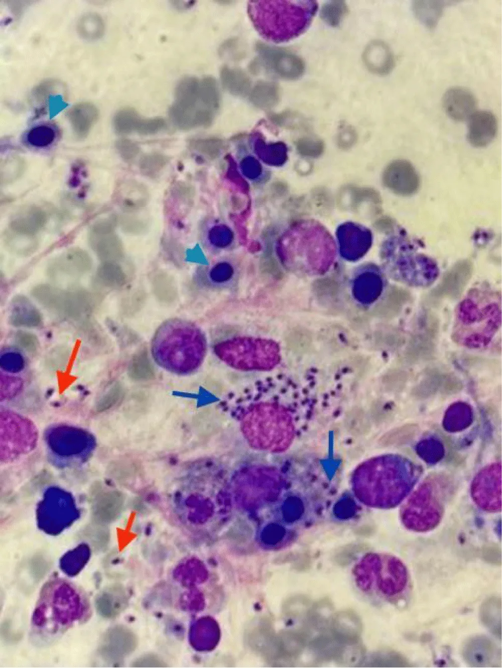
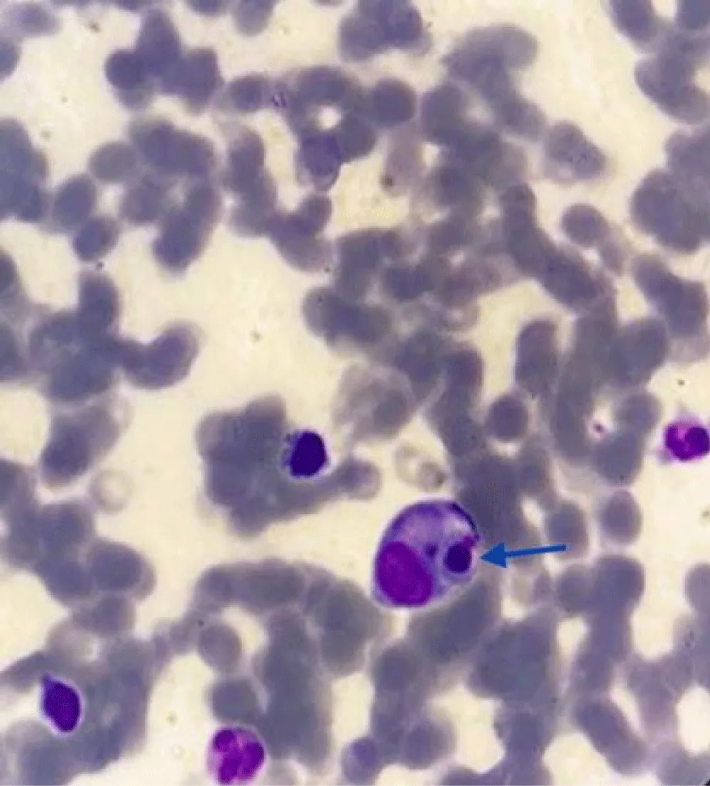
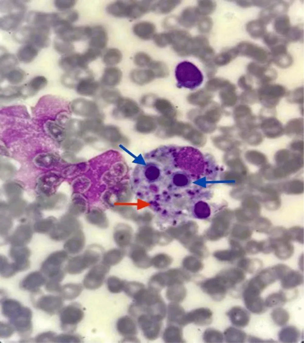
On the 4th day of ICU admission, cell count started to drop again, and CBC showed WBC of 1010, hemoglobin of 8.2, and platelets of 13000. Bone marrow aspiration was done, and it showed a particulate and hemodiluted bone marrow sample containing trilineage hematopoietic elements. Erythroid cells exhibited normoblastic maturation (28%), and myeloid cells showed terminal differentiation with an M: E ratio of 1.86:1. Megakaryocytes appeared normal. Lymphocytes comprised 15% of the nucleated cells, and plasma cells accounted for 3%. Multiple intracellular and extracellular Leishman-Donovan (LD) bodies were observed, along with histiocytes showing hemophagocytosis. There was no increase in blast count (Figure 1). Final Diagnosis of bone marrow aspiration was visceral leishmaniasis with secondary Hemophagocytic Lymphohistiocytosis (HLH). Serum RK39 qualitative antibody test became positive, Serum Triglyceride was 418, and Serum Ferritin was 1500.
The patient was promptly managed according to HLH protocols. Treatment initiated with amphotericin B and dexamethasone. Blood pressure stabilized with the support of vasopressors, and pulmonary edema was managed through diuresis. After six days of ICU admission, the patient was successfully weaned off the mechanical ventilator and transferred from the ICU to the medical ward.
Over the course of one week, there was a noticeable improvement in the patient’s cell counts. The patient was subsequently discharged from the hospital and continued follow-up at the medical clinic. Two months post-discharge, laboratory results indicated that cell counts, organ function, ferritin, and triglyceride levels had all returned to normal ranges, demonstrating a positive response to the treatment and effective management of HL (Table 1).
Erythroid precursor cells (arrowhead) and myeloid cells. Multiple intracellular Leishmania parasites, LD bodies (blue arrow). Extracellular parasites (red arrow) this typically happens when delicate macrophages rupture in the process of slide preparation (Figures 2,3).
Discussion
Hemophagocytic lymphohistiocytosis : Non-malignant histiocytosis encompasses a range of disorders characterized by lesions that impact the immune regulation of the mononuclear phagocyte system and/or dendritic cells. An example is Langerhans Cell Histiocytosis, which primarily affects dendritic cells, whereas HLH predominantly involves macrophages [2]. HLH can be divided into two distinct conditions, which can be challenging to differentiate.
Primary HLH (Familial hemophagocytosis): This is a hereditary, autosomal recessive disorder typically presenting in pediatric patients. Diagnosis relies on clinical, laboratory, and histopathological findings. The 2004 HLH diagnostic criteria (Table 1) are useful for diagnosing this condition. However, due to its high mortality rate, clinicians may need to start treatment even if not all criteria are strictly met [2,3].
Secondary Hemophagocytic Lymphohistiocytosis (HLH): Secondary HLH is a severe condition resulting from intense immunological activation triggered by infections, malignancies, or rheumatologic disorders. This form of HLH is often underdiagnosed as it may not be included in the initial differential diagnosis. Conditions such as sepsis, Disseminated Intravascular Coagulation (DIC), and thrombotic thrombocytopenic purpura (TTP) can present with overlapping clinical features, potentially leading to misdiagnosis. Key diagnostic indicators, such as elevated ferritin and Lactate Dehydrogenase (LDH) levels, along with specific bone marrow findings, are critical for distinguishing secondary HLH and should prompt further investigation [4].
HLH associated with leishmania infection: Secondary HLH due to leishmania infection is rare, representing only about 1% of all leishmaniasis cases [5]. The exact mechanisms through which Leishmania infection triggers HLH remain unclear. It is believed that hyperactivation of macrophages or histiocytes leads to impaired recognition and phagocytosis of normal blood cells, due to compromised infection control [6].
Treatment approach: The management of secondary Hemophagocytic Lymphohistiocytosis (HLH) focuses on several key areas:
- Stabilization and support: Initial treatment includes stabilizing the patient and providing necessary organ support.
- Trigger identification and management: It is essential to identify and address underlying triggers such as infections or malignancies.
- Modulating the immune response: Recent studies emphasize the importance of regulating T cell dysregulation in HLH. In the case of Visceral Leishmaniasis (VL)-associated HLH, treatment with liposomal amphotericin has been effective. Unlike primary HLH, which often requires etoposide or cyclosporine, VL-associated HLH did not necessitate these treatments. Dexamethasone was used initially in severe cases but was gradually tapered as patients improved. Remarkably, the study reported excellent outcomes with no deaths or relapses during a 40-month follow-up period [7].
Overall, secondary Hemophagocytic Lymphohistiocytosis (HLH), particularly when associated with leishmania infection, presents significant diagnostic and treatment challenges. Early recognition and targeted therapy are crucial for improving patient outcomes and preventing severe complications (Table 2).
Cardiac manifestations of visceral leishmaniasis: While visceral leishmaniasis typically affects various organs throughout the body, involvement of the heart is rare. Cardiovascular manifestations are less well-documented compared to other symptoms of the disease. Early detection of cardiac involvement is challenging but essential to prevent severe complications. Leishmania infection has been associated with myocarditis and inflammatory cardiomyopathy [8].
In a canine model, leishmania infection induces a strong inflammatory response, characterized by widespread infiltration of mononuclear immune cells in the myocardium [9]. This inflammatory reaction can result in fatal arrhythmias and acute heart failure. Additional cardiac manifestations include pericarditis and pericardial effusion, which in severe cases can progress to cardiac tamponade (Figure 4).
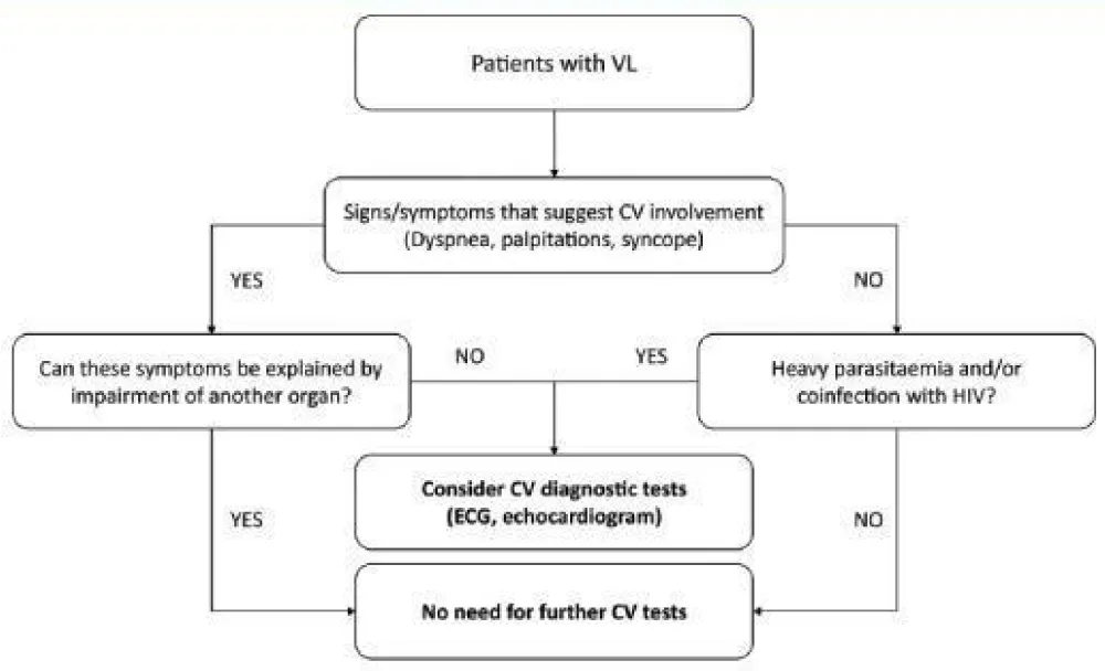
Future research should explore the use of biomarkers for screening myocardial injury in individuals with Visceral Leishmaniasis (VL). Given that VL is prevalent in underdeveloped regions, the application of expensive imaging techniques, such as cardiac magnetic resonance imaging, has not been extensively studied for their diagnostic value. Advances in imaging technology could potentially enhance early detection of cardiovascular involvement in these patients [10].
The primary objective in treating VL is to eliminate the parasite, as cardiac complications typically improve with appropriate supportive care when necessary [11]. Currently, there are no specific treatments for cardiovascular issues associated with leishmaniasis; management focuses on standard care for myocarditis or severe pericarditis [12].
Conclusion
Leishmania can present with rare and potentially fatal complications, requiring a high index of suspicion. Diagnostic criteria for HLH and myocarditis may not always need to be strictly met before initiating treatment. Exaggerated immune responses to the parasite can lead to conditions such as HLH, where normal blood cells are consumed by overactive immune cells. Similarly, strong immune responses can cause myocardial and pericardial damage. Early initiation of antiprotozoal therapy and prompt administration of steroids for secondary Hemophagocytic Lymphohistiocytosis (HLH), along with supportive care, are essential to preventing devastating outcomes.
- Haftom M, Petrucka P, Gemechu K, Nesro J, Amare E, Hailu T, et al. Prevalence and Risk Factors of Human Leishmaniasis in Ethiopia: A Systematic Review and Meta-Analysis. Infect Dis Ther. 2021;10(1):47-60. Available from: https://doi.org/10.1007/s40121-020-00361-y
- The Writing Group of the Histiocyte Society. Histiocytosis syndromes in children. Lancet. 1987;1(8526):208-209. Available from: https://pubmed.ncbi.nlm.nih.gov/2880029/
- Henter JI, Arico M, Elinder G, Imashuku S, Janka G. Familial hemophagocytic lymphohistiocytosis. Primary hemophagocytic lymphohistiocytosis. Hematol Oncol Clin North Am. 1998;12:417–433. Available from: https://doi.org/10.1016/s0889-8588(05)70520-7
- Henter JI, Horne A, Aricó M, Egeler RM, Filipovich AH, Imashuku S, et al. HLH-2004: Diagnostic and therapeutic guidelines for hemophagocytic lymphohistiocytosis. Pediatr Blood Cancer. 2007;48(2):124-131. Available from: https://doi.org/10.1002/pbc.21039
- Neycheva S, Oparanov B, Kamburova A, Karalilova R, Stoeva V. Hemophagocytic Lymphohistiocytosis Triggered by Leishmaniasis: A Case Report and Literature Review. Am J Case Rep. 2021;22:e933012. Available from: https://doi.org/10.12659/AJCR.933012
- Morimoto A, Uchida K, Chambers JK, Sato K, Hong J, Sanjoba C, et al. Hemophagocytosis induced by Leishmania donovani infection is beneficial to parasite survival within macrophages. PLoS Negl Trop Dis. 2019;13(11):e0007816. Available from: https://doi.org/10.1371/journal.pntd.0007816
- Mottaghipisheh H, Kalantar K, Amanati A. Shokripour M, Shahriari M, Zekavat OR, et al. Comparison of the clinical features and outcome of children with hemophagocytic lymphohistiocytosis (HLH) secondary to visceral leishmaniasis and primary HLH: a single-center study. BMC Infect Dis. 2021;21:732. Available from: https://doi.org/10.1186/s12879-021-06408-w
- Caforio AL, Pankuweit S, Arbustini E, Basso C, Gimeno-Blanes J, Felix SB, et al. Current state of knowledge on etiology, diagnosis, management, and therapy of myocarditis:a position statement of the European society of cardiology working group on myocardial and pericardial diseases. Eur Heart J. 2013;34(33):2636–2648. Available from: https://doi.org/10.1093/eurheartj/eht210
- Martínez-Hernández L, Casamian-Sorrosal D, Barrera-Chacón R, Cuesta-Gerveno JM, Belinchón-Lorenzo S, Gómez Nieto LC, et al. Comparison of myocardial damage among dogs at different stages of clinical leishmaniasis and dogs with idiopathic chronic kidney disease. Vet J. 2017;221:1–5. Available from: https://doi.org/10.1016/j.tvjl.2016.11.015
- Farina JM, García-Martínez CE, Saldarriaga C, Pérez GE, Melo MB, Wyss F, et al. Leishmaniasis and Heart. Arch Cardiol Mex. 2022;92(1):85-93. Available from: https://doi.org/10.24875/acm.20000508
- Nunes MC, Júnior MH, Diamantino AC, Gelape CL, Abreu Ferrari TC. Cardiac manifestations of parasitic diseases. Heart. 2017;103:651–658. Available from: https://doi.org/10.1136/heartjnl-2016-309870
- Puerto-Alonso JL, Molina-Ruano FJ, Gómez-Soto F, Gómez-Rodríguez F. Visceral leishmaniasis with cardiac affectation in an immunocompetent patient. Med Clin. 2006;127:519. Available from: https://doi.org/10.1157/13093277


 Save to Mendeley
Article Alerts
Subscribe to our articles alerts and stay tuned.
 This work is licensed under a Creative Commons Attribution 4.0 International License.