Archives of Clinical Nephrology
Ochoa syndrome: An overlooked diagnosis – A case report
Mário Nicolau Barros Jacobino1*, Patrícia Barros Aquino Jacobino2, Matheus Saraiva Lopes2 and Francisco Arisneto Avelino Fontenele Júnior2
2Differential Integral College-Facid Wyden (Daefid-facid), Federal University of Piaui, Brazil
Cite this as
Barros Jacobino MN, Aquino Jacobino PB, Lopes MS, Fontenele Júnior FAA (2023) Ochoa syndrome: An overlooked diagnosis – A case report. Arch Clin Nephrol 9(1): 013-016. DOI: 10.17352/acn.000065Copyright License
© 2023 Barros Jacobino MN, et al. This is an open-access article distributed under the terms of the Creative Commons Attribution License, which permits unrestricted use, distribution, and reproduction in any medium, provided the original author and source are credited.The Urofacial Syndrome or Ochoa is a very rare clinical condition, and is unknown by a large part of the medical community; it is characterized by an inverted facial expression, resulting from abnormal contraction of facial and ocular muscles, especially when smiling, in addition to the presence of urinary abnormalities. Patients with this syndrome are at a higher risk of developing urinary incontinence, changes in the bladder, vesicoureteral reflux, hydroureteronephrosis, and predisposition to severe urinary infections, in addition to chronic kidney disease. This article presents a case of a 22-year-old female, resident of Piauí/Brazil, who presented at the age of 5, the first symptoms of the disease mainly related to the urinary tract (such as urinary frequency), in addition to the sign of inverted face, in which the patient presents the inverted smile characteristic of the disease when commanded to smile, associated with nocturnal lagophthalmos. The patient evolved at 12 years of age, with end-stage chronic kidney disease and a need for renal replacement therapy. This is one of the rare cases of the disease, in which the patient presents the complete characteristics of the inverted smile pathology and complications in the urinary tract. The inverted facial expression is an easily recognized sign, and it is a very characteristic finding of the disease, not finding explanations of morphological alterations or lesions. therefore, it is evident that early diagnosis with the institution of appropriate treatment, avoids possible damage to the urinary tract from childhood, allowing better management and quality of life in these patients.
Introduction
Ochoa Syndrome is a very rare clinical condition, characterized by inverted facial expression, resulting from abnormal contraction of the corners of the mouth and eyes, especially when smiling or crying, and urinary abnormalities. patients with this syndrome are at greater risk of developing urinary incontinence, megacystosis, vesicoureteral reflux, hydroureteronephrosis, urosepsis, and chronic kidney disease [1].
Ochoa syndrome can also be called urofacial syndrome, as it is characterized by the presence of functional obstructive uropathy in association with the inversion of facial expressions when smiling. In addition, it affects both sexes, with consanguineous parents having a greater chance of having children with the pathology [2].
More recent studies on the syndrome show that mutations of autosomal recessive inheritance occur resulting from biallelic pathogenic variants with evidence of mutations in the gene Heparanase 2 (HPSE2), located in 10q23-q24, and in the gene LRGI2, located in 1p13.2 [1,3-5]. Despite not being well defined and clarified about the exact biological role of HPSE2 in patients with the orofacial syndrome, its presence suggests a strong relationship in the genesis of the syndrome [4].
Diagnosis is based on investigations of the urinary tract that presents with characteristic abnormalities and physical examination that reveals facial movement typical of the condition. therefore, recognizing the disease and its pathophysiology is essential for early diagnosis and prevention of complications such as recurrent urinary tract infections and Chronic Kidney Disease (CKD) [1,6].
Case presentation
A young woman, 22 years old, residing in the city of Barras, state of Piauí/Brazil, presented the first symptoms of the disease at the age of 5: urinary frequency and persistent urinary discomfort, which were treated only with symptomatic medications, in addition to urinary tract infections. recurrent Urinary Tract Infections (UTI), occurring at least twice in six months or three times in one year, were treated with empiric cephalexin and antibiotic prophylaxis also with cephalexin.
There was an increase in the frequency and worsening of symptoms at 7 years of age, mainly related to episodes of recurrent UTIs. From 9 to 12 years of age, she was frequently hospitalized for symptoms of dysuria, urinary urgency, and urinary frequency. At the age of 12, she underwent an ultrasound of the kidneys and urinary tract, which showed bilateral hydronephrosis, that is, an alteration due to incomplete and ineffective emptying of the bladder. During this period of time, there was a gradual decrease in diuresis, concomitant with a worsening of renal function, which led to the establishment of Stage V Chronic Kidney Disease, which is when the Glomerular Filtration Rate (GFR) is less than 15ml/min and the kidney is unable to maintain its basic functioning.
At the time, she was hospitalized and treated for UTI, and after a few days, she was discharged with a recommendation for the use of Ampicillin (UTI prophylaxis) and intermittent bladder catheterization. After a few days, she returned to the hospital, as she had evolved with a new episode of dysuria, frequency, and oliguria, she was hospitalized again, staying 14 days under treatment for UTI.
After a few months, he returned to the office again with dysuria, frequency, oliguria, and the appearance of anasarca. Renal function tests were performed, which revealed urea of 237, creatinine of 7.6, metabolic acidosis, anemia (Hb 6.5) and positive urine culture for Klebsiella resistant to Ampicillin, Cefadroxil, Cephalotin, Ampicillin (UTI) sulbactan and Sulfamethoxazole + Trimetropin and urinalysis (EAS) with haemoglobin, numerous bacteria, and severe pyuria. Given the test results, she was admitted to the health service in the Intensive Care Unit (ICU), where she received packed red blood cells for anemia, started treatment for UTI with ceftazidime, and underwent her first hemodialysis session. She remained in the ICU for 6 days and was transferred to the Hospital ward. During her stay at the site, she underwent hemodialysis on alternate days, she underwent 14 days of treatment with ceftazidime with negative urinary symptoms, and received the second concentrate of red blood cells. There were improvements in the signs and symptoms and she was discharged after 17 days in hospital, continuing to undergo hemodialysis on an outpatient basis.
He is currently 22 years old and has been undergoing hemodialysis for ten years. The recommended dry weight for the patient is 39.5 kg, however, due to poor adherence to dietary measures and water restriction, she attends hemodialysis sessions weighing approximately 43 to 44 kg. In addition to the dialysis treatment, she uses angiotensin receptor antagonists, alpha-2 adrenergic agonists, and peripheral vasodilators, respectively: Losartan (50 mg, twice a day) and Clonidine (0.15 mg, twice a day), both used to maintain stable systemic blood pressure and Hydralazine (50 mg, twice a day) also used to decrease blood pressure levels.
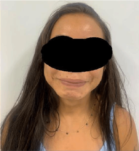
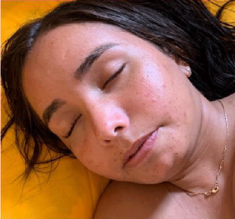
Physical examination showed an inverted face sign (Figure 1). It occurs when observing the face and asking the patient to smile, the presence of the inverted smile is characteristic of the disease. Furthermore, he has nocturnal yogophthalmia (Figure 2). Clinical sign in which the patient keeps the eye slightly open during sleep.
The patient’s diagnosis was clinical since she presented the pathognomonic sign of the disease, the inverted facial expression or inverted smile, characterized by the facial presentation of crying when the individual with the pathology smiles; It should be noted that, at the time of diagnosis, about 15 years ago, due to the socioeconomic condition involved, genetic tests were not available.
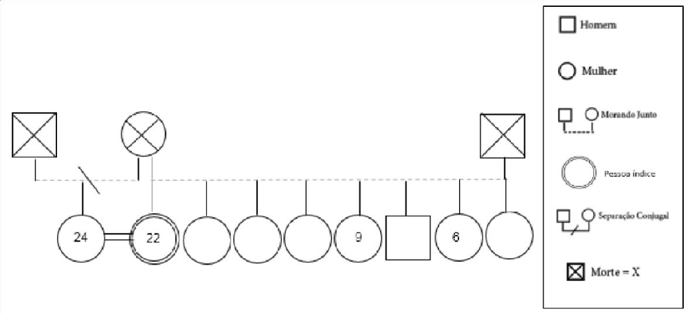
He has 8 siblings, 1 male, 7 females (1 half-sister on his mother’s side) (Figure 3). Of these, four cases of Urofacial Syndrome (Ochoa’s) have already been diagnosed in the family, including the patient, diagnosed at the age of 7, two sisters diagnosed since birth, and the half-sister who, although she is older, for being practically asymptomatic from the urological point of view, the diagnosis was confirmed only after the patient. The patient denies knowledge about parental consanguinity (both parents are already deceased).
In addition, the patient continues with joint follow-up from nephrology and urology to verify the feasibility of kidney transplantation, with or without the need for prior bladder enlargement. He is undergoing tests and follow-ups to assess the viability of a kidney transplant.
Discussion
First described by Bernardo Ochoa in the 1960s, the inverted facial smile allows the early detection of this syndrome and precedes the appearance of urological symptoms [2]. Age may or may not correlate with the severity of disease symptoms, as pathological findings may go unnoticed on prenatal ultrasounds. Thus, patients may have severely compromised renal function from birth or after a few years. However, it is well known that the delay in diagnosis can culminate in greater renal repercussions, proving the importance of early diagnosis [7].
The two classic components (characteristic smile and impaired bladder emptying) in patients with Ochoa Syndrome are usually present in individuals with this pathology (Chart 1). Despite the variability, bladder dysfunction follows the same behavior in all clinical presentations.
Clinical cases bring in their presentation the signs and symptoms that may be present in each patient, such as craniofacial abnormalities characterized by an inverted smile [8,9], bladder dysfunction [10,11], and nocturnal lagophthalmos [12].
This is one of the rare cases reported in the state of Piauí, with a patient presenting the complete characteristics of the inverted face pathology and complications in the urinary tract. Our case is similar to that described in another study, regarding the characteristic presentation of peculiar facial expressions and chronic renal failure and associated complications [6]. The inverse facial expression has functional characteristics, being a pathognomonic finding of the disease, not finding explanations of morphological alterations or injuries [10].
In one study it was found that for 25 families affected by the disease, 21 of them have a mutation in one of the two genes associated with the disease, and 15% may not have any of the affected genes [13]. Mutation of the HPSE2 gene, mapped to chromosome 10q23-q24 that encodes the heparanase 2 protein or the LRIG2 gene that encodes the protein that promotes epidermal growth factor signaling, is related to aberrant bladder innervations. Furthermore, these genes act on the facial nerve, which is closely related to the inverted smile, that is, the characteristic grimace when smiling [14].
Mutations in the HPSE2 gene have already been studied in mice, proving the relationship with bladder dysfunction, with excessive formation of fibrotic tissue from the accumulation of collagen deposits, resulting in organ remodeling, more common in the detrusor [15].
In another study conducted in mice involving the presence or absence of homozygous mutations in the study group, when evaluating the immunohistochemistry, a prevalence of nerve fibers towards the detrusor and a decrease in those directed to the bladder outflow tract were observed, common findings in the bladder. hypercontractile, which is how it appears in Urofacial Syndrome [16].
Patients with Urofacial or Ochoa Syndrome almost always have alterations in urinary emptying due to sphincter-detrusor dyssynergia, consequently, high bladder pressures and/or post-voiding residues occur that promote vesicular reflux, hydronephrosis, UTI and renal scarring with subsequent development of CKD. Furthermore, a trabeculated bladder with thickened walls can be found [17].
As for the findings of lagophthalmos, it is accompanied by persistent ocular symptoms and exposure to keratopathy [18]. It can be a clinical characteristic observed by parents, guardians, or people who live with it for a long time, but it can also be manifested in such a light way, not being found or found again. For this reason, its true frequency in patients with the syndrome is still unknown [7].
There is a high chance that the patient will progress to terminal CKD, mainly due to late diagnosis, and it is important to note that there are few cases of successful transplants in these patients, as there is an increased risk of post-transplant UTI [18,19]. In less severe situations, bladder enlargement is recommended, either with or without continent external urinary diversion. The option of intermittent bladder catheterization is recommended when the objective is to prevent damage to the renal graft [18].
All images are published with authorization and informed consent from the patient, who filled out the appropriate terms. Ethical norms in carrying out this study were followed. It has no conflict of interest.
Conclusion
One of the causes of the rarity of this syndrome is its lack of knowledge on the part of the medical community, neglecting its diagnosis and the consequent institution of adequate early treatment. Therefore, it is important to evaluate and, in case of clinical suspicion, carry out genetic research, when available, including close family members, for proper follow-up, in order to prevent the disease from developing into serious complications, such as end-stage chronic kidney disease.
Therefore, it is evident that early diagnosis with the institution of adequate treatment, avoids possible damage to the urinary tract from childhood, allowing better management and quality of life in these patients.
- Newman WG, Woolf AS. Urofacial Syndrome. In: Adam MP, Mirzaa GM, Pagon RA, Wallace SE, Bean LJH, Gripp KW, Amemiya A, editors. GeneReviews®. Seattle (WA): University of Washington, Seattle; 2013 Aug 22 [updated 2018 Jun 7].1993–2023. PMID: 23967498.
- Ochoa B. Can a congenital dysfunctional bladder be diagnosed from a smile? The Ochoa syndrome updated. Pediatr Nephrol. 2004 Jan;19(1):6-12. doi: 10.1007/s00467-003-1291-1. Epub 2003 Nov 25. PMID: 14648341.
- Skálová S, Rejtar I, Novák I, Jüttnerová V. The urofacial (Ochoa) syndrome--first case in the central European population. Prague Med Rep. 2006;107(1):125-9. PMID: 16752812.
- Pang J, Zhang S, Yang P, Hawkins-Lee B, Zhong J, Zhang Y, Ochoa B, Agundez JA, Voelckel MA, Fisher RB, Gu W, Xiong WC, Mei L, She JX, Wang CY. Loss-of-function mutations in HPSE2 cause the autosomal recessive urofacial syndrome. Am J Hum Genet. 2010 Jun 11;86(6):957-62. doi: 10.1016/j.ajhg.2010.04.016. Erratum in: Am J Hum Genet. 2010 Jul 9;87(1):161. Fisher, Richard B. PMID: 20560209; PMCID: PMC3032074.
- Mahmood S, Beetz C, Tahir MM, Imran M, Mumtaz R, Bassmann I, Jahic A, Malik M, Nürnberg G, Hassan SA, Rana S, Nürnberg P, Hübner CA. First HPSE2 missense mutation in urofacial syndrome. Clin Genet. 2012 Jan;81(1):88-92. doi: 10.1111/j.1399-0004.2011.01649.x. Epub 2011 Mar 10. PMID: 21332471.
- Sutay NR, Kulkarni R, Arya MK. Ochoa or Urofacial syndrome. Indian Pediatr. 2010 May;47(5):445-6. doi: 10.1007/s13312-010-0067-5. PMID: 20519791.
- Osorio S, Rivillas ND, Martinez JA. Urofacial (ochoa) syndrome: A literature review. J Pediatr Urol. 2021 Apr;17(2):246-254. doi: 10.1016/j.jpurol.2021.01.017. Epub 2021 Jan 24. PMID: 33558177.
- Schmidt W, Schroeder TM, Buchinger G, Kubli F. Genetics, pathoanatomy and prenatal diagnosis of Potter I syndrome and other urogenital tract diseases. Clin Genet. 1982 Sep;22(3):105-27. doi: 10.1111/j.1399-0004.1982.tb01422.x. PMID: 7151297.
- Rondon AV, Leslie B, Netto JM, Freitas RG, Ortiz V, Macedo Junior A. The Ochoa urofacial syndrome: recognize the peculiar smile and avoid severe urological and renal complications. Einstein (Sao Paulo). 2015 Apr-Jun;13(2):279-82. doi: 10.1590/S1679-45082015RC2990. Epub 2015 May 1. PMID: 25946049; PMCID: PMC4943824.
- Ochoa B. The urofacial (Ochoa) syndrome revisited. J Urol. 1992 Aug;148(2 Pt 2):580-3. doi: 10.1016/s0022-5347(17)36659-4. PMID: 1640526.
- Velez-Tejada P, Niño-Serna L, Serna-Higuita LM, Serrano Gayubo AK, VelezEcheverri C, Vanegas-Ruiz JJ, et al. Evolution of pediatric patients diagnosed with hydronephrosis who consulted the San Vicente Fundacion University Hospital, Medellÿn, Colombia, between 1960 and 2010. Iatreia. 2019; 27:147e54.
- Mermerkaya M, Suer E, Oztürk E, Glpÿnar O, Goké M, Yalcÿndag FN, et al. Lagoftalmo noturno em crianças com síndrome urofacial (Ochoa): um novo sinal. Eur J Pediatr. 2013; 173:661e5. doi: 10.1007/s00431-013-2172-7.
- Stuart HM, Roberts NA, Burgu B, Daly SB, Urquhart JE, Bhaskar S, Dickerson JE, Mermerkaya M, Silay MS, Lewis MA, Olondriz MB, Gener B, Beetz C, Varga RE, Gülpınar O, Süer E, Soygür T, Ozçakar ZB, Yalçınkaya F, Kavaz A, Bulum B, Gücük A, Yue WW, Erdogan F, Berry A, Hanley NA, McKenzie EA, Hilton EN, Woolf AS, Newman WG. LRIG2 mutations cause urofacial syndrome. Am J Hum Genet. 2013 Feb 7;92(2):259-64. doi: 10.1016/j.ajhg.2012.12.002. Epub 2013 Jan 11. PMID: 23313374; PMCID: PMC3567269.
- Roberts NA, Hilton EN, Lopes FM, Singh S, Randles MJ, Gardiner NJ, et al. Lrig2 e Hpse2, mutantes na síndrome Urofacial, padrões de nervos na bexiga urinária. Rim Int. 2019; 95:1138–52.
- Guo C, Kaneko S, Sun Y, Huang Y, Vlodavsky I, Li X, Li ZR, Li X. A mouse model of urofacial syndrome with dysfunctional urination. Hum Mol Genet. 2015 Apr 1;24(7):1991-9. doi: 10.1093/hmg/ddu613. Epub 2014 Dec 15. PMID: 25510506; PMCID: PMC4355027.
- Penna FJ, Presbítero JS. DRC e problemas de bexiga em crianças. Adv Chron Rim https: 2011; 18:362e9.
- Latkany RL, Lock B, Speaker M. Nocturnal lagophthalmos: an overview and classification. Ocul Surf. 2006 Jan;4(1):44-53. doi: 10.1016/s1542-0124(12)70263-x. PMID: 16671223.
- Gomes DM. Transplante renal em receptor com Síndrome de Ochoa e Insuficiência Renal Crônica - relato de caso. J Bras Transpl. 2011 Jun-Jul; 14:14951540.
- Nicanor FA, Cook A, Pippi-Salle JL. Early diagnosis of the urofacial syndrome is essential to prevent irreversible renal failure. Int Braz J Urol. 2005 Sep-Oct;31(5):477-81. doi: 10.1590/s1677-55382005000500012. PMID: 16255797.
Article Alerts
Subscribe to our articles alerts and stay tuned.
 This work is licensed under a Creative Commons Attribution 4.0 International License.


 Save to Mendeley
