Archives of Clinical Gastroenterology
Acute pancreatitis: A rare cause of ogilvie syndrome
Maroua Michouar1*, Abderrahmane Jallouli1, Oussama Nacir1, Fatima Ezzahra Lairani1, Adil Ait Errami1, Sofia Oubaha2, Zouhour Samlani1 and Khadija Krati1
2Departement of Physiology, Faculty of Medicine, Cadi Ayyad University, Marrakech 40000, Morocco
Cite this as
Michouar M, Jallouli A, Nacir O, Lairani FE, Errami AA, et al. (2023) Acute pancreatitis: A rare cause of ogilvie syndrome. Arch Clin Gastroenterol 9(3): 012-014. DOI: 10.17352/2455-2283.000118Copyright License
© 2023 Michouar M, et al. This is an open-access article distributed under the terms of the Creative Commons Attribution License, which permits unrestricted use, distribution, and r eproduction in any medium, provided the original author and source are credited.Acute colonic pseudoocclusion or Ogilvie syndrome is an acute expansion of the previously healthy colon, occurring in the absence of an obstructive lesion. Pathological circumstances such as pancreatitis or neuroleptic medication have been recognized as predisposing. The incidence of this digestive complication varies from 0.5% to 1%. The aim of our work is to recall, through a case of acute colon pseudo-obstruction in a patient with acute pancreatitis, the clinical and paraclinical data of this syndrome and its physiopathological bases, as well as its therapeutic management. The clinical picture was characteristic of our patient: significant abdominal meteorism. The abdominal scan showed an enormous colonic dilation without mechanical obstacles. The evolution was favourable after colonoscopic exsufflation. Ogilvie syndrome is a rare occurrence that can lead, without effective treatment, to cecal perforation with a poor prognosis. It is, therefore, necessary to establish the diagnosis early and especially to carry out strict radiological surveillance after the implementation of medical treatment, in which colonoscopy is a valuable contribution.
Introduction
Ogilvie syndrome, or acute colonic pseudoobstruction (POCA), corresponds to dilation of all or part of the colonic frame and rectum with the risk of cecal perforation without an intrinsic mechanical obstacle or extrinsic inflammatory process; this definition excludes mechanical dilations prior to an organic obstacle, those occurring in the context of severe acute, ischemic or cryptogenic colitis (toxic and ileal megacolon). Reflex (accompanying peritonitis) in patients predisposed by their history, pathology, or surgery [1]. H. Ogilvie (1887-1971) was the first to describe this entity in 1948 in two patients with neoplastic infiltration of the celiac and mesenteric plexuses [2]. Most cases of Ogilvie syndrome have been published in European and North American journals, but are extremely rare in Morocco. Its pathophysiology remains imprecise and is currently not fully elucidated, its genesis is multifactorial and the hypothesis of neurological dysfunction of the para-sympathetic system with paralysis of the muscles of the colon that allows itself to be distended passively, without any increase in endoluminal pressures is the mechanism accepted today. This is a rare pathology, mainly encountered during digestive surgery, referring to a patient who underwent surgery for a colectomy and presented with Ogilvie syndrome.
Case presentation
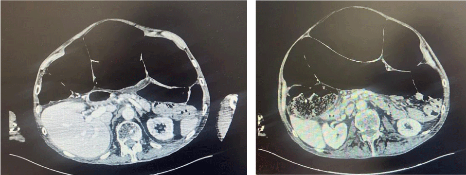
The patient was a 43-year-old man, mechanic, type 2 diabetic on insulin, hypertensive on amlodipine at a dose of 10 mg/day, never operated, overweight with a body mass index (BMI) = 37 kg/m2, his social history was remarkable for cocaine use, but no history of smoking or alcohol. who presented to the emergency room for abrupt epigastralgia, for two days, triggered by a copious, cramp-type meal of moderate intensity, fixed without irradiation, aggravated by diet, relieved by the pre-flexed position, associated with early post-prandial vomiting, without other associated digestive manifestations, in particular, no externalized upper or lower digestive hemorrhage or transit disorders or other associated digestive or extra-digestive signs, all evolving in a context of apyrexia and an alteration in the general condition consisting of asthenia and anorexia. His admission constants included a blood pressure of 133/68 mmHg, a heart rate of 77 beats per minute, a respiratory rate of 22 cycles per minute, apyretic 36.6 °C, and an oxygen saturation of 98% of the ambient air. The physical examination was remarkable for a more pronounced diffuse abdominal tenderness in the epigastrium, without abdominal defense or contracture, a normal pleuropulmonary and cardiovascular examination. The biological evaluation showed hyperleukocytosis at 15080/L. Its inflammatory markers were elevated with C-reactive protein at 32 mg/L with hyperferritinemia at 1120 ng/mL. Hyper-lipasemia of 779 IU/l. Renal function was correct with creatinine at 9 mg/L and urea at 0.22 g/L. The liver test was without abnormalities (ALT at 26 IU/l, AST at 21 IU/l, GGT at 90 IU/l, PAL at 90 IU/l), an abdominal CT scan was performed and showed a swollen pancreas at the expense of his pancreas Cephalic portion measuring 34 mm anterior-posterior in diameter, increases homogeneously after injection of the injection product with infiltration of peri-pancreatic fat without a flow of necrosis, pneumoperitoneum or effusion peritoneal. The liver was homogeneous, normal in size, and with regular contours. A dilation of the intrahepatic and extrahepatic bile ducts was observed, with a maximum diameter of 12 mm without a clearly detectable computational image on this examination. This aspect was compatible with stage C pancreatitis of the Balthazar classification. The diagnosis of acute pancreatitis was accepted and the treatment consisted of stopping the diet, intravenous rehydration with injectable analgesics with gastric protection by Omeprazole at an intravenous dose of 20mg/day. A complementary ultrasound as part of the etiological evaluation of pancreatitis revealed a gallbladder in semi-repetitive multi-lithiasis as well as a dilation of the intrahepatic bile ducts and the main biliary tract measuring 13 mm in diameter and the seat of multiple hyperechogenic stones at the lower biliary tract, generating a posterior shadow cone. Our patient has never had high serum calcium or jaundice or a history of alcohol consumption or hypertriglyceridemia. The etiology of our patient’s acute pancreatitis was biliary. On his 3rd day of hospitalization, the patient complained of worsening epigastric pain with minimal abdominal distension without stopping materials or gases, a control abdominal CT scan was performed which showed significant colonic distension with no mechanical obstacles detected. This expansion measured 12 cm at the level of the cecum, without notable complications, in particular, no perforation (Figures 1,2). It should be noted that the Computed Tomography (CT) of the initial abdomen did not show colonic dilation.
The patient then benefited from gastrointestinal evacuation upstream and downstream with osmotic laxatives and a permanently aspirated nasogastric tube. Coloscopic decompression was performed on this patient to prevent perforation following intestinal ischemia, the procedure was performed without oral laxatives or intestinal preparations with minimal air insufflation, gas aspiration decompressed the colon, and mucosal viability was assessed while slowly removing the colonoscope. The evolution was favourable and the patient was discharged on day 8. The radioclinical control at 3 and 6 months was unremarkable.
Discussion
Acute colonic pseudoobstruction is a motility disorder characterized by massive expansion of the colon in the absence of anatomical or mechanical damage that obstructs the flow of intestinal contents. This form of adynamic ileus is also called Ogilvie’s syndrome. It is named after the British surgeon Sir William Heneage Ogilvie (1887 - 1971), who first reported it in 1948 [1]. The prevalence of colonic pseudoobstruction is difficult to know, but the disease almost always occurs in hospitalized patients. It is commonly found in patients undergoing major surgeries, patients with advanced malignancies, and patients with spinal trauma. It is generally associated with surgical procedures that require prolonged bed rest. Thus, the development of colonic pseudo-obstruction is common in orthopedic procedures such as total hip replacement (up to 1.5% of cases) and after total knee replacement (2.3%) [3]. The incidence is higher among inpatients with mental disabilities reaching up to 18.5% [4]. In 95% of patients, it is associated with an underlying disease. The most frequent associations are: non-operative trauma, infection (pneumonia, sepsis, appendicitis, pancreatitis, sepsis), cardiac (myocardial infarction, heart failure), obstetric or gynecological disease (cesarean section, normal vaginal delivery), abdominal surgery, neurological (Parkinson’s disease, spinal cord injury, multiple sclerosis, Alzheimer’s disease), orthopedic surgery, medical conditions (cancer, respiratory failure, kidney failure) and surgical conditions (urology, thoracic, neurosurgery) [5], a metabolic cause (diabetes, para-neoplastic syndrome, hepatopathy, hyperuricemia, hypothyroidism) [6], pancreatitis [7], a drug cause (calcium channel blocker, antidepressant, anticholinergic), a toxic cause such as drugs and insecticides [8-11]. The effectiveness of gastrointestinal motility drugs such as metoclopramide, urecholin, and cisapride is not proven. Cisapride is being withdrawn from the market because of these cardiac side effects (rhythm disorders). It is neostigmine that is most often used after conservative treatment has failed. A few studies have suggested that neostigmine may be an effective treatment. That of Pannec is very interesting and demonstrates its effectiveness [12]. Decompression colonoscopy is currently the third-line treatment after conservative and drug treatment with prostigmine has failed or if there is a contraindication to it. The success rate is 80%, with a recidivism rate estimated at 15% [13-15]. It has a double interest in allowing both colonic decompression and the elimination of possible mechanical obstruction. The recurrence rate can be reduced by leaving an intra-colic tube. It can be slid onto the guide wire during the colonoscopy [16-18]. The mortality of medically treated patients is 14% to 30%. This remains important even when the treatment has been done properly [5]. However, it should be considered that Ogilvie syndrome often concerns weakened individuals due to intercurrent pathologies such as burns and sepsis, whose treatment is a priority.
Conclusion
The etiopathogenesis of Ogilvie syndrome has not yet been elucidated. An imbalance between sympathetic and parasympathetic innervation remains the most likely path in a population of patients who are most often debilitated. Poor prognostic factors are represented by age, ischemia, cecal perforation, and the time before colonic decompression. Conservative medical treatment remains the treatment of choice when the diagnosis is made early.
- Maloney N, Vargas HD. Acute intestinal pseudo-obstruction (Ogilvie's syndrome). Clin Colon Rectal Surg. 2005 May;18(2):96-101. doi: 10.1055/s-2005-870890. PMID: 20011348; PMCID: PMC2780141.
- OGILVIE H. Large-intestine colic due to sympathetic deprivation; a new clinical syndrome. Br Med J. 1948 Oct 9;2(4579):671-3. doi: 10.1136/bmj.2.4579.671. PMID: 18886657; PMCID: PMC2091708.
- Nelson JD, Urban JA, Salsbury TL, Lowry JK, Garvin KL. Acute colonic pseudo-obstruction (Ogilvie syndrome) after arthroplasty in the lower extremity. J Bone Joint Surg Am. 2006 Mar;88(3):604-10. doi: 10.2106/JBJS.D.02864. PMID: 16510828.
- Khalid K, Al-Salamah SM. Spectrum of general surgical problems in the developmentally disabled adults. Saudi Med J. 2006 Jan;27(1):70-5. PMID: 16432597.
- Vanek VW, Al-Salti M. Acute pseudo-obstruction of the colon (Ogilvie's syndrome). An analysis of 400 cases. Dis Colon Rectum. 1986 Mar;29(3):203-10. doi: 10.1007/BF02555027. PMID: 3753674.
- PALEY RG, MITCHELL W, WATKINSON G. Terminal colonic dilation following intractable diarrhea in a diabetic. Gastroenterology. 1961 Oct;41:401-7. PMID: 14483326.
- Teh SH, O'Riordain DS, O'Connell PR. Colonic pseudo-obstruction following acute pancreatitis. Ir J Med Sci. 1998 Jan-Mar;167(1):41-2. doi: 10.1007/BF02937554. PMID: 9540300.
- Bear R, Steer K. Pseudo-obstruction due to clonidine. Br Med J. 1976 Jan 24;1(6003):197. doi: 10.1136/bmj.1.6003.197. PMID: 1247772; PMCID: PMC1638445.
- Faulk DL. Approach to patients with intestinal pseudo-obstruction. Gastroenterology. 1978; 74: 1320.
- Faulk DL, Anuras S, Gardner GD, Mitros FA, Summers RW, Christensen J. A familial visceral myopathy. Ann Intern Med. 1978 Nov;89(5 Pt 1):600-6. doi: 10.7326/0003-4819-89-5-600. PMID: 717927.
- Sriram K, Schumer W, Ehrenpreis S, Comaty JE, Scheller J. Phenothiazine effect on gastrointestinal tract function. Am J Surg. 1979 Jan;137(1):87-91. doi: 10.1016/0002-9610(79)90016-3. PMID: 581534.
- Dive A. Gastrointestinal motility disorders in critical patients. Réanimation. 2008; 17:454-461.
- Jetmore AB, Timmcke AE, Gathright JB Jr, Hicks TC, Ray JE, Baker JW. Ogilvie's syndrome: colonoscopic decompression and analysis of predisposing factors. Dis Colon Rectum. 1992 Dec;35(12):1135-42. doi: 10.1007/BF02251964. PMID: 1473414.
- Lavignolle A, Jutel P, Bonhomme J, Cloarec D, Cerbelaud P, Lehur PA, Galmiche JP, Le Bodic L. Syndrome d'Ogilvie: résultats de l'exsufflation endoscopique dans une série de 29 cas [Ogilvie's syndrome: results of endoscopic exsufflation in a series of 29 cases]. Gastroenterol Clin Biol. 1986 Feb;10(2):147-51. French. PMID: 3754523.
- Hudziak H. Ogilvie syndrome: Therapeutic management. Hepatogastroenterologist. 1999; 2:29-31.
- Nano D, Prindiville T, Pauly M, Chow H, Ross K, Trudeau W. Colonoscopic therapy of acute pseudoobstruction of the colon. Am J Gastroenterol. 1987 Feb;82(2):145-8. PMID: 3812419.
- Groff W. Colonoscopic decompression and intubation of the cecum for Ogilvie's syndrome. Dis Colon Rectum. 1983 Aug;26(8):503-6. doi: 10.1007/BF02563739. PMID: 6688216.
- Rex DK. Acute colonic pseudo-obstruction (Ogilvie's syndrome). Gastroenterologist. 1994 Sep;2(3):233-8. PMID: 7987621.
Article Alerts
Subscribe to our articles alerts and stay tuned.
 This work is licensed under a Creative Commons Attribution 4.0 International License.


 Save to Mendeley
