Archives of Clinical Gastroenterology
Primary peritoneal hydatidosis revealed by an abdominal mass in a case report
Erguibi Driss, Fadwa Azhari*, Hajri Amal, Boufettal Rachid, Saad Rifki El Jay and Farid Chehab
Cite this as
Driss E, Azhari F, Amal H, Rachid B, El Jay SR, et al. (2023) Primary peritoneal hydatidosis revealed by an abdominal mass in a case report. Arch Clin Gastroenterol 9(1): 001-003. DOI: 10.17352/2455-2283.000115Copyright License
© 2023 Driss E, et al. This is an open-access article distributed under the terms of the Creative Commons Attribution License, which permits unrestricted use, distribution, and r eproduction in any medium, provided the original author and source are credited.Hydatid cyst is caused by the parasite Echinococcus granulosus commonly found in temperate regions. Primary peritoneal hydatidosis is a rare entity. Diagnosis can be made with ultrasound and contrast-enhanced CT of the abdomen and pelvis and serology.
Eosinophilia is a strong indicator of hydatid cysts as a differential diagnosis. Open excision of the cyst combined with medical treatment remains the treatment of choice.
Introduction
Peritoneal hydatidosis is one of the most serious complications of hydatid disease. It is favored by the location of the cyst, its size and the high intracystic pressure. Traumatic rupture is most often iatrogenic during surgery for a hydatid cyst of the liver (HFC).
The rarity of peritoneal localization represents about 5% to 16% of all hydatid cyst localizations [1], prompted us to report the case of an elderly patient hospitalized with an isolated firm abdominal mass.
The treatment of peritoneal hydatidosis is mainly surgical. The aim is to treat the peritoneal cysts as well as the primary hydatid cyst in the same operation, if it exists in the case of secondary HP, or to eradicate the peritoneal cysts in the case of exceptional HP.
We report a case of primary peritoneal hydatidosis and discuss the diagnostic and therapeutic means.
Observation
The patient was 19 years old, without any particular pathological history, presenting with pain in the left iliac fossa without any transit disorder or externalized digestive hemorrhage, all evolving in a context of conservation of the general state.
The clinical examination on admission found a conscious patient stable on the HD and respiratory plan, apyretic. Abdominal examination: Renal mass straddling the hypogastrium and left iliac fossa measuring 10 cm in major axis without inflammatory signs opposite. The rest of the somatic examination was unremarkable.
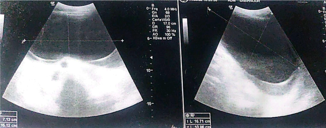
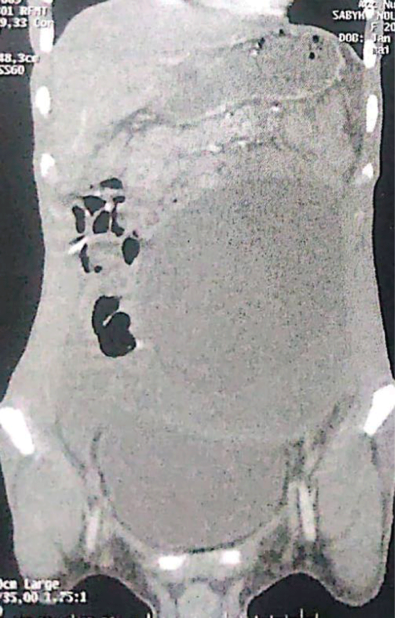
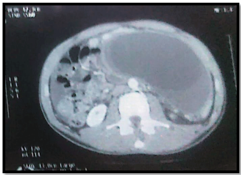
Abdominal ultrasound showed an abdominal mass of cystic appearance (Figure 1), complemented by a CT scan showing a retrovesical cystic lesion, extending to the left flank, with a double thick, hypodense liquid component with a thin wall measuring 168 mm x 69 mm x 213 mm in height. Hydatid serology: negative (Figures 2,3).
After preoperative preparation, the patient underwent resection of an encapsulated peritoneal cystic mass with drainage of the Douglas cul de sac by Salem probe.
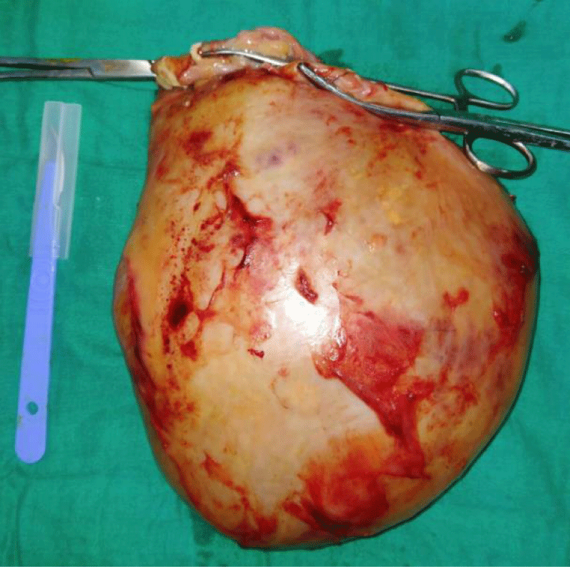
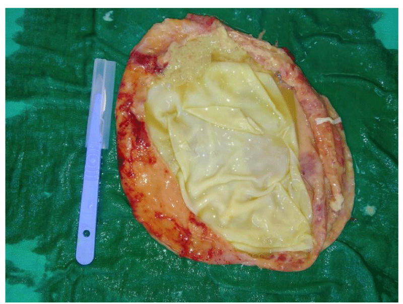
The surgical exploration had objectified the presence of an encapsulated peritoneal cystic mass of 20 cm of large axis adherent to the gallbladder and epiploon (adhesiolysis done), without ascites (Figures 4,5)
The immediate postoperative follow-up was simple with the resumption of transit on Day 1. Food was authorized on day 1. The patient was declared discharged at D3 postoperatively under anthelmintic and antiprotozoal treatment.
The patient was seen in consultation with good evolution.
Discussion
Hydatidosis is a cosmopolitan anthropozoonosis. In Morocco, this disease is endemic. It affects humans of all ages and sexes [2].
Peritoneal hydatidosis is due to the seeding of the peritoneal serosa by the larval form of the dog taenia: Echinococcus granulosus. It is a rare parasitic condition. It represents about 5 to 16% of all hydatid cyst locations. Peritoneal hydatidosis is most often secondary to the rupture or fissuring of hepatic hydatid cysts, but can also be primary but exceptionally [3].
The main symptoms of the disease are abdominal pain, which differs according to the location of the cyst, although all abdominal locations are possible, as well as abdominal fullness, dyspepsia, anorexia and vomiting. In the most frequent cases, a firm abdominal mass is found on clinical examination, rarely responsible for a compression syndrome [4] as in our case: palpation of a firm mass occupying the umbilical region and the hypogastrium.
The diagnosis of hydatid cysts should be evoked especially in endemic areas with a history of animal husbandry or pet ownership, whenever a cystic mass is felt in the abdominal cavity [5].
The differential diagnosis of these cystic intra-abdominal masses includes pancreatic cyst, mesenteric cyst, gastrointestinal duplication cyst, ovarian cyst, lymphangioma, intra-abdominal abscess, loculated ascites and hematoma [6].
Ultrasound remains an essential and sometimes sufficient examination to make a positive diagnosis of hydatid cyst; it has a sensitivity of about 90% to 95%. The CT scan allows a more precise diagnosis, especially in the peritoneal location [7].
Immunoelectrophoresis is a more sensitive test, but ELISA is more specific. ELISA can also be used postoperatively to monitor patient recurrence [8].
The treatment of peritoneal hydatidosis is mainly surgical. The aim is to treat the peritoneal cysts as well as the primary hydatid cyst in the same operation, if it exists in the case of secondary HP, or to eradicate the peritoneal cysts in the case of exceptional HP [9].
Curative surgery is not always feasible, but medical treatment is still important in the treatment of HP, either preoperatively in order to sterilize the cysts or reduce their size before the invasive procedure, or postoperatively in order to avoid recurrences and prevent a possible secondary HP, which is difficult to cure [10].
Several authors have supported the use of albendazole as an adjunct to surgery in inoperable cases in the literature [11].
Conclusion
Primary peritoneal hydatidosis is a rather rare entity of hydatid disease.
The clinical picture is very polymorphic and the positive diagnosis is essentially based on epidemiological, clinical and paraclinical arguments.
Surgery is the mainstay of treatment for peritoneal hydatidosis, based on radical or conservative methods that must act on both the peritoneal hydatidosis and the primary visceral hydatid cyst.
Prevention remains the best therapeutic method.
- Abbassi P, Aboussad P, Ali PAB, Yazidi PA, Belaabidia P, Bouskraoui P. Professeurs D’enseignement Superieur. 115.
- Seifeddine B, Amel C, Ghofrane T, Dhouha B, Lassaad G, Rached B, Taher KM. Rupture spontanée d’un kyste hydatique du foie dans la cavité péritonéale avec une membrane proligère intacte: à propos d’un cas et revue de la littérature [Spontaneous rupture of hydatid cyst of liver in the peritoneal cavity with intact proligerous membrane: about a case and literature review]. Pan Afr Med J. 2018 Jun 26;30:174. French. doi: 10.11604/pamj.2018.30.174.15054. PMID: 30455803; PMCID: PMC6235496.
- Jaouahar AA. Exceptional Peritoneal Hydatidosis, Apropos of a Case with Literature Review. 24 May, 2020. http://ao.um5.ac.ma/xmlui/handle/123456789/18023
- Massoud W, Saheb N, Iliescu B, Kreitmann L, Chabenne J, Campeggi A, Molinie V, Baumert H. Kyste hydatique rétropéritonéal géant [Giant retroperitoneal hydatid cyst]. Prog Urol. 2009 Jun;19(6):442-5. French. doi: 10.1016/j.purol.2009.01.002. Epub 2009 Mar 3. PMID: 19467467.
- Hegde N, Hiremath B. Primary peritoneal hydatidosis. BMJ Case Rep. 2013 Aug 2;2013:bcr2013200435. doi: 10.1136/bcr-2013-200435. PMID: 23912661; PMCID: PMC3762414.
- Sable S, Mehta J, Yadav S, Jategaokar P, Haldar PJ. "Primary omental hydatid cyst": a rare entity. Case Rep Surg. 2012;2012:654282. doi: 10.1155/2012/654282. Epub 2012 Sep 23. PMID: 23050190; PMCID: PMC3461611.
- Kamaoui I, Maaroufi M, Squalli N, Lamhadri M, Tizniti S. TROP12 Primary peritoneal hydatid cyst: a differential diagnosis not to be overlooked. J Radiol. 1 oct 2005; 86(10):1588.
- Madi AE, Bouabdallah Y. Peritoneal hydatidosis in an adolescent. Pan Afr Med J. 13 March, 2014;18. http://www.panafrican-med-journal.com/content/article/18/210/full/
- Daali M, Hssaida R, Zoubir M, Hda A, Hajji A. Hydatidose péritonéale. A propos de 25 cas marocains [Peritoneal hydatidosis: a study of 25 cases in Morocco]. Sante. 2000 Jul-Aug;10(4):255-60. French. PMID: 11111243.
- Liu Y, Wang X, Wu J. Continuous long-term albendazole therapy in intraabdominal cystic echinococcosis. Chin Med J (Engl). 2000 Sep;113(9):827-32. PMID: 11776080.
- Horton RJ. Albendazole in treatment of human cystic echinococcosis: 12 years of experience. Acta Trop. 1997 Apr 1;64(1-2):79-93. doi: 10.1016/s0001-706x(96)00640-7. PMID: 9095290.
Article Alerts
Subscribe to our articles alerts and stay tuned.
 This work is licensed under a Creative Commons Attribution 4.0 International License.


 Save to Mendeley
