Archives of Clinical Gastroenterology
To study the indications of cholecystectomy, types of surgery and complications of surgery in a tertiary care hospital in Nepal
Narayan Prasad Belbase1*, Sagar Khatiwada2, Nishnata Koirala2, Sushim Bhujel2, Hari Prasad Upadhaya2 and Bikash Shah2
Cite this as
Narayan Prasad B, Sagar K, Nishnata K, Sushim B, Hari Prasad U, et al. (2022) To study the indications of cholecystectomy, types of surgery and complications of surgery in a tertiary care hospital in Nepal. Arch Clin Gastroenterol 8(1): 020-024. DOI: 10.17352/2455-2283.000107Copyright License
© 2022 Narayan Prasad B, et al. This is an open-access article distributed under the terms of the Creative Commons Attribution License, which permits unrestricted use, distribution, and r eproduction in any medium, provided the original author and source are credited.Background: Cholelithiasis is a chronic recurrent disease of the hepatobiliary system and cholecystectomy is one of the commonly performed surgeries. This study intends to evaluate the demographic of patients with cholelithiasis, its various mode of presentation, surgical intervention, and its outcome.
Methods: This was a quantitative retrospective cross-sectional study conducted in the College of Medical Sciences- a Superspeciality Tertiary Care Teaching Hospital in Bharatpur, Chitwan in central Nepal. All patients undergoing laparoscopic or open cholecystectomy in the department of GI and General Surgery at the College of Medical Sciences from 1st May 2017 to April 30, 2021were included in the study. Study tools were records of the patients retrieved from the medical record section.
Results: A total of 355 patients data were analyzed. The mean age of the study samples was 46.43 +/- 16.47 years. Female predominance was seen at 76.18% with M: F ratio of 1:3.18. The most common presenting symptoms were pain abdomen (70.4%), bloating of the abdomen (63.9%), and fatty dyspepsia (46.8%). Acute cholecystitis was seen in 14.36%, biliary pancreatitis in 6.2%, and gallbladder perforation in 2.25% cases. Laparoscopic cholecystectomy was done in 269(83.94%), open cholecystectomy in 57(16.05%), and laparoscopic converted into open cholecystectomy 29 in (9.73%). Intra-operative complications like gallbladder perforation and controlled bleeding were seen in 10.14% and 4.23% of cases. Post-operative complications like bile leak and major bile duct injury were seen in 6.19% and 1.40% of cases. Regarding management of bile leak, conservative management was successfully done in 54.54% of cases while they were managed with ERCP in 27.27%, hepaticojejunostomy in 13.63%, and with T-tube repair in 4.5% cases. The overall mortality was 0.8%.
Introduction
The prevalence of gall bladder stones varies widely in different parts of the world. In India estimated to be around 4% whereas in the western world it is 10% [1]. In Nepal overall prevalence of gallstone disease was found to be 4.87% [2].
Although symptomatic and complicated stones represent only 20% of all gallstones, they lead to clinically relevant morbidity and complications as well as high costs of medical care.
The classic presentation of symptomatic gallstones is a patient with recurrent right upper quadrant pain (sometimes epigastric), that is related to fatty food intake, and most likely at night. This pain comes from an impacted stone in the cystic duct [3].
Sometimes, the initial presentation of gallstones may be acute cholecystitis, in some cases, the stone becomes impacted in the common bile duct, causing its obstruction and the development of cholestasis. Another serious complication is acute pancreatitis which can happen due to transient obstruction of the main pancreatic duct at the ampulla of Vater. Sometimes, the stone can form a fistula from the gallbladder directly into the duodenum [3].
After a physical examination is done, ultrasonography is considered to be the method of choice in diagnosing cholelithiasis and cholecystitis. It has high sensitivity and specificity and can diagnose even small stones. It can also detect dilation of the bile duct, and/or gallbladder wall thickening [3].
Cholecystectomy can be performed by laparoscopy, by a small incision (< 8 cm in length), or by open operation, and several meta-analyses indicate surgical procedures as the gold standard for the treatment of symptomatic gallstones. Laparoscopic cholecystectomy (LC), or alternatively, small incision cholecystectomy, are both safe with a similar mortality rate ranging from 0.1% to 0.7% [4].
Laparoscopic cholecystectomy is the most commonly performed abdominal surgery in industrialized countries. The rate of conversions to laparotomy for uninflamed gallbladder disease ranges from 2% to 15%, and in cases of acute cholecystitis, from 6% to 35% [5].
Postoperative bile leaks following cholecystectomy may arise from the liver bed (i.e., ducts of Luschka), cystic duct stump, or injury to the biliary tree. Overall, they are relatively uncommon, occurring in 1% to 11% of all cases [6]. In addition, bile duct strictures and biliary leakages are severe long-term complications after LC. These injuries are associated with high morbidity, mortality, and prolonged hospitalization [7].
Currently, endoscopic procedures are most frequently used in the management of postoperative iatrogenic bile duct injury (IBDI). There are several endoscopic techniques available, e.g. biliary stent placement, biliary sphincterotomy, and nasociliary drainage [8]. Nonetheless, if major IBDI occurs, i.e. complete dissection of the common bile duct (CBD), surgical management is required to resolve this issue [8]. Several surgical options exist including primary repair over a T-tube, a choledochoduodenostomy, or a hepaticojejunostomy. A Roux en Y hepaticojejunostomy (HJ) is the preferred method for repairing high-grade bile duct injuries, however, it is associated with increased morbidity and mortality [9]. In a report of a French experience with 253 hepaticojejunostomies for bile duct repair, Iannelli, et al. identified morbidity and mortality rates of 19.7% and 1.6%, respectively [10].
Due to the increased incidence of gallstones and their variable presentations, this study was undertaken to evaluate the demographic of patients with cholelithiasis, its various mode of presentation, surgical intervention, and its outcome at a tertiary care hospital in central Nepal.
Methods
This was a quantitative retrospective cross-sectional study conducted in the College of Medical Sciences- a Superspeciality Tertiary Care Teaching Hospital in Bharatpur, Chitwan in central Nepal. All patients undergoing laparoscopic or open cholecystectomy in the department of GI and General Surgery at the College of Medical Sciences from 1st May 2017 to April 30, 2021were included in the study. Census sampling (retrospective retrieval of data from medical record files) was done. This study was conducted after ethical approval from the Institutional review committee in July 2021. A total of 415 files were retrieved. From the files patients’ age, sex, mode of presentation, Ultrasound of abdomen findings, type of surgery performed, intra-operative findings, intraoperative complications, need to convert to open surgery, post-operative complications, and their management was retrieved.
Inclusion criteria
All patients above 15 years of age who underwent laparoscopic or open cholecystectomies.
Exclusion criteria
Patients with concomitant CBD stones, patients with acalculus cholecystitis, patients with Gallbladder polyp, patients with a diagnosis of carcinoma gallbladder, and those patients whose data were missing from record files.
Collected data were entered on a preformed proforma with patients meeting all inclusion and exclusion criteria. Appropriateness of data entry was under the guidance of the primary investigator.
Data management and analysis
Collected data was entered into the SPSS data software version20.0 and analyzed accordingly. For descriptive statistics categorical variables were described using frequency and percentage and illustrated using appropriate graphs or charts, continuous variables were described using mean with SD or median with IQR.
Results
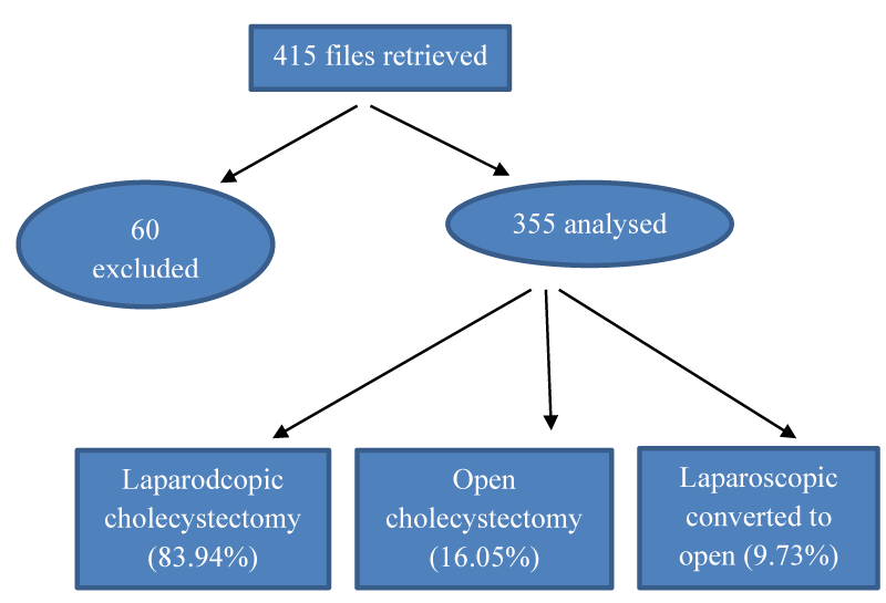
A total of 415 cases of cholecystectomy files were reviewed. Files of 60 patients were excluded from the study as either their USG findings or operative findings were missing. A total of 355 cases were included in the final analysis. The findings of this study are summarized in Tables 1-4 and Figures 1-2. The mean age of the study samples was 46.43 +/- 16.47 years. Female predominance was seen at 76.18% with M: F ratio of 1:3.18.
The most common presenting symptoms were pain abdomen (70.4%), bloating of the abdomen (63.9%), and fatty dyspepsia (46.8%). Right upper abdominal tenderness was found in 16.3% of patients and twenty-two patients (6.2%) had an attack of biliary pancreatitis. Multiple calculi were seen in 69.9% cases and single calculi in 30.1% cases.
The different surgeries performed were laparoscopic cholecystectomy 269(83.94%), open cholecystectomy 57(16.05%), and laparoscopic converted into open cholecystectomy 29(9.73%). The intraoperative complications were gallbladder perforation with or without bile and stone spillage (10.14%), minor bleeding from calot’s triangle or liver bed (4.23%), and bile duct injury (0.56%).
Postoperatively surgical site infection, minor bile leak (<300ml/day), and major bile leak(>300ml/day) were seen in 33(9.29%), 15(4.22%), and 5(1.40%) cases respectively. Of the 15 bile leaks seen, 9 were seen in laparoscopic cholecystectomy and 4 were seen in open cholecystectomy. Among 5 major bile leaks, all were seen in patients undergoing laparoscopic cholecystectomy.
The overall mortality was 3(0.8%) and all were in patients with gall bladder perforation. All the surgical site infections and 12 cases of minor bile leak were managed conservatively and thus were presumed to be Strasberg A injuries.
Discussion
Gallstone disease is a common public health issue that affects people all over the world, especially adults. In the present study, the mean age of the study population was 46.43+/_16.47 years. In a study done by Mohan CP, et al. [11] in India the mean age was 48+/-15.21 years and in Joshi HN, et al. [12] study from Nepal it was 44.51+/-14.57 years.
Cholelithiasis is more commonly seen in females and this was seen in our study with 76.18% of female patients. A similar trend was seen in a study done by Bhattacharjee PK, et al. [13] (75.05%) and Samal SS, et al. [14] (70.3%).
The common mode of presentation of gallstone disease in our study was pain upper abdomen (70.4%), bloating of the abdomen (63.9%) and fatty dyspepsia (46.8%). In a study done by Saxena PK, et al. [15], pain upper abdomen was seen in 78.9% of cases, and bloating of the abdomen and flatulent dyspepsia were seen in 50.3% of cases whereas in a study done by Samal SS, et al. [14] pain abdomen was seen in 80% cases and dyspepsia and flatulence were seen in 70% cases.
In symptomatic patients with cholelithiasis other than biliary colic (60-70%), cholelithiasis can present as acute cholecystitis in 15-20% cases and gall stone pancreatitis in 10-15% cases [16]. According to different studies, an incidence rate of 3.3–5.9% is reported for acute and chronic perforations of the Gallbladder [17]. In the present study acute cholecystitis was seen in 14.36%, gallstone pancreatitis in 6.2%, and gall bladder perforation in 2.25% cases.
Mirizzi syndrome is a rare complication with a reported incidence of 0.05-2.7% [18]. The incidence of Mirizzi’s syndrome in our study was 1.41%.
Ultrasonography of the abdomen is a common modality of diagnosing cholelithiasis. In the present study, multiple stones were seen in 69.9% and single calculi in 30.1% cases. In a similar study by Sharadha B, et al. [19], multiple stones were seen in 75.56% and solitary stones in 24.44% cases, and in a study by Bansal A, et al. [20], multiple stones were seen in 63.5%% and solitary stones in 36.5%.
Gallbladder perforation (GP), which is a common intraoperative complication during cholecystectomy, has been reported to occur with a high incidence of 10%-33% [21]. The reported incidence of bleeding in lap cholecystectomy can be up to 2% (reported range, 0.03% to 10%) [22]. The rate of conversion reported in today’s literature is 2-15%. In cases with acute inflammatory processes reported rates of conversion increase up to 35% [23]. In the present study looking at intraoperative and postoperative complications gallbladder perforation, with or without bile and stone spillage was seen in 36 (10.14%), minor bleeding from calot’s triangle or liver bed in 15(4.23%), and rate of conversion was 9.73%.
Wound infection (port site in laparoscopic and surgical incision site in open cholecystectomy) was seen in 9.29% of cases in this study which is in range when compared with similar studies done by Samal SS, et al. [14] and Ranjan R, et al.[24] (4.68% and 13.33%).
Postoperative bile leaks following cholecystectomy may arise from the liver edge (i.e., ducts of Luschka), cystic duct stump, or injury to the biliary tree. Overall, they are relatively uncommon, occurring in 1% to 11% of all cases [6]. But bile leak in the cohort of elective cholecystectomy ranged between 0.1% to 0.5% in open cholecystectomy and 0.5% to 3% in laparoscopic cholecystectomy [8.] There is a wide range (0.5%-1.5%) of incidence of bile duct injury during laparoscopic cholecystectomy compared to 0.3% for open cholecystectomy [25].
In a study of post-cholecystectomy bile leakage in 60 patients by Kumar S, et al. [26] bile leakage from the Gallbladder bed, duct of Luschka and minor accessory duct, cystic duct, common hepatic duct, common bile duct, and aberrant hepatic duct in 56.67%, 16.67%, 10.0%, 10.0% and 6.67% patients respectively. Conservative management was successful in 53.33% of patients, ERCP ± stenting in 16.67% patients, PTBD in 3.3% patients, and Hepaticojejunostomy in 25.0% of patients. In a study by El-Kabeer MMM, et al. [27] in Egypt the incidence of bile leak was 2% in which the source of bile leak was gallbladder bed, duct of Anuschka and the minor accessory duct was 70%, from CBD it was 10%, from CHD it was 10% and from cystic duct stump leak was 10%. In the same study conservative treatment was successful in70% of cases of bile leak. ERCP management was done in 20% of cases and exploratory laparotomy and hepaticojejunostomy were done in 5% of cases. Similarly from another study conducted in Nepal by Pandit N, et al. [28]. The incidence of bile leak was 18/2300 (0.78%). The source of bile leak in this study as per Strasberg’s classification system was as: Strasberg’s type A (52.9%), type D (5.9%), and type E (41.1%). In this study 8(47%) of cases of bile leak were managed conservatively, and 2(11.11%) were managed by laparotomy and lavage. In 3(16.67%) patients Bile duct injury was detected intraoperatively out of which 2(11.11%) patients were managed with hepaticojejunotomy and 1(5.55%) patient was managed with end to end repair of the bile duct. Delayed repair of bile duct with laparotomy and hepaticojejunotomy was done in 5(27.78%) patients.
In the current study total of 20 (5.63%) bile leaks were seen postoperatively out of which 15 were minor bile leaks (<300ml/24 hrs) and 5 were major bile leaks (>300ml/24 hrs). We had more bile leaks than in other studies as we included cholecystectomy in acute cholecystitis and even perforated gallbladder.
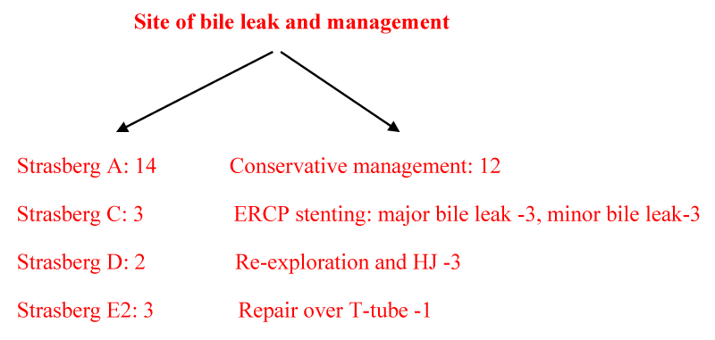
Since we have a protocol of keeping abdominal drain in all patients following cholecystectomy all patients with bile leak were observed for a minimum of 5 days with the addition of injection octreotide ranging from 800-1200 microgram in slow intravenous infusion. Out of 20 bile leaks, 12(60%) settled over a mean duration of 3.5 days. So they were presumed to be Strasberg type A injuries. Two patients with major bile leak developed biliary peritonitis on the 3rd and 4th postoperative days and were taken for exploratory laparotomy. They had Strasberg E2 injury so Roux-en-Y hepaticojejunostomy was performed. The rest 6 patients with bile leaks (3 with minor bile leak and 3 with major bile leak) were subjected to ERCP and ERCP stenting after 5 days of conservative management. All 6 patients settled with ERCP stenting. Among the 3 minor bile leaks 2 Strasberg A injury and 1 Strasberg C injury was found. Among the 3 cases of major bile leak, 2 had Strasberg C injury, and 1 had Strasberg D injury. Two cases of bile duct injury were detected intra-operatively. One case had Strasberg E2 injury and was managed with hepaticojejunostomy and the other case had Strasberg D injury and was managed with T-tube repair. So taking into consideration both intraoperatively detected and postoperatively detected bile leaks we had a total of 22(6.19%) bile leaks out of which 14(63.64%) cases of Strasberg A injury, 3(13.64%) cases of Strasberg C injury, 2(9.09%) patient had Strasberg D injury and 3(13.64%) patients had Strasberg E2 injury. So in actuality, we had 5 major bile duct injuries i.e in 1.41% of cases. In a nutshell, 54.54% of bile leaks were managed conservatively, 27.27% managed with ERCP,13.63% managed with hepaticojejunotomy and 4.5% managed with T-tube drainage.
Postoperative mortality following cholecystectomy, even after the introduction of laparoscopic cholecystectomy, according to different population-based studies has shown to be in the range of 0.1%-0.7%. Risk factors like increased age and acute admissions with underlying comorbidities are associated with high mortality [29]. In the current study the overall mortality was 0.8% and its incidence was high in patients with acute cholecystitis and gallbladder perforations.
Conclusion
Other than chronic abdominal pain, bloating of the abdomen and fatty dyspepsia, acute cholecystitis, biliary pancreatitis, and gallbladder perforation were the indications for surgery in 14.36%,6.2%, and 2.25% cases respectively. Laparoscopic surgery was the preferred method of cholecystectomy. The majority of minor bile leaks can be managed conservatively. Hepaticojejunostomy is the treatment for major bile duct injury beyond E1injury. Lateral CBD injury detected intraoperatively can be managed with T-tube repair and those detected postoperatively settles with ERCP stenting.
Limitations
This was a single-centered study so the results cannot be generalized to all populations of Nepal.
- Bagdai AK, Sutaria A (2020) A Clinical Study Of Cholelithiasis Presentation And Management In Tertiary Care Hospital. NJIRM 11: 17-20. Link: https://bit.ly/37KXYRa
- Panthee MR, Pathak YR, Acharya AP, Mishra C, Jaisawal RK (2007) Prevalence of Gall Stone Disease in Nepal: Multi Center Ultrasonographic Study. PMJN 7: 45-50. Link: https://bit.ly/3ETKPkQ
- Al-Saad MH, Alawadh AH, Al-Bagshi AH, Ali MHA, Alshehab AA, et al. (2018) Surgical Management of Cholelithiasis. EJHM 70: 1416-1420. Link: https://bit.ly/3vk7uUl
- Portincasa P, Ciaula AD, Bonfrate L, Wang DQ (2012) Therapy of gallstone disease: What it was, what it is, what it will be. World J Gastrointest Pharmacol Ther 3: 7-20. Link: https://bit.ly/3LoyBU3
- Abraham S, Rivero HG, Erlikh IV, Griffith LF, Kondamudi VK (2014) Surgical and Nonsurgical Management of Gallstones. Am Fam Physician 89: 795-802. Link: https://bit.ly/3xSM0jj
- Shawhan RR, Porta CR, Bingham JR, McVay DP, Nelson DW, et al. (2015) Biliary Leak Rates After Cholecystectomy and Intraoperative Cholangiogram in Surgical Residency. Mil Med 180: 565-569. Link: https://bit.ly/3vlBEqz
- Kaman L, Behera A, Singh R, Katariya RN (2004) Management of major bile duct injuries after laparoscopic cholecystectomy. Surg Endosc 18: 1196–1199. Link: https://bit.ly/3EUFDgY
- Renz BW, Bösch F, Angele MK (2017) Bile Duct Injury after Cholecystectomy: Surgical Therapy. Visc Med 33: 184-190. Link: https://bit.ly/399ORKa
- Ismael HN, Cox S, Cooper A, Narula N, Aloia T (2017) The morbidity and mortality of hepaticojejunostomies for complex bile duct injuries: a multi-institutional analysis of risk factors and outcomes using NSQIP. HPB (Oxford) 19: 352-358. Link: https://bit.ly/3F6dpjr
- Iannelli A, Paineau J, Hamy A, Schneck AS, Schaaf C, et al. (2013) Primary versus delayed repair for bile duct injuries sustained during cholecystectomy: results of a survey of the Association Francaise de Chirurgie. HPB 15: 611-666. Link: https://bit.ly/3MwuKV0
- Mohan CP, Kabalimurthy J, Jayavarma R (2018) A clinical study on cholelithiasis and management. JMSCR 6: 766-770.
- Joshi HN, Singh AK, Shrestha D, Shrestha I, Karmacharya RM (2020) Clinical Profile of Patients Presenting with Gallstone Disease in University Hospital of Nepal. Kathmandu Univ Med J 18: 256-259. Link: https://bit.ly/3MwuZiS
- Bhattacharjee PK, Halder SK, Rai H, Ray RP (2015) Laparoscopic Cholecystectomy: A Single Surgeon’s Experience in some of the Teaching Hospitals of West Bengal. Indian J Surg 77: S618–S623. Link: https://bit.ly/37L5GuJ
- Samal SS, Behera MR, Das S, Hotta A, Sahu S (2020) A Study on Prevalence, Clinical Presentation, and Management of Gall Stone Diseases in Southern Odisha. J Evid Based Med Health 7: 1628-1632. Link: https://bit.ly/398APbJ
- Saxena PK, Golandaj VK, Malviya VK (2019) Epidemiological study in operated patients with cholelithiasis and analysis of risk factors. Int J surg orthopedics 5: 340-345. Link: https://bit.ly/39pejf7
- Vogt DP (2002) Gallbladderdisease: An update on diagnosis and treatment. Cleve Clin J Med 69: 977-984. Link: https://bit.ly/399Uhow
- Ponten JB, Selten J, Puylaert JBCM, Bronkhorst MWGA (2015) Perforation of the gallbladder: a rare cause of acute abdominal pain. Journal of Surgical Case Reports 1–2. Link: https://bit.ly/3Oz83Bv
- Bashir M, MalikAA, Nazeer NUI, ZargarSA (2018) Clinical Profile and management of Mirizzi's syndrome-ten years Experience in a tertiary Care Hospital in a developing Country. JMSCR 6: 552-560. Link: https://bit.ly/3vgpGhH
- Sharadha B, Srinivas D (2017) Clinical Study of Cholelithiasis. Int J Sci Stud 5: 210-214. Link: https://bit.ly/3vJR3Qd
- Bansal A, Akhtar M, Bansal AK (2014) A clinical study: prevalence and management of cholelithiasis. Int Surg J 1: 134-139. Link: https://bit.ly/37NGX98
- Brockmann JG, Kocher T, Senninger NJ, Schürmann GM (2002) Complications due to gallstones lost during laparoscopic cholecystectomy. Surg Endosc 16: 1226-1232. Link: https://bit.ly/3OJMH4y
- Kaushik R (2010) Bleeding complications in laparoscopic cholecystectomy: Incidence, mechanisms, prevention and management. J Min Access Surg 6: 59-65. Link: https://bit.ly/3kik40a
- Radunovic M, Lazovic R, Popovic N, Magdelinic M, Bulajic M, et al. (2016) Complications of Laparoscopic Cholecystectomy: Our Experience from a Retrospective Analysis. J Med Sci 4: 641-646. Link: https://bit.ly/3vhluOP
- Ranjan R, Sinha KK, Chaudhary M (2018) A comparative study of laparoscopic (LC) vs. open cholecystectomy (OC) in a medical school of Bihar, India. Int J Adv Med 5: 1412-1416. Link: https://bit.ly/3EPLtA7
- Sharma S, Behari A, Shukla R, Dasari M, Kapoor VK (2020) Bile duct injury during laparoscopic cholecystectomy: an indian e-survey. Ann Hepatobiliary pancreat Surg 24: 469-476. Link: https://bit.ly/37L4CXK
- Kumar S, Singh RK, Mandal M, Prasad U (2019) Non-surgical management of Post cholecystectomy bile leakage: Experience from tertiary care centre in Bihar. Int J Med Health Res 5: 114-116. Link: https://bit.ly/3Kl66oI
- El-Kabeer MMM, EL-Rahman AEA, Hassan AM (2021) Biliary Leak After Laparoscopic Cholecystectomy; Incidence and Management. Egypt J Hosp Med 82: 746-754. Link: https://bit.ly/3rVUUZl
- Pandit N, Yadav TN, Awale L, Deo KB, Dhakal Y, et al. (2020) Current Scenario of Postcholecystectomy Bile Leak and Bile Duct Injury at a Tertiary Care Referral Centre of Nepal. Minim Invasive Surg 2020: 4382307. Link: https://bit.ly/3kmkuT2
- Sandblom G, Videhult P, Guterstam YC, Svenner A, Sadr-Azodi O (2015) Mortality after a cholecystectomy: a population-based study. HPB 17: 239-243. Link: https://bit.ly/3vjxSO6
Article Alerts
Subscribe to our articles alerts and stay tuned.
 This work is licensed under a Creative Commons Attribution 4.0 International License.


 Save to Mendeley
