Archives of Clinical Gastroenterology
Phytobezoar Simulated Oblitoma as Cause of Bowel Obstruction: A Case Report
Jama Stanley1, Gomez Nestor2*, Garcia Gabriel3, Betancourth Teresa4 and Emily Rivas5
1Attending Physician of General Surgery of the Teodoro Maldonado Carbo IESS Hospital, Guayaquil, Ecuador
2Attending Physician of the Kennedy Clinic, Graduate Professor of the University of Guayaquil, Guayaquil, Ecuador
3Resident Doctor of Emergency of the Leon Becerra Hospital, Milagro, Ecuador
4General Doctor, Guayaquil, Ecuador
5Biological Health Sciences Student at the University of South Florida, Tampa, USA
Cite this as
Stanley J, Nestor G, Gabriel G, Teresa B, Rivas E. Phytobezoar Simulated Oblitoma as Cause of Bowel Obstruction: A Case Report. Arch Clin Gastroenterol. 2024;10(3):023-026. Available from: 10.17352/2455-2283.000124Copyright License
© 2024 Stanley J, et al. This is an open-access article distributed under the terms of the Creative Commons Attribution License, which permits unrestricted use, distribution, and r eproduction in any medium, provided the original author and source are credited.Bezoars are collections of non-digestible organic material, which after being ingested, accumulate over time in the stomach or small intestine. The most common presentation of the bezoar is in the form of phytobezoar, which is by the accumulation of undigested particles of plant origin. Phytobezoars are hard masses of undigested plant materials that accumulate in the stomach and small intestine. We present the case of a 42-year-old patient with a history of cholecystectomy who presented an intestinal sub-occlusion with suspected oblitoma. The tomography was reported as oblitoma because the image was suggestive. We performed exploratory laparotomy with the presence of a tumor mass in the sub-occluded ill area, one meter from the ileocecal valve ileal. The surgical decision was tumor resection plus manual end-to-end anastomosis. The pathology results were for phytobezoar.
Introduction
Bezoar is the translation of the Arabic word “badzehr”, or the Turkish word “panzehr” which means antidote. The bezoars are secondary from many causes such as alterations of gastric emptying, previous gastric surgeries, excessive consumption of high-fiber foods, bad chewing, ingestion of solid materials difficult to digest, alterations in intestinal motility, etc. The most common bezoars are the phytobezoars. Phytobezoars are responsible for only 0.4 - 4% of all intestinal obstructions [1].
The etiology is diverse; they can develop due to a diet rich in vegetables with poor chewing, poor digestion factors due to gastric hypomotility and hypochlorhydria, gastric stasis, retention of plant fibers, diabetic gastroparesis, hypothyroidism or muscular dystrophy [2]. The 80% of intestinal obstructions occur in the small intestine caused by adhesions, neoplasms, or hernias. Approximately 4.3% of intestinal occlusion symptoms are due to some type of bezoar [3].
Intestinal obstruction is a very common cause of hospitalization and the main cause is the presence of secondary adherences from previous surgeries, internal hernias, or neoplasms. Less than 4% of the cases of occlusion are the result of a type of bezoar, but due to diagnostic failures, generally, its treatment is belated, which increases morbidity and mortality.
The initial handling is conservative in most cases, but in others, the bezoar is located beyond the duodenum, which implies a surgical treatment.
The objective of this work is to inform about the case of a patient who presented intestinal sub-occlusion, due to a phytobezoar simulating an oblitoma with suggestive images in x-rays and tomographies, which was solved through laparotomy plus tumor resection plus manual primary end-to-end anastomosis.
Presentation of the case
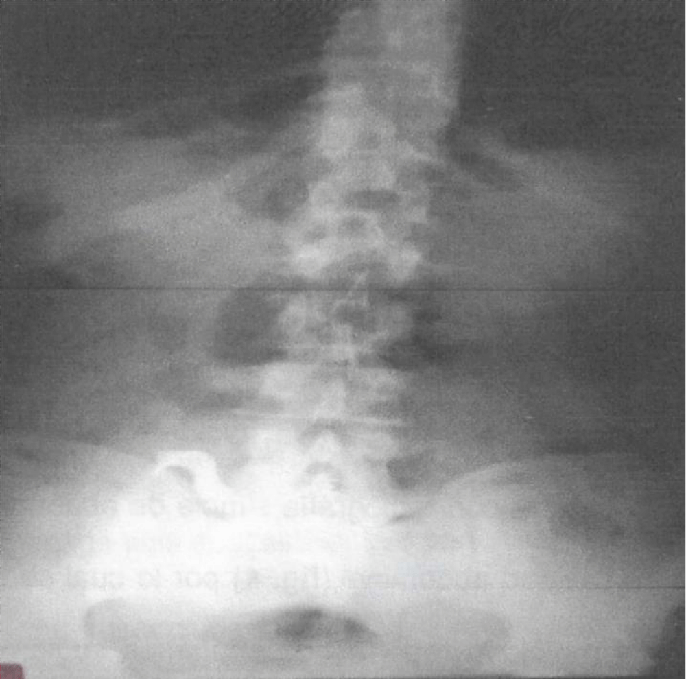
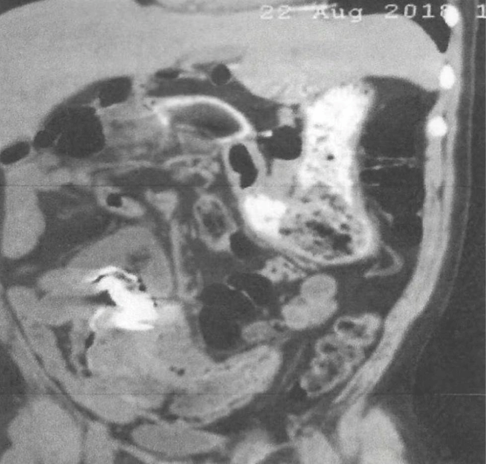
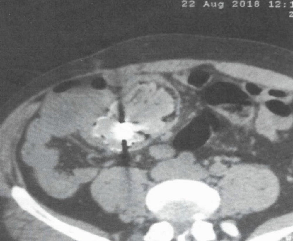
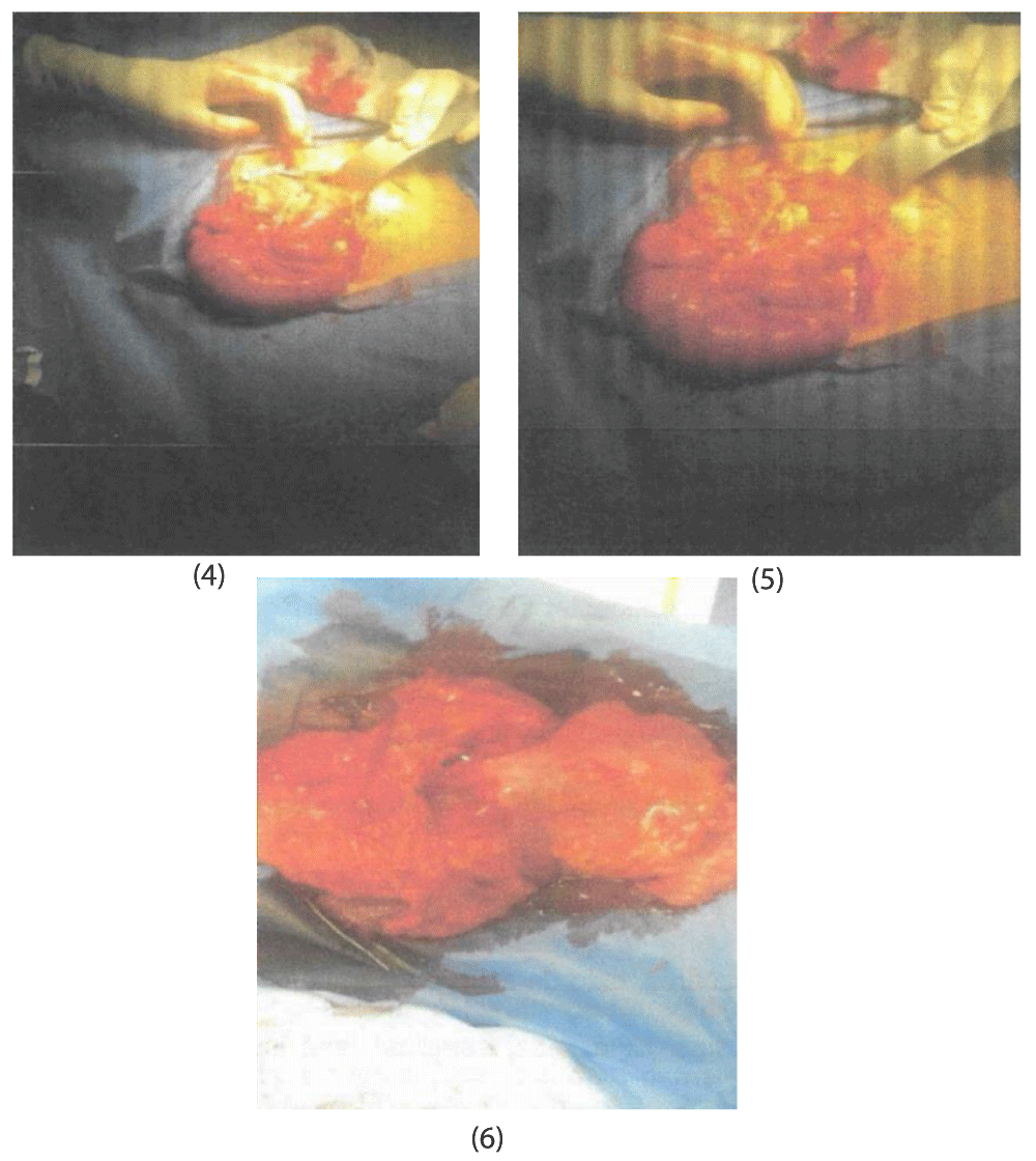
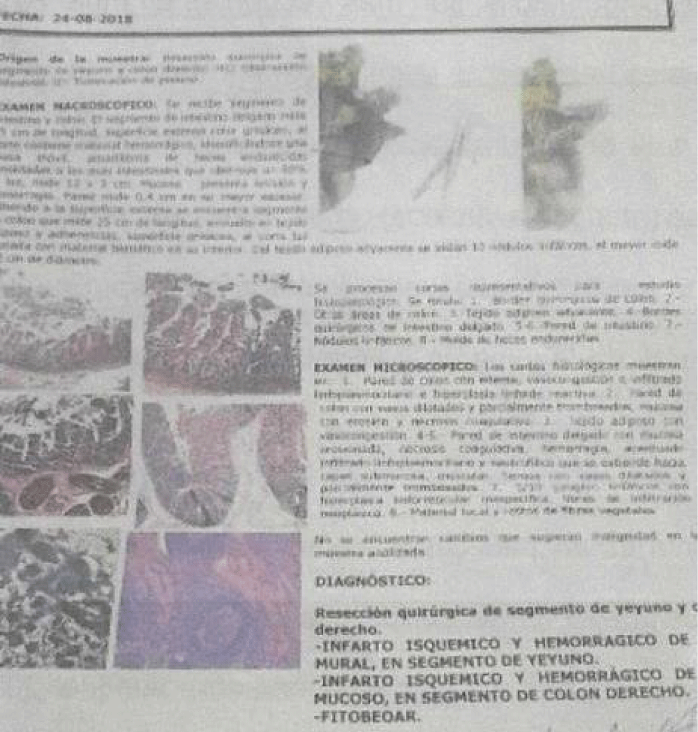
Masculine patient 45 years old, with a history of open cholecystectomy eight months earlier. He came for presenting a clinical picture of four months of evolution characterized by cramping pain in the right hemi-abdomen, which included the epigastrium and mesogastrium, which has gradually increased in intensity, without irradiation, accompanied by nausea and occasional vomiting that alters his diet, until tolerate only liquids with progressive evacuation disorders plus weight loss of approximately 15 pounds, he comes with a simple abdominal X-ray whose image suggests the presence of a foreign body in the abdominal cavity, (Picture 1) for which an abdominopelvic tomography is performed, whose report from the radiologist reports that images suggest an apparent foreign body in the cavity that obstructs almost 90% of the intestinal lumen, (Pictures 2,3), which is why it was decided to be admitted to the Leon Becerra Hospital of the MSP in the city of Milagro and programmed for surgery, an exploratory laparotomy was performed, finding a jejunal tumor with attachment of several jejunoileal loops and the right colon. Tumor resection and compromised jejunal loops plus right hemicolectomy, omentectomy, and primary manual end-to-end ileon transverse anastomosis were performed (Pictures 4,5) with excellent postoperative evolution. The histopathological study report revealed ischemic infarction (Picture 7) in the jejunum segment and the presence of phytobezoar in the right colon segment (Picture 6). At the moment the patient is asymptomatic with an adequate weight and evolving well.
Discussion
The bezoars are an agglomerate of vegetable, animal, or textile material, retained in the gastrointestinal tract and with the inability to progress through it. Most take place in the stomach, however, they can occur in many other places in the digestive tract. Bezoar is the translation of the Arabic word “badzehr” or the Turkish word “panzehr”, which means antidote. The bezoar stones were appreciated until the XVIII century, as a cure for many sicknesses. On the ascension of James I to the English throne in 1662, the crown jewels lay on a bezoar stone on a gold pedestal. Dr. Rudolph Matas, in 1914, was one of the first to publish about the bezoars, placing emphasis on trichobezoars [1]. Vaughan and collaborators, in 1968 were the first ones to describe the Rapunzel Syndrome, characterized by the presence of a hairball in the gastric chamber and a “hair tail” of hair towards the jejunum [4].
They are different types of bezoars, according to the material from which it is formed, such as phytobezoars (vegetable fibers, dried fruit, seeds), which are the most common in adults with alterations in gastric acidity, gastric emptying or edentulous; trichobezoars (hair) are rare and occur more in patients with mental retardation and psychiatric disorders; They are more frequent in women between 10 and 19 years of age (90%), of which only half have a history of trichophagia, they are usually dark in color due to protein denaturation and brilliance due to mucus sequestration [1,5,6]; lactobezoars (dairy products), most common in premature or low birth weight infants, and pharmacobezoars (drugs). In our case, it was a patient with a high intake of legumes and vegetables with insufficient chewing, which was what the patient reported.
Intestinal obstruction is a very common condition in emergencies and can be due to multiple etiologies. Most of the cases correspond to intestinal subocclusions, they are generally resolved with conservative management, although 30% of the cases require surgical treatment. Postoperative adhesions are the cause of 60% of cases, while bezoars only account for 4% or less, but they generally do not respond to conservative methods, they normally correspond to populations that ingest a greater amount of food that induces the formation of bezoars [2,3]. Predisposing risk factors for developing a bezoar include previous abdominal surgery, specifically gastric surgery. Patients with previous antrectomy have a higher risk than the rest of the population of developing a bezoar and the prevalence observed is 10% to 25% [3,4,7]. The most common occlusion site is the distal ileum [2,7].
The clinical diagnosis is sometimes not so clear due to the insidious evolution, occasional cramping pain, nausea, sometimes altered defecation habits, and occasionally Hyporexia. Upper digestive endoscopy is the study of choice for the diagnosis of a gastric bezoar. A transit with water-soluble material also plays a vital role in the diagnosis of intestinal occlusion secondary to a bezoar [2,7]. Computed tomography has a sensitivity of 81% to 96% and a specificity of 96% for establishing the diagnosis; It also allows us to differentiate the bezoar from a neoplasm, indicating its size, shape, and location. The diagnosis is based on finding dilation and an increase in the caliber of the loops (> 2.5 cm) above the transition point. The typical image of a bezoar is usually an ovoid or rounded mass, with a density similar to that of soft tissue, containing air in its interstice and peripherally outlined by contrast material in the dilated intestinal loop at the site of obstruction [4,6].
There are several non-surgical treatment options such as the use of endoscopic baskets, lithotripsy equipment, acetylcysteine, laser, washing with carbonated drinks, paraffin, and cellulose, among others, especially for gastric bezoars can be resolved by nonsurgical methods, not so, intestinal bezoars.
Treatment of small intestine bezoars is traditionally surgical due to the high failure rate of endoscopic treatments. Surgical treatment can be by laparotomy or laparoscopy, both under general anesthesia. Laparoscopic treatment is an effective alternative, offering the patient the benefits of a short hospital stay, less scarring, less postoperative pain, shorter surgical time, and a decrease in the number of postoperative adhesions compared to laparotomy. The laparoscopic approach should be selected based on the location of the bezoar, its size, the patient’s surgical history, and the probable percentage of free field to manipulate the loops dilated by the obstruction. The dimension of the loop distension may increase the risk of perforation during laparoscopic manipulation [9-11]. Laparoscopic surgery has been used in the extraction of bezoars by performing gastrotomy, crushing it in the intestinal lumen to manually milk it to the cecum, or by combining it with mini-laparotomy to manually exteriorize the site of obstruction and perform enterotomy [10,12,13]. The other option is laparotomy plus enterostomy and removal of the phytobezoar plus intestinal resection and anastomosis as in our case, which also presented an extensive granulomatous reaction that produced adhesions of jejunoileal loops with the right colon that simulated a malignant tumor. It is important to remember that the presence of bezoars is associated with complications such as ulcerations, perforation, intussusception, and obstruction, with a morbidity and mortality of 30% [10].
Conclusion
We believe that in patients with images suggestive of a foreign body with a clinical picture of intestinal subobstruction and a history of previous abdominal surgery, phytobezoar should also be considered as a cause of intestinal subobstruction, in addition to patients with a history of insufficient chewing and excessive intake of legumes and vegetables, although clinically they can have an insidious evolution, are very important in the diagnosis, the studies of tomographic images to define an adequate treatment.
Ethical considerations
We declare that standard guidelines for the case have been meticulously followed. Despite a loss of contact with the patient, we have ensured that all personal information has been maintained with strict confidentiality and security.
- Cruz RJ, Ramirez LC, Ramos RJ, O'Farril HM. Mechanical intestinal occlusion due to phytobezoar. Rev Cubana Cir. 2016;55(1):67-73. Available from: https://www.medigraphic.com/cgi-bin/new/resumen.cgi?IDARTICULO=65245
- Lacoma Latre EM, Sánchez Lalana E, Saudí Moro S. Intestinal phytobezoar. Diagnostic Image. 2017;8(1):21-23.
- Hernández-Vera FX, Hugo-Guerrero V, Cosme-Reyes C, Belmonte-Montes C. Fitobezoar oclusión intestinal. Rev Gastroenterol Mex. 2010;75(3):348-352. Available from: https://pubmed.ncbi.nlm.nih.gov/20959190/
- Liauw JJ, Cheah WL. Laparoscopic management of acute small bowel obstruction. Asian J Surg. 2005;28(3):185-188. Available from: https://pubmed.ncbi.nlm.nih.gov/16024312/
- Ho T, Koh D. Small-bowel obstruction secondary to bezoar impaction: A diagnostic dilemma. World J Surg. 2007;31(5):1072-1078. Available from: https://doi.org/10.1007/s00268-006-0619-y
- Ulusan F, Koc Z, Törer N. Small bowel obstructions secondary to bezoars. Ulus Travma Acil Cerrahi Derg. 2007;13(4):217-221. Available from: https://pubmed.ncbi.nlm.nih.gov/17978897/
- Teng HC, Nawawi O, Ng K, Yik YI. Phytobezoar: an unusual cause of intestinal obstruction. Biomed Imaging Interv J. 2005;1:1-4. Available from: https://doi.org/10.2349/biij.1.1.e4
- Watt C, Harner J. Bezoars causing acute intestinal obstruction. Ann Surg. 1947;126:56-61.
- Zamir D, Goldblum C, Linova L, Polychuck I, Reitblat T, Yoffe B. Phytobezoars and trichobezoars: A 10 year experience. J Clin Gastroenterol. 2004;38(10):873-876. Available from: https://doi.org/10.1097/00004836-200411000-00007
- Palanivelu C, Rangarajan M, Senthilkhumar R, Madankumar M. Trichobezoars in the stomach and ileum and their laparoscopy-assisted removal: a bizarre case. Singapore Med J. 2007;48(1):37. Available from: https://pubmed.ncbi.nlm.nih.gov/17304375/
- Kan J, Huang TJ, Heish JS. Laparoscopy-assisted management of jejunal bezoar obstruction. Surg Laparosc Endosc Percutan Tech. 2005;15(5):297-298. Available from: https://doi.org/10.1097/01.sle.0000183263.09347.07
- Lin Ch, Tung Ch, Peng Y, Chow WK, Chang CS, Hu WH. Successful treatment with a combination of endoscopic injection and irrigation with Coca-Cola for gastric bezoar-induced gastric outlet obstruction. J Chin Med Assoc. 2008;71(1):49-52. Available from: https://doi.org/10.1016/s1726-4901(08)70073-x
- Yau KK, Siu WT, Law BKB, Cheung HY, Ha JP, Li MK. Laparoscopic approach compared with conventional open approach for bezoar-induced small-bowel obstruction. Arch Surg. 2005;140(10):972-975. Available from: https://doi.org/10.1001/archsurg.140.10.972
- Kyo Young S, Byung Jo Ch, Seung Nam K, Park CH. Laparoscopic removal of gastric bezoar. Surg Laparosc Endosc Percutan Tech. 2007;17(1):42-44. Available from: https://doi.org/10.1097/01.sle.0000213765.86170.77
Article Alerts
Subscribe to our articles alerts and stay tuned.
 This work is licensed under a Creative Commons Attribution 4.0 International License.


 Save to Mendeley
