Global Journal of Medical and Clinical Case Reports
Cutaneous phaeohyphomycosis of foot web by Curvularia lunata
Mohammed Rasheeduddin* and Visalakshi P
Cite this as
Rasheeduddin M, Visalakshi P (2017) Cutaneous phaeohyphomycosis of foot web by Curvularia lunata. Glob J Medical Clin Case Rep 4(3): 074-075. DOI: 10.17352/2455-5282.000053A case of cutaneous phaeohyphomycosis of the foot affecting interdigital spaces between toes in a 31 year old immunocompetent male with no history of diabetes is illustrated. Fugal elements were found in direct microscopic examination of skin scrapping. The etiologic agent was identified as Curvularia lunata based on macromophology and microscopic examination of pure culture growth. The patient was prescribed with a topical cream containing sertaconazole nitrate and oral itraconazole by the treating dermatologist. Nondermatophyte infections clinically mimic dermatophytosis, and microscopically resemble dermatophytes. Therefore, a correct identification of etiologic agent is important for appropriate treatment.
Case Report
Dematiaceous fungi are frequently considered as ubiquitous saprobes inhabiting plants and residing in the soil. However, these generalized assumptions are now incorrect as several etiologic agents occupy specific ecological niches that contribute to our understanding of their opportunistic/pathogenic potential. Curvularia is one of the important members of these melanized fungi and [1], is a causative agent of phaeohyphomycosis which is an emerging mycotic infection of humans where the tissue morphology of the causative organism is mycelial. This separates it from other clinical types of disease involving brown-pigmented fungi where the tissue morphology of the organism is a grain (mycotic mycetoma) or sclerotic body (chromoblastomycosis) [2].
A case of cutaneous phaeohyphomycosis of the foot affecting interdigital spaces between toes in a 31 year old immunocompetent male with no history of diabetes is illustrated. Cutaneous scaling lesions were prominent between webs of foot. Lesions were irregularly marginated, pruritic and darker than the surrounding skin. It was clinically suspected as intertriginous tinea pedis by a referring dermatologist.
Direct examination of skin scrapping in 10% potassium hydroxide mount revealed branching septate hyaline hyphae with some circular to oval structures of distorted morphology. Dermatophyte test medium (DTM) (HiMedia, India) without dermato supplement, and, with and without actidione used for culture. Inoculated slopes of DTM incubated aerobically in biological oxygen demand (BOD) incubator at 250C + 10C.
On DTM, dermatophytes produce alkaline metabolites, which causes the phenol red pH indicator to change the color of the medium from yellow to pink-red, whereas in case of Non-Dermatophytes media remains yellow in color [3].
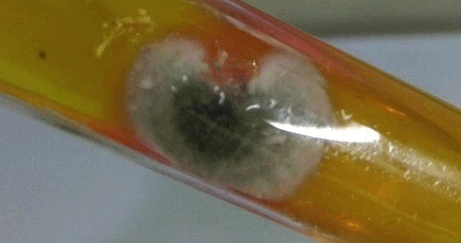
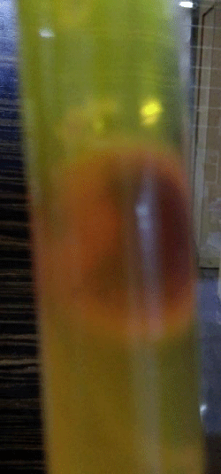
Macromorphology: Fast growing colony, fully matured within 5 days, suede-like, greenish grey at the centre, effuse and grayish white towards the periphery, with a fimbriate margin (Figure 1), reverse brownish black (Figure 2). The color of the medium remains yellow. No growth observed on media containing cycloheximide till 30 days.
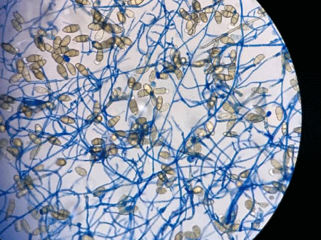
Micromorphology: Branched septate, hyaline (stained blue with lactophenol cotton blue) to brown hyphae, large pale brown macroconidia with no more than four transverse septa, the subterminal cell curved, swollen, larger and darker than the remaining, characteristic of Curvularia lunata were seen (Figure 3) in a stand-alone laboratory. The patient was prescribed with a topical cream containing sertaconazole nitrate and oral itraconazole by the treating dermatologist.
Although rarely pathogenic, Curvularia species have been reported to cause a variety of human systemic, subcutaneous, and cutaneous mycoses [4]. It is interesting to note that Curvularia species form dark pigmented colonies on the culture media whereas in tissues the fungal elements are hyaline to brown, with varying morphologic features [5]. Nondermatophyte infections clinically mimic dermatophytosis, and microscopically resemble the dermatophytes [4]. Therefore, a correct identification of etiologic agent is important for appropriate treatment.
- Sanjay G R, Deanna A S (2010) Melanized Fungi in Human Disease. Clin Micro Rev 23: 884–928. Link: https://goo.gl/LCvgBQ
- David E, Stephen D, Helen A, Rosemary H, Robyn B (2007) Curvularia Boedijn. In Descriptions of Medical Fungi, 2nd ed, Mycology Unit, Women’s and Children’s Hospital, School of Molecular & Biomedical Science, University of Adelaide, Adelaide, Australia. P.57.
- D.T.M. Agar Base (Dermatophyte Test Agar Base) M188, HiMedia Technical Data Sheet https://himedialabs.com/TD/M188.pdf. Link:
- Jorge O L, Neicy M.J (1998) Dermatomycosis of the toe web caused by Curvularia lunata. Rev Inst Med Trop S.Paulo 40: 327-328. Link: https://goo.gl/3m1FNB
- Kwon-Chung K J, Bennett J E (1992) Phaeohyphomycosis (Chromomycosis, phaeosporotrichosis, cerebral chromomycosis). In: Kwon-Chung K.J, Bennett J.E. Medical Mycology. Philadelphia, Lea & Febiger. P.620-677.

Article Alerts
Subscribe to our articles alerts and stay tuned.
 This work is licensed under a Creative Commons Attribution 4.0 International License.


 Save to Mendeley
