International Journal of Vascular Surgery and Medicine
Traumatic Dissection of the Innominate Artery Revealed by an Ischemic Stroke in a Young Patient: A Case Report
1Cardiovascular Surgery Department, Abidjan Heart Institute, Abidjan, Ivory Coast, Côte d’Ivoire
2Cardiovascular Surgery Department, Donka University Hospital, Conakry, Guinea, Côte d’Ivoire
3Thoracic Surgery Department, Abidjan Heart Institute, Abidjan, Ivory Coast, Côte d’Ivoire
4Surgical Intensive Care Department, Abidjan Heart Institute, Abidjan, Ivory Coast, Côte d’Ivoire
5Thoracic and Cardiovascular Surgery Department, Bouaké University Hospital, Bouaké, Ivory Coast, Côte d’Ivoire
6Medical Imaging Department, Abidjan Heart Institute, Abidjan, Ivory Coast, Côte d’Ivoire
Author and article information
Cite this as
Diane M, et al. Traumatic Dissection of the Innominate Artery Revealed by an Ischemic Stroke in a Young Patient: A Case Report. Int J Vasc Surg Med. 2026; 12(1): 6-10. Available from: 10.17352/2455-5452.000049
Copyright License
© 2026 Diane M, et al. This is an open-access article distributed under the terms of the Creative Commons Attribution License, which permits unrestricted use, distribution, and reproduction in any medium, provided the original author and source are credited.Introduction: This clinical case highlights the diagnostic challenge posed by blunt traumatic injuries involving the arteries of the supra-aortic trunks in young patients, and the implications of their endovascular treatment.
Case Presentation: We report the case of a 25-year-old woman admitted for the diagnostic and therapeutic management of a left hemothorax associated with a sudden ischemic stroke, following a high-speed road traffic accident involving abrupt deceleration. A computed tomography angiography of the supra-aortic trunks, performed 10 days after the trauma, revealed a dissection limited to the innominate artery with no signs of rupture, associated with thrombotic occlusion of the right common carotid artery, the left subclavian artery, and the left vertebral artery, without ischemia of the left upper limb. Treatment consisted of the endovascular placement of a covered stent via an endovascular procedure, which improved cerebral perfusion, and pleural drainage, which allowed for adequate pulmonary re-expansion.
Conclusion: The therapeutic approach to these arterial injuries in the context of multiple trauma is discussed on a case-by-case basis.
BTOAI: Blunt Thoracic Outlet Arterial Injuries; BCVI: Blunt Traumatic Cerebrovascular Injuries
Introduction
Traumatic dissections involving the supra-aortic trunks and their branches in their extracranial segments are the leading cause of ischemic stroke in young adults (accounting for 20% of ischemic strokes in this population) and can result in significant neurological sequelae [1,2]. They most commonly occur in the context of high-velocity polytrauma [1]. Arterial dissection of the supra-aortic trunks is defined as the formation of a hematoma within the wall of the main trunks arising from the aortic arch, splitting the parietal layers. This results in occlusion, stenosis, or aneurysmal dilation of the vessel [3]. Initial symptoms are often poor, masked by other craniofacial injuries, meaning that diagnosis is often delayed until neurological deficits become apparent [1]. These findings clearly highlight the issue of early diagnosis of these lesions and their treatment, which presents a real prognostic challenge for the patient. The endovascular approach in patients with multiple trauma is decided on a case-by-case basis. We report a rare case of traumatic dissection of the innominate artery in a patient with multiple trauma, occurring as part of blunt injuries to the supra-aortic trunks complicating an ischemic stroke, which was successfully treated by endovascular surgery.
Case presentation
A 25-year-old shopkeeper, with no significant medical history, was involved in a high-speed road traffic accident on 14 February 2026. She was a passenger, not wearing a helmet, on the back of a motorcyclist travelling at high speed, which resulted in a collision with a large lorry. The impact caused her to fall, followed by an immediate loss of consciousness lasting approximately five minutes. The initial assessment of her injuries by emergency services revealed:
- diffuse bilateral cerebral edema and a Glasgow Coma Scale score of 14/15, with no ischaemic injury (Figure 1A) and no focal neurological deficit;
- a massive left hemothorax and bilateral pulmonary contusion (Figure 1B) with hypoxaemic respiratory distress; peripheral oxygen saturation was 84%;
- unifocal fractures of the posterior arches of the 1st, 2nd, 3rd and 4th left ribs (Figure 1C).
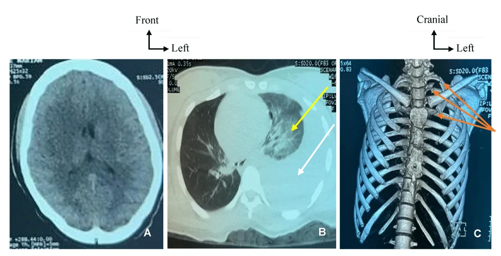
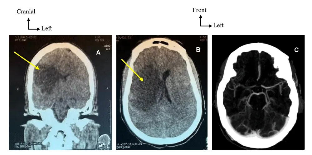
The ISS score was assessed at 21. The patient was subsequently admitted to the intensive care unit, where left pleural drainage and temporary mechanical ventilation were performed. Her condition improved favourably, and she was extubated on the third day of her hospitalization. On the fourth day of hospitalization, left-sided hemiparesis developed suddenly without any impairment of consciousness. A cerebral CT scan revealed (Figure 2) an infarct in the territory of the right middle cerebral artery without hemorrhagic reorganization (Figures 2A and 2B). Antithrombotic treatment consisting of acetylsalicylic acid had been initiated. With regard to the chest, the pleural drain was removed on the fifth day of hospitalization, followed by the recurrence of hemothorax on the tenth day of hospitalization. The patient was then transferred to the Department of thoracic and cardiovascular surgery at the Abidjan Heart Institute 10 days after the injury for further management of her chest trauma. On admission, the patient’s reassessment revealed: a Glasgow Coma Scale score of 15/15; anisocoria with reactive pupils; and flaccid, complete pyramidal syndrome, not proportional to the left side of the body, predominantly affecting the upper limbs and face; hemodynamic and ventilatory stability. There was an absence of a radial pulse in the left upper limb, with a significant systolic blood pressure gradient of 10 mmHg between the two upper limbs. The transthoracic cardiac Doppler ultrasound was normal. Following these clinical findings, a CT angiography of the supra-aortic trunks performed on 25 February 2026 revealed:
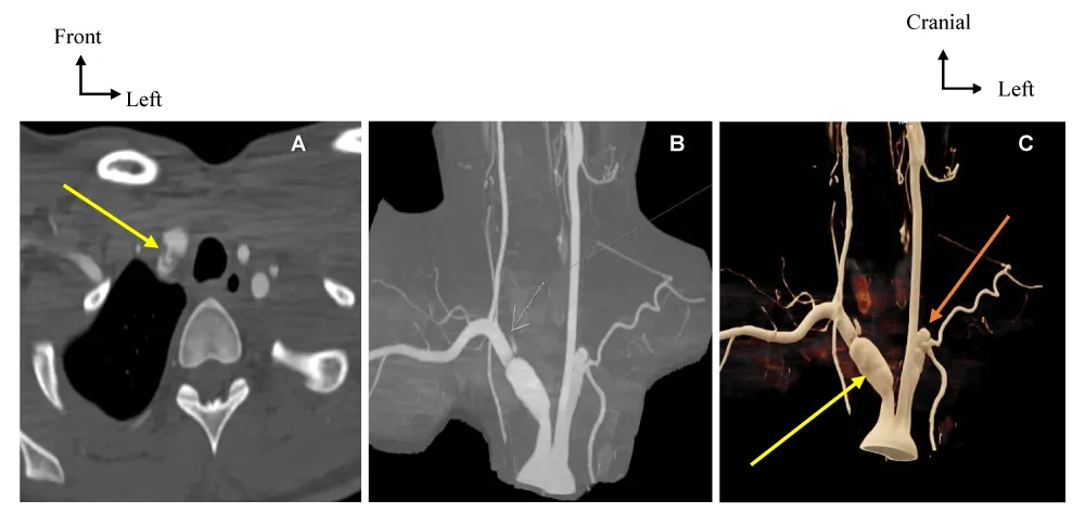
- a dissection of the innominate artery extending to its bifurcation, measuring 16,2 mm in anteroposterior diameter, without retrograde extension into the ascending aorta or the aortic arch (Figures 3A, 3B and 3C)
- a thrombotic occlusion of the right internal carotid artery (Figures 3B and 3C);
- stenosis of the left vertebral artery;
- occlusion of the left subclavian artery (Figures 3B and 3C).
The antithrombotic treatment was discontinued, and the hemothorax was drained again, which cleared the left pleural cavity.
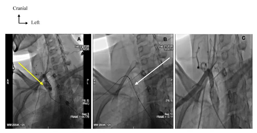
In view of the established cerebral ischemia arising from multiple trauma, an angiography was indicated with a view to endovascular treatment and was carried out on 27 February 2026. The endovascular procedure was performed under local anesthesia combined with light sedation. A retrograde puncture of the right common femoral artery was performed, with a 6 French introducer sheath inserted. Following the injection of 3,500 IU of sodium heparin, a Terumo Stiff 0.035×260 guidewire was inserted, followed by catheterization of the innominate artery and the right subclavian artery using a hydrophilic Cobra catheter. Intraoperative angiography confirmed the lesions identified on the CT angiography, as well as an extension of the dissection at the origin of the right subclavian artery (Figure 4A), with right vertébro-subclavian shunting. We then proceeded to place and deploy a Bentley 7x37x120 covered stent at the level of the innominate artery, extending it by a few millimetres into the right subclavian artery (Figure 4B), which eliminated the entry point and the intimal flap, as well as improving blood flow in the right vertebral artery (Figure 4C). However, the right internal carotid artery remained occluded. Bilateral cerebral arteriography was performed with satisfactory results. The dose of contrast medium used was 70 mL. The procedure concluded with the removal of the angioplasty equipment, followed by the application of a compression dressing. We did not use a cerebral protection filter system.
Discussion
Blunt thoracic outlet arterial injuries (BTOAI), together with blunt traumatic cerebrovascular injuries (BCVI), are relatively rare but potentially severe complications of high-velocity trauma. BTOAI involves damage to the innominate artery, the subclavian and axillary arteries, whilst BCVI involves damage to the carotid and vertebral arteries [4,5]. Their incidence is low, estimated at between 1% and 3% of all closed injuries [2,5]. However, this incidence may be underestimated due to initially asymptomatic clinical presentations, which are often masked by craniofacial and thoracic injuries [6]. BTOAI and BCVI are most often caused by an indirect mechanism [7], in which the arteries of the neck or thoracic passage may undergo longitudinal stretching, torsion, or shearing in relation to the cervical spine or the sternoclavicular joints, leading to parietal lesions [3,8,9]. In our context, the mobility difficulties faced by people due to road traffic could explain the proliferation and recurrent use of two-wheeled vehicles by most workers in the informal sector, with no gender disparity. Furthermore, the pathophysiological mechanisms underlying cerebral ischemia in these lesions involve the formation of a thrombus within the vascular lumen following activation of the coagulation system by endothelial rupture, with the potential for distal emboli on the one hand; and the extension of the intramural hematoma, compressing the vascular lumen and leading to severe stenosis or occlusion [1,3,7]. The most obvious hypothesis in our case is that of an indirect impact to the thoracic-cervical region, involving a combination of these complex pathophysiological mechanisms as the cause of the cerebral ischemic manifestations [2,10].
For the arteries of the thoracic canal, the most common clinical presentation is ischemia of the limb on the affected side, accompanied by hypotension in the thoracic limbs, resulting in a gradient of 10 mmHg or more, mediastinal widening, and ischemic steal syndrome distal to the thrombosis [5,10]. Carotid artery involvement is suspected in the presence of the classic triad of headache, Claude-Bernard-Horner syndrome, and the onset of neurological deficit (ischemic stroke) [3]. However, it should be noted that in around 80% of patients with traumatic cerebrovascular injuries, there is a latency period of varying duration between the time of the injury and the onset of the first neurological symptoms. This period can last an average of 72 hours [2,11]. In our case, the latency period was 96 hours. This latency period could be explained by a compensatory mechanism of cerebral perfusion via the circle of Willis and the self-regulatory capacity of cerebral circulation. Furthermore, the fact that therapeutic efforts were initially focused on pleuropulmonary and oedematous cerebral lesions contributed to a delay in diagnosis. No signs of ischemia of the upper limbs were observed, despite stenosis of the left subclavian artery and a pressure gradient of 10 mmHg. This could be explained by extensive collateral circulation supplying blood to the left upper limb and the presence of right vertébro-subclavian shunting. With regard to the severity of BTOAI and BCVI, the Denver scale (Table 1) is the most widely used. It defines five grades of increasing severity [7]. In our case, the CT findings allowed the severity of the arterial involvement to be classified as grade II for the innominate artery, grade IV for the right internal carotid artery, and grade I for the left subclavian artery. The management of these injuries can be complex [3]. In the acute phase, there is now a consensus on the administration of antithrombotic therapy for a period of 3 to 6 months [2,7]. However, the majority of patients with traumatic cerebral vascular injury have concomitant multisystemic injuries which initially contraindicate the initiation of antithrombotic treatment [6]. The risk-benefit ratio (stroke prevention versus the risk of hemorrhage) must be assessed on a case-by-case basis for each patient [12]. Endovascular surgery using minimally invasive techniques is a treatment option, particularly in patients with multiple trauma who are unstable and unable to undergo a cervico-sternotomy or receive heparin therapy due to severe head or chest injuries that are likely to bleed [7,8]. However, this approach has given rise to some controversy. Early studies suggested the use of stents for grade II lesions with significant luminal narrowing, as well as for grade III lesions, in order to reduce the risk of embolism and rupture by decreasing blood flow within the pseudoaneurysm. Although initial results were favourable, more recent studies have highlighted a potentially high risk of complications due to in-stent thrombosis, particularly in the absence of antiplatelet therapy, and suggest that stenting should only be considered in cases where lesions are persistent or worsening. Consequently, the guidelines of the Eastern Association for the Surgery of Trauma (EAST) recommend against the use of routine stenting as an adjunct to antiplatelet therapy in adult patients with Grade II or III BCVIs to reduce the risk of stroke [13]. In our patient, the presence of a recurrent left-sided hemothorax that contraindicates the continuation of the antithrombotic treatment initially started upon the discovery of the ischaemic stroke initial antithrombotic treatment, the presence of an occlusion in two of the four vascular axes supplying the brain, with a right vertébro-subclavian shunting, indicated endovascular treatment involving the placement of a covered stent. This endovascular procedure was carried out once the hemothorax had resolved. This treatment improved cerebral perfusion and limited the extent of the innominate artery dissection. The remainder of the management consisted of rehabilitation through motor physiotherapy, and the antithrombotic treatment was gradually reintroduced. Post-traumatic dissection of the innominate artery is associated with significant morbidity and mortality, at around 6.8% and 3%, respectively [3]. Our patient was reviewed six months later, reporting good functional recovery from the motor deficit, with segmental muscle strength improving from 0/5 to 3/5 on the left side of the body, as well as a gradual return to walking. It should nevertheless be noted that rehabilitation and spontaneous recovery may have contributed to this outcome.
Conclusion
BTOAI and BCVI are rare, but require early diagnosis due to the significant risk of ischemic stroke. Their management relies on antithrombotic therapy in the acute phase. Although there is some controversy surrounding endovascular treatment, the variety of treatment options in the context of multisystemic lesions means that the therapeutic approach must be discussed on a case-by-case basis.
Acknowledgements
Literature review: Mohamed Diane, Kouadio Ismaël Nguessan, Alassane Binate, Kouame Samuel Abro
Writing of the article: Mohamed Diane, Zole Cedrick Doh, Assoumou Lucien Asseke
Images: Mohamed Diane, Kesse Marc-Antoine Brou
Revision and correction: Kwadjau Anderson Amani, Afif Ghassani, Kouakou Gregoire Ayegnon
Surgical management of the patient: Afif Ghassani, Mohamed Diane, Kwadjau Anderson Amani, Raphaël Ouede, Jean Calaire Degre, Ibrahim Junior Yeo, Koutoua Eric Katche, Dominique Sarra Arriko, Jeanne Iris Degni, Ahou Sarrah Carrelle Assie, Yoboua Aimé Kirioua-Kamenan, Kouassi Antonin Souaga
Anesthesia and reanimation of the patient: Zole Cedrick Doh.
Ethics
The patient’s informed consent was obtained for the publication of this clinical case report, and for the use and dissemination of the clinical images. The information collected has been treated as confidential and used strictly for scientific purposes.
Institutional approval was obtained.
- Bouzat P, Payen JF. Lésions traumatiques des vaisseaux du cou. Congrès SFAR. Société Française d'Anesthésie et de Réanimation; 2013. Available from: https://sfar.org/search/Lésions
- Stone DK, Viswanathan VT, Wilson CA. Management of blunt cerebrovascular injury. Curr Neurol Neurosci Rep. 2018;18(98):1-9. Available from:https://doi.org/10.1007/s11910-018-0906-7
- Guillon B, Desal H. Dissections artérielles cervicales: modalités diagnostiques et thérapeutiques. mt Cardio. 2005;1(4):364-73.
- McCullough MA, Cairns AL, Shin J, Appelbaum RD, Couture DE, Nunn AM, et al. Above the clavicle: a simplified screening method for asymptomatic blunt cerebral vascular injury. Am Surg. 2023;89(1):79-83. Available from:https://doi.org/10.1177/00031348211011141
- Shalhub S, Starnes BW, Hatsukami TS, Karmy-Jones R, Tran NT. Repair of blunt thoracic outlet arterial injuries: an evolution from open to endovascular approach. J Trauma Acute Care Surg. 2011;71(5):E114-21. https://doi.org/10.1097/TA.0b013e31822cf359
- Murphy PB, Severance S, Holler E, Menard L, Savage S, Zarzaur BL. Treatment of asymptomatic blunt cerebrovascular injury (BCVI): a systematic review. Trauma Surg Acute Care Open. 2021;6(1):e000668. Available from:https://doi.org/10.1136/tsaco-2020-000668
- Rutman AM, Vranic JE, Mossa-Basha M. Imaging and management of blunt cerebrovascular injury. Radiographics. 2018;38(2):542-63. Available from: https://doi.org/10.1148/rg.2018170140
- Ben Omrane S, Ben Hammamia M, Ben Mrad M, Kaouel K, Daoued Z, Khayati A. Dissection traumatique du tronc artériel brachiocéphalique. À propos d'un cas. J Mal Vasc. 2014;39(1):73-6. Available from: https://doi.org/10.1016/j.jmv.2013.11.005
- Huang CL, Kao HL. Endovascular management of post-traumatic innominate artery transection with pseudo-aneurysm formation. Catheter Cardiovasc Interv. 2008;72(4):569-72. Available from: https://doi.org/10.1002/ccd.21660
- Hirose H, Gill IS. Blunt injury of the innominate artery: a case report and review of literature. Ann Thorac Cardiovasc Surg. 2004;10(4):218-23.
- Bruns BR, Tesoriero R, Kufera J, Sliker C, Laser A, Scalea TM, et al. Blunt cerebrovascular injury screening guidelines: what are we willing to miss? J Trauma Acute Care Surg. 2014;76(3):691-5. Available from:https://doi.org/10.1097/TA.0b013e3182ab1b4d
- McNutt MK, Kale AC, Kitagawa RS, Turkmani AH, Fields DW, Baraniuk S, et al. Management of blunt cerebrovascular injury (BCVI) in the multisystem injury patient with contraindications to immediate anti-thrombotic therapy. Injury. 2018;49(1):67-74. Available from:https://doi.org/10.1016/j.injury.2017.07.036
- Kim DY, Biffl W, Bokhari F, Brakenridge S, Chao E, Claridge JA, et al. Evaluation and management of blunt cerebrovascular injury: a practice management guideline from the Eastern Association for the Surgery of Trauma. J Trauma Acute Care Surg. 2020;88(6):875-87. Available from:https://doi.org/10.1097/TA.0000000000002668


 Save to Mendeley
Article Alerts
Subscribe to our articles alerts and stay tuned.
 This work is licensed under a Creative Commons Attribution 4.0 International License.