International Journal of Dermatology and Clinical Research
A rare case report of piloleiomyomas
Amar Uttamrao Surjushe1*, Anand Saraswat2, Vaishali Atram3 and Neha Mundada4
2Assistant Professor, Department of Dermatology, Venereology and Leprosy, Shri Vasantrao Naik Government Medical College, Yavatmal, Maharashtra, India
3Registrar, Department of Dermatology, Venereology and Leprosy, Shri Vasantrao Naik Government Medical College, Yavatmal, Maharashtra, India
4Dermatologist Skin Clinic, Department of Dermatology, Venereology and Leprosy, Shri Vasantrao Naik Government Medical College, Yavatmal, Maharashtra, India
Cite this as
Surjushe AU, Saraswat A, Atram V, Mundada N (2022) A rare case report of piloleiomyomas. Int J Dermatol Clin Res 8(1): 005-006. DOI: 10.17352/2455-8605.000043Copyright
© 2022 Surjushe AU, et al. This is an open-access article distributed under the terms of the Creative Commons Attribution License, which permits unrestricted use, distribution, and reproduction in any medium, provided the original author and source are credited.Leiomyoma is a rare benign tumor of smooth muscle. In the word leiomyomas, “Leio” means “smooth”, “myo” means ‘muscle’, and “oma” means ‘tumor’. It may arise from smooth muscles of the skin, uterus, bladder, urethra. Depending upon the origin of skin smooth muscles cutaneous leiomyomas are of three main types: 1. Piloleiomyoma or leiomyoma cutis or pilar leiomyoma derived from arrector pili muscle; 2. Genital leiomyoma or dartoic myoma is derived from smooth muscle of the scrotum, vulva, and areola of the nipple, and 3. Angioleiomyoma is derived from the media of blood vessels. We report one such rare case of multiple piloleiomyomas.
Case history
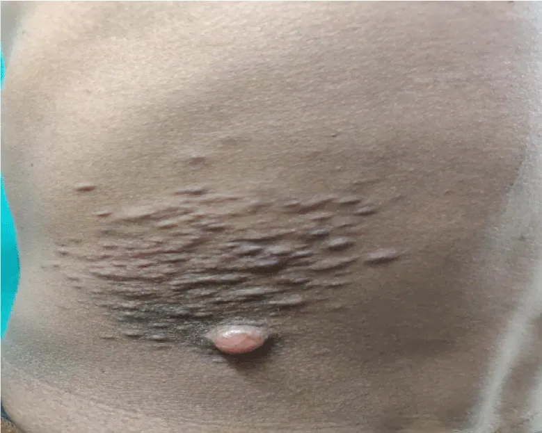
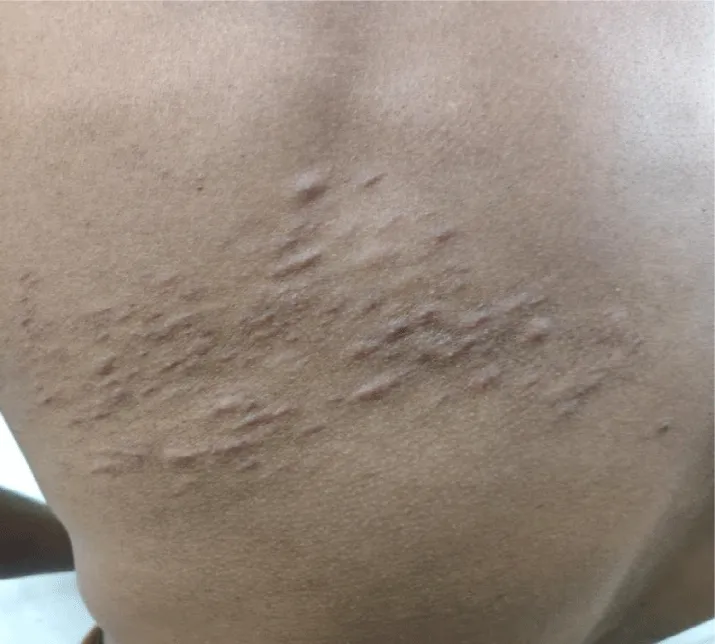
A 38 years-old-male patient presented with multiple raised lesions over the left side of the lower back, upper back, right arm, and right shoulder for 10 years. There was a history of pain in lesions aggravated by touching. There was no history of antecedent trauma and no history of ulceration, discharge or bleeding from the lesions. No history is suggestive of any other systemic involvement. He denied any history of similar complaints in the family or first-degree relatives. His birth and development history was normal. Cutaneous examination revealed multiple skin-colored papules and nodules on the right shoulder arranged in clusters. Multiple skin-colored papules and nodules coalescing to form plaque were present over the left side of the upper and lower back (Figure 1a,1b). Deep dermal tenderness was present. A provisional diagnosis of Leiomyoma cutis or Piloleiomyoma was cosidered with differential diagnosis of Neurofiroma, Eccrine spiradenoma, Dermatofibroma, Angiolipoma [1].
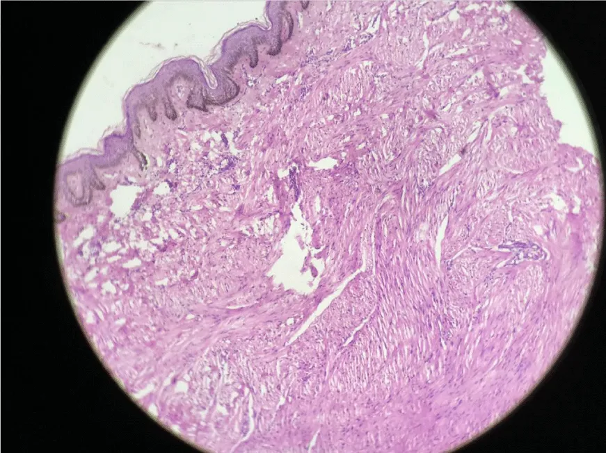
All routine investigations including haemogram, blood sugar, liver function, and renal function tests were within normal limits. Skin excision biopsy from a nodule on the back shows normal epidermis while the dermis showed circumscribed and non-encapsulated tumor composed of bundles of smooth muscle cells in an interlacing pattern. Cells have abundant eosinophilic cytoplasm with plump to spindle nuclei with blunt ends suggestive of benign mesenchymal tumor leiomyoma. There were areas of nuclear palisading seen (Figure 2). The patient was symptomatically treated with tablet Nifedipine 10 mg TDS for pain and referred to a plastic surgeon in view of multiple leiomyomas for further management.
Discussion
Piloleiomyomas are a type of cutaneous leiomyoma and are benign tumors arising from the pilomotor muscle. The first case of multiple leiomyomas arising from the skin near the areola was reported by Rudolf Virchow in 1854 in a 32-year-old male [2]. The exact incidence of these uncommon neoplasms is not known. Uterine leiomyomas are the most common pelvic neoplasm and the second most commonest location of leiomyomas after the uterus is the skin. Cutaneous leiomyomas account for 75% of extra-uterine leiomyomas. El Salvador reported the incidence of piloleiomyoma more than that of genital leiomyoma while Yokoyama R et al reported genital leiomyoma more than that of piloleiomyoma [3,4]. They usually occur in early adult life but can occur at any age from birth. They have been reported in identical twins, siblings and also have a familial background. Patients with multiple piloleiomyomas have a germline mutation of the fumarate hydratase gene located on chromosome 1q42.3-43 and are frequently associated with uterine leiomyomatosis [5]. They occur in both sexes equally.
Clinically, piloleiomyoma presents as dermal reddish-brown, brown, pink, or skin-colored papules or nodules; either as solitary or multiple lesions. When multiple they are distributed in linear, clustered, along Blaschko’s lines, or widespread. The most common sites of involvement are the extremities and trunk especially the shoulder as seen in our case. The characteristic of most of the piloleiomyomas is pain which is either spontaneous or can be provoked by touch, cold, or by emotional disturbances. The exact mechanism of pain is not known; but the contraction of smooth muscles, compression of nearby nerves, or an increased number of nerve bundles are possible proposed etiologies [6]. Histopathologicaly piloleiomyomas show tumors composed of numerous bundles of eosinophilic spindle-shaped cells with central cigar-shaped nuclei. Special staining with Masson’s trichrome stains smooth muscles red and immunohistochemistry can be done using antibodies against smooth muscle actin and desmin. Mitotic figures are rare in piloleiomyomas [7]. Treatment includes surgical excision of solitary or limited piloleiomyomas. Other treatment modalities include the use of calcium channel blockers, gabapentin, and the use of Co2 laser ablation which may provide relief from pain [8]. The main aim of presenting this case is because of its rarity.
- Burns T, Breathnach S, Cox N, Griffiths C (2010) Rook's Textbook of Dermatology. Eight Edition. Wiley-Blackwell Publisher 1: 4432. Link: https://bit.ly/3iO1iNy
- Stout AP (1937) Solitary cutaneous and subcutaneous leiomyoma. Am J Cancer 29: 435-469. Link: https://bit.ly/3LnmUMX
- Orellana-Diaz O, Hernandez-Perez E (1983) Leiomyoma cutis and leiomyosarcoma; a 10-year study and a short review. J Dermatol Surg Oncol 9: 283-287. Link: https://bit.ly/38cyCM6
- Yokoyama R, Hashimoto H, Daimaru Y, Enjoji M (1987) Superficial leiomyomata. A clinicopathologic study of 34 cases. Acta Pathol Jpn 37: 1415-1422. Link: https://bit.ly/3uBsXGV
- Alam NA, Bevan S, Churchman M, Barker K, Barclay E, et al. (2001) Localization of a gene (MCULI) for multiple cutaneous leiomyomata and uterine fibroids to chromosome 1q42.3-43. Am J Hum Genet 68: 1264-1269. Link: https://bit.ly/3uCGkqb
- Alam NA, Barclay E, Rowan AJ, Tyrer JP, Calonje E, et al. (2005) Clinical features of multiple cutaneous and uterine leiomyomatosis: an underdiagnosed tumor syndrome. Arch Dermatol 141: 199–206 . Link: https://bit.ly/3qLbevs
- Raj S, Calonje E, Kraus M, Kavanagh G, Newman PL, et al. (1997) cutaneous pilar leiomyoma: clinicopathologic analysis of 53 lesions in 45 patients. Am J Dermatopathol 19: 2-9. Link: https://bit.ly/3NwIf8x
- Christenson LJ, Smith K, Arpey CJ (2000) Treatment of multiple cutaneous leiomyomata with CO2 laser ablation. Dermatol Surg 26: 319-322. Link: https://bit.ly/3DuibpV
Article Alerts
Subscribe to our articles alerts and stay tuned.
 This work is licensed under a Creative Commons Attribution 4.0 International License.


 Save to Mendeley
