International Journal of Dermatology and Clinical Research
Incontinentia Pigmenti: an unusual and fast presentation
Eduardo Duarte Sobrosa*, Bruno Evangelista de Toledo* and Ana Daniela Izoton de Sadovsky*
Bruno Evangelista de Toledo, Federal University of Espírito Santo, Brazil, E-mail: [email protected]
Ana Daniela Izoton de Sadovsky, Federal University of Espírito Santo, Brazil, E-mail: [email protected]
Cite this as
Sobrosa ED, de Toledo BE, de Sadovsky ADI (2018) Incontinentia Pigmenti: an unusual and fast presentation. Int J Dermatol Clin Res 4(1): 011-012. DOI: 10.17352/2455-8605.000028Incontinentia pigmenti is a rare X-linked neuroectodermal dysplasia estimated to occur in approximately 1:50,000 births. It’s first clinical manifestations are unique cutaneous lesions comprehending four different stages that emerge throughout the first year of life and persist until adolescence. One third of these patients develop central nervous system and ocular manifestations, causing great disability. The authors report the case of a patient with vesicular cutaneous lesions within 5 days after birth, with a fast and uncommon evolution. Although rare, the early identification of this disorder can prompt thorough investigation of associated comorbidities and adequate familiar and medical assistance.
Abreviation
IP: Incontinentia Pigmenti
Introduction
Incontinentia Pigmenti (IP) is a dominant, X-linked neurocutaneous dysfunction with presumed male lethality. It is usually diagnosed in female newborns based on dermatological changes, which divide the disease into four phases, occurring concomitantly or sequentially, usually associated with teeth, nails and hair abnormalities. Despite being an uncommon condition, one third of the patients develops ocular and/or neural symptoms/signs, which are the mais causes of disability in these patients.
Case Description
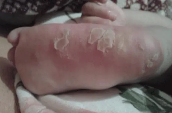
M.K.D.N., 4 years, patient of HUCAM (Vitória-ES, Brazil). She was born at term, C-section, without complications. In the first day of life, presented alopecia in the parietal region and generalized hyperemic patches. She was discharged after 48 hours. On day 4, new lesions appeared, and increased hyperemia and growth of the old ones. On the 5th day, vesicles appeared mainly in limbs and extremities. Those lesions affected all skin until the 4th month. In the 5th month, verrucous plaques developed at the vesicle sites. The lesions coexisted up to the 1st year. After healing, the skin lesions became hyperpigmented, bluish-gray, with the “appearance of Chinese figures”. At 3 years old, there were already hypopigmented-scarring macules. Vesicles always result from fever and/or when infection occurs. There were no neurologic, ocular or ungueal manifestations. The clinical suspicion and the classic evolution of the skin lesions allowed the diagnosis without confirmatory histopathology, even though the presentation was faster than usual. It presented with a milder and faster evolution, contributing to a better outcome. Also noteworthy to mention the consequences of a appropriate familial care in the favorable evolution of this patient.
Discussion and Conclusion
Incontinentia pigmenti(IP) or Bloch-Sulzberger syndrome is a dominant neurocutaneous disorder caused by a mutation in the IKBKG gene, located in the q28 portion of the X chromosome [1-4], and is associated with several immune, inflammatory and cellular apoptosis-related pathways [1]. Cases of the disease in males have been associated with somatic mosaicism and Klinefelter’s syndrome, usually lethal in male foetuses [1,3,4].
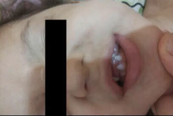
The main clinical manifestation of IP, and the only major criterion for its diagnosis, is cutaneous involvement characterized by four different stages [1,3], (Stage 1) vesicular or inflammatory, characterized by papules, vesicles and pustules that accompany the distribution of Blaschko lines, especially in extremities, presenting at birth or within the first 2 weeks of life up to 4 months of age ; (Stage 2) verrucous, identified by verrucous plaques and papules, between 2 and 6 weeks of age, concomitantly or not to the vesicular stage; (Stage 3) hyperpigmented, characterized by linear and swirling brownish macules, appearing in early childhood and disappears progressively during adolescence, and may persist into adulthood; (Stage 4) atrophic or hypopigmented, identified by atrophic and hypopigmented macules, especially at the extremities, beginning in adolescence and persisting during adulthood [2-4] (Figures 1-7).
This patient evolved with all stages of the cutaneous manifestation, but in an accelerated and uncommon way, presenting characteristic lesions of the fourth stage at 3 years of age.
Other manifestations described in IP include alopecia (28% to 38% of patients), dental modifications (17% to 34%), including hypodontia, microdontia, delayed eruption and abnormal forms, as well as ocular manifestations (17%) (1,3,4). Ocular lesions are highly incapacitating, secondary to ischemia of the retina and persist throughout the life, requiring early investigation and treatment [3,5].
Central nervous system abnormalities are the major cause of disability in cases of IP, occurring in 30% of cases [1] and are usually severe (62%) [3,6]. The most frequent are mental retardation, motor abnormalities (plegias) and epilepsy [6].
We emphasize the importance of recognizing this rare genetic syndrome to stimulate appropriate family and medical care, providing a better quality of life and better prognosis to these patients.
- Levy ML (2018) Incontinentia pigmenti: Link: https://goo.gl/CaZNMV
- Poziomczyk CS, Recuero JK, Bringhenti L, Maria FD, Campos CW, et al. (2014) Incontinentia pigmenti. Anais brasileiros de dermatologia 89: 26-36. Link: https://goo.gl/riC2ag
- Greene-Roethke C. (2017) Incontinentia Pigmenti: A Summary Review of This Rare Ectodermal Dysplasia with Neurologic Manifestations, Including Treatment Protocols. J Pediatr Health Care 31: e45-e52. Link: https://goo.gl/5e3FNN
- Minic S, Trpinac D, Obradovic M (2014) Incontinentia pigmenti diagnostic criteria update. Clin Genet 85: 536-542. Link: https://goo.gl/3XFfwm
- Swinney CC, Han DP, Karth PA (2015) Incontinentia Pigmenti: A Comprehensive Review and Update. Ophthalmic Surg Lasers Imaging Retina 46: 650-657. Link: https://goo.gl/3PdK5K
- Narayanan MJ, Rangasamy S, Narayanan V (2015) Incontinentia pigmenti (Bloch-Sulzberger syndrome). Handb Clin Neurol ss132: 271-280. Link: https://goo.gl/pHBpwn
Article Alerts
Subscribe to our articles alerts and stay tuned.
 This work is licensed under a Creative Commons Attribution 4.0 International License.






 Save to Mendeley
